Title
[症例報告]左レンズ核線状体動脈から発生した動静脈奇
形,動脈瘤合併の1手術例
Author(s)
久田, 均; 六川, 二郎; 宮城, 航一; 高良, 英一; 中田, 宗朝; 金
城, 則雄; 堀川, 恭偉; 銘苅, 晋
Citation
琉球大学医学会雑誌 : 医学部紀要 = Ryukyu medical
journal, 8(2): 81-91
Issue Date
1985
URL
http://hdl.handle.net/20.500.12001/2361
Ryukyu Med. J., 8(2): 81-91, 1985.
左レンズ核線状体動脈から発生した
動静脈奇形・動脈癖合併の1手術例
久田 均 六川 二郎 中田 宗朝 金城 則雄 宮城 航- 高良 英一 堀川 恭偉 銘苅 晋 琉球大学医学部脳神経外科学教室 は じ め に 脳内出血で発症した左大脳基底核部の動静脈 奇形(以下AVMと略)と動脈癖の合併した1 治験例を報告する。基底核部のAVM 動脈癌 合併例は稀であり,渉猟した限りでは大田ら の報告した1例のみであるJ) AVMに合併 しておこる脳動脈痛の発生メカニズムと手術適 応について,若干の文献的考察を加えて報告す る. 症 例 症 例:38才 男性 主 訴:右不全片麻埠 既往歴:心疾患・高血圧の既往はない. 家族歴:特記することはない 現病歴:昭和58年12月3日,突然の意識障 害・右完全片麻痔・全失語で発症した.他医に おいて脳内出血の診断のもとに保存的治療を受 けた. 1週間後には簡単な単語の理解が可能と なり, 1ヵ月後には独歩可能となったが,なお 右不全片麻樺および失語症が残ったので,昭和 59年5月リ-ビリテ-ションを目的として他病 院へ転院した.そのさいCT-scan で左大脳基 底核部にcontrast mediumでenhanceされる 病変部が認められたので,精査を目的として当 料-紹介,入院となった. 入院時現症:体格,栄養は中等度.右利き. 右不全片麻樺.言語了解,発語,書字は中等度 障害され読字はおおむね良好であり,既往歴か らみて全失語からの回復過程にあるものと考え られた.その他,理学的な異常は認められなか った. 臨床検査所見:特に異常はない. 頭部単純撮影:特に異常所見はない. 頭部CT-scan:単純CT-scanでは,左大脳 基底核部に低吸収城があり左シルヴィウス裂が 関大し,脳内出血の陳旧性変化と判断した Fig. 1左).造影CT-scanでは低吸収城の内側すな わち内包の外側に9mmX18mm大の楕円形の高吸 収城が造影された(Fig. 1右). 左額動脈撮影(昭和59年5月21日施行, Fig. 2, 3 :左大脳基底核部に,レンズ核線状体動派(lenticulostriate arteries, LSA )の一枝か ら流入し basal vein, anastomotic mesence-phahc vein, internal cerebral vein, insular
vein,の4本-流出する10mmX 7mmのnidusを もつAVMを認めた(模式図:Fig. 4).また, nidusの外下方に接してLSAを母動脈とする直 径約4mmの動脈痛の存在を認めた.その後,鍋 2ヵ月間に計3度左頚動脈撮影をおこなってこ の合併の確認につとめた AVMの性状,動脈 痛の大きさおよび両者の位置関係に変化は認め られなかった. 手術所見:く昭和59年`7r月24日, 1左前頭側頭関 頭により根治術を行った.島回皮質の約1cmの 深さで古い血腫腔に達し,まずその直下に動脈 痛が,ついで動脈癌の内側にAVMのnidusが それぞれ独立して見られた.動脈癌はネック・ クリップした後摘出し, AVMのnidusは剥離 操作中出血したため焼灼凝固した. 病理組織学的所見(Fig. 5; :摘出した動脈癌 の内膜は肥厚して硝子様変性を示し,さらに内弾 性板は断裂している.中膜は平滑筋細胞が梢失し て線維化しており,外膜との境界は不明瞭である.
82 久田 均 ほか
術後経過:手術後一過性に片麻埠・失語が増 歩可能となり,失語もほぼ術前のレベルに回復 要したものの合併症なく経過し, 1ヵ月後には した.術後の左額動脈撮影では,動脈癌 AVM 顔面を含む右半身の軽い麻棒は残したものの独 はもはや造影されなかった(Fig. 6).
Fig. 1 Left: Plain CT-scan. A low density area is seen at the left putaminal region. Right: Contrasトenhanced CT-scan. An enhanced area locates lateral to the internal capsule.
レンズ核線状体動脈から発生したAVM
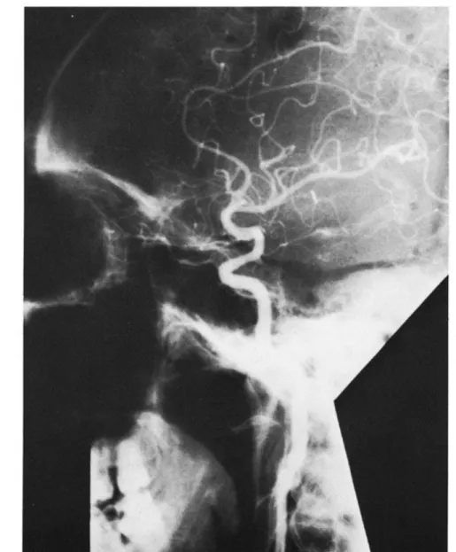
Fig.2A-PviewofleftCAG.ThereareanidusofAVM(A).feedingartery(詛)and J*-n*ォiォサ・,r^."/.^¥A_:j
久EQ 均 ほか
Fig. 3 Lateral viewof left CAG. The draining veins (
artery (
) are clearly seen. A feeding one of the lenticulostriate arteries) runs parallelly along with the middle
レンズ核線状体動脈から発生したAVM 85
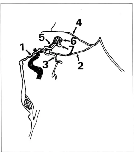
Fig. 4 Schematic drawing of left CAG. 1: insular vein, 2: basal vein, 3: anastomotic me-sencephalic vein, 4: internal cerebral vein, 5: feeding artery, 6: nidus of AVM, 7: aneurysm.
86 久田 均 ほか
Fig. 5 Histology of aneurysm. (elastic tissue van Gieson stain, × 25).The imtimaof aneurysm (A) is hypertrophic and of hyalinedegeneration. The internal elastic lamina( B) is fragment-ed, and smooth muscle celles of the media (C ) disappear.
レンズ核線状体動脈から発生したAVM
88 久田
考 療
AVMに動脈痛が合併する頻度は, Paterson and McKissockによると110例中4例(3.6%),2> Perret and Nishioka によると490例中37例 (7.6%),3) Suzukiらによると140例中9例(6.4 %)で4)さ程高いものではないf.しかし, LSA 末梢における動脈癌を合併したAVMの症例は 大田らが報告した1例のみである1).もともと, 真性脳動脈癌自身が単独で大脳基底核部穿通枝 動脈末梢に発生したという報告も見られないヲ) 私どもの症例では,病理組織学的に, (参内弾 性板の断裂, ②中模筋層の綿維化, ③内膜の硝 子様変性・肥厚が認められた. (9②は真性嚢状 動脈痛に認められる所見であり6)7) ③はAVMに も認められる所見である竺)9)すなわち AVMで は病理組織学的に動脈でも静脈でもない硝子化 した血管が認められ(McCormick9'),また硝子 化した内膜のnoduleが内腔へ突出した像がし ばしば認められる(Bailey8').本症例の動脈癌 は,断裂はしているが内弾性板を有し,また内 膜・中膜・外膜の3層構造を有して明らかな動 脈の形態をとっていて AVMの1部であると は考えられない.また,手術所見上も動脈癌と AVMはそれぞれ独立してLSAから生じてい た.以上の事実から,本症例の動脈痛は真性嚢 状動脈癌であると判断した. AVM・動脈癌合併例における動脈癌の発生メ カニズムについては, 1)先天性血管異常説(Con-genital)10' 2) AVMによる血流量増大説11) 3)同時発生説(coincident)がある王2) Arietti らは,頭蓋内AVM 脳動脈癖・脳血管のびま ん性の異常・腹部大動脈痛を合併した特異なア ルコール中毒患者1例を報告し,その発生は congenitalなものであろうとした王o) Shenkin らは AVMの流入動脈に発生した動脈癌が AVM摘出後著しく縮小した例をあげ,その発 生にAVMによる血流量の増大が大きく関与し ていたであろうと推論しておりさ1)現在でも多 くの支持を得ている王)4)他方, Boyd-Wilsonは, 一般人剖検群における脳動脈癌発生率と AVM 症例における脳動脈痛発生率を比較してその間 均 ほか に大きな差がないことから AVM症例におけ る脳動脈痛の合併はcoincidentであるとした12) しかし,流入動脈に動脈痛が発生した症例では 血行動感の変化がその発生に何らかの影響を与 えるという可能性を否定してはいない.本症例 では,元来真性嚢状動脈癖が発生することはほ ぼないと考えられるLSA末梢に動脈痛が存在 しており,その発生にはAVMによる血流量の 増大が大きく関与したであろうと推察される.
Perret and Nishiokaによれば, AVMの再 出血率は23%であり,初回出血から2度目の出 血までの間隔は数日から20年以上とまちまちで ある.3)また,Forsterらによれば, 1度出血した 例ではその後の4年間に4例に1例は出血を生
じる危険があり,それに伴って症状も悪化する という王3)他方 Norlen and Olivecronaによれ ば,脳動脈癌の再出血率は20-50%であり,そ の死亡率は70-80%ときわめて高率である14) 本症例では,出血が脳動脈癌とAVMのどちら に由来するものであったかの判断はできなかっ たが,いずれからの出血にしろ再出血の危険性 は両病変によって倍増すると想定されるので, 手術に踏み切った. AVM 脳動脈痛合併例の治療に関する記載 は少ない号)ll)両病変のうちAVMが病態生理学 的に主役をなすと考えられるので,その治療は AVMに準ずるものと考えられる.基底核部を 含めて内包・脳幹部のAVMは手術適応はない という意見があり15)そのため手術以外に se-lective arterial embolizationや16)stereotac-tic radiosugeryが報告されている17)しかし, 基底核部のAVMに対しては良好な手術結果を 得たとする報告がある王8)金らは,脳深部領域に AVMが存在する場合,脳内血腫が存在しかつ 神経症候の重篤な場合手術適応があると報告し ている!9)本症例ではCT-scan上AVM 脳動脈 癖の外側に陳旧憧出血病変が存在し,さらに両 者が内包より外側で被殻部領域に存在すること から,手術侵襲とその結果は被殻出血における ものと同程度であろうと推測した. また, AVM 脳動脈壇合併例における手術 方法としては, ①AVMをまず治療する11)か,
レンズ核線状体動脈から発生したAVM ②両者を同時に治療する4)かの2つの対立意見 がある.当症例では,脳内出血の既往があり, その出血源がAVM 脳動脈癌のいずれからか 判定し難いこと.しかも流入動脈と母動脈が同 一であることから両者に対して同時に手術を行 った. 結 語 私どもは,左大脳基底核部のレンズ核線状体 動脈末梢に生じた動静脈奇形と動脈壇が合併し た1例で,関頭手術により両者をともに根治し えたので,文献的考察を加えて報告した.要点 は以下の如くである. 1.この部に脳動静脈奇形と動脈痛が合併し た症例は極めて稀である. 2.脳動脈癖の発生機序として,脳動静脈奇形 による血流量増大が大きく関与していたものと 推測した. 3.両者が大脳深部領域に存在する場合,内 包より外側であれば手術適応があると考える. 文 献 1)大田英則,伊藤善太郎,中島健二,深沢仁,上 村和夫:脳動静脈奇形に合併したレンズ核線状 体動脈の一例,脳神経, 8 : 181-185, 1980. 2) Paterson, J. H. and McKissock, W: A clinical
survey of intracranial angiomas with special reference to their mode of progression and surgical treatment : A report of 110 cases., Brain, 79 : 233-266, 1956.
3) Perret, G. and Nishioka, H∴ Report on the cooperative study of intracranial aneurysms and subarachnoid hemorrhage. Section VI. Arteriovenous malformations. An analysis of 545 cases of crani0-cerebral artenovenous malformations and fistulae reported to the cooperative study., J. Neurosurg., 25 : 467-490, 1966.
4) Suzuki, J. and Onuma, T. : Intracramal aneurysms associated with artenovenous
89
malformations., J. Neurosurg., 50 : 742-749, 1979.
5) Suzuki. J., Hori, S. and Sakurai, Y. Intracranial aneurysms in the neurosurgical clinics in Japan., J. Neurosurg., 35 : 34-39, 1971.
6)大根田玄寿:脳出血の病理,文考堂,東京,1974.
7) Sahs, A. L∴ Observations onthe pathology of saccular aneurysms., J. Neurosurg., 24 : 792-806, 1966.
Bailey, O. T. : The vascular component of congenital malformatons in the central nervous system., J. Neuropath.exp.Neurol., 20 : 170-184, 1961.
9) McCormick, W. F. : The pathology of
vascular ("arteriovenous") malformations., J, Neurosurg., 24 : 807-816, 1966.
Arieti, S. and Gray, E. W. : Progressive multiform angiosis. Association of a cerebral angioma, aneurysms and other vascular changes in the brain., Arch. Neurol. Psychiat, 51 : 182-189, 1944.
ll) Shenkin, H. A., Jenkins, F. and Kim, K. : Arteriovenous anomaly of the brain as-sociated with cerebral aneurysm. Case report., J. Neurosurg., 34 : 225-228, 1971. 12) Boyd-Wilson, J. S. : The association of
cerebral angiomas with intracranial aneur-ysms., J. Neurol. Neurosurg. Psychiat, 22 :
218-223, 1959.
13) Forster, D. M. C, Steiner, L. and Hakanson, S. : Arteriovenous malformations of the brain. A long-term clinical study., J.
Neur-osurg., 37 : 562-570, 1972.
14) Norlen, G. and Olivecrona, H. : The treat-ment of aneurysms of the circle of Willis., J. Neurosurg., 10 : 404-415, 1953.
15)小沼武英,鈴木二郎:脳動静脈奇形の外科的療 法, 88例の統計と手術成績,そのfollow-up,脳 外, 2:29-37,1974.
16) Luessenhop, A. J. and Presper, J. H. : Surgical embolization of cerebral arteriovenous
90 久田 均 ほか
malformations through internal carotid and vertebral arteries., J. Neurosurg., 42 : 443-451, 1975.
17) Backlund, E. -0. : Stereotactic Radiosugery in Intracranial Tumours and Vascular Mai-formations. : Advances and Technical Standards in Neurosugery. Vol. 6., Krayen-biihl, H., Springer-Verlag., Wien, New York,
1979.
Juh丘sz, J. : Surgical treatment of arter-iovenous angiomas localized in the corpus callosum, basal ganglia and near the brain stem., Acta. Neurochir, 40 : 83-101, 1978.
19)金是仁,斎藤勇,有竹康一,馬杉則彦,佐野圭 司: ParaventricularAVM,第四回脳卒中の外 科研究会講演集: 1-9, 1975.
91
Arteriovenous malformation combined with
an aneurysm arising from one of the
left medial lenticulostriate arteries.
A case report
Hitoshi Kuda, Jiro Mukawa, KOuichi Miyagi, Eiichi Takara, Munetomo Nakata, Norio Kinjyo,
Kyoi Horikawa and Susumu Mekaru
Department of Neurosurgery, University of the Ryukyus School of Medicine,
I Key words : Basal ganglia, Lenticulostriate arteries, Artenovenous malformation, Aneurysm
Abstract
A case of arteriovenous malformation combined with an aneurysm in the left basal ganglia is presented.
A 38-year-old male patient was admitted in our hospital because of right hemiparesis and aphasia. He had history of left putaminal hemorrhage six months prior to the admission, and had since been treated conservatively. Left carotid angiogram demonstrated an AVM combined with an aneurysm, both arising from one of the medial lenticulostriate arteries. In order to prevent rebleeding, both lesions were surgically operated. The nidus of AVM was electrically coagulated, and the aneurysm was resected at the neck. Histological examination revealed that the intima of aneurysm was hypertrophic associated with hyaline degeneration. The internal elastic lamina was fragmented,
and the smooth muscles of the media disappeared and were displaced by fibrosis. Postoperative angiogram showed no abnormal vessels any more.
It is suggested that increased blood flow might be the main factor to develop an aneurysm in such an AVM case. The hazard of rebleeding from such lesions is to indicate, in case possible, radical treatment of both lesions.