総
説
脳小血管病の臨床研究のトレンドと課題
―
1 型脳小血管病を中心に―
藥師寺祐介
1)*
要旨:脳小血管病(cerebral small vessel disease,以下 SVD と略記)とは,脳微細血管劣化に伴う効率的な 脳内微小循環・代謝・ネットワーク維持の困難な状態,及びそれらによる認知・身体機能低下状態を指す.高血 圧性細動脈障害を基盤とした 1 型 SVD は,白人に比べ東アジア人に多い.近年,脳小血管負荷を包括的に半定量 化するための取り組み,すなわち MRI マーカー評価に基づいた SVD スコア化が注目されている.この概念は「脳 小血管負荷に実用的な閾値はあるか?」という新たな疑問を生んだ.SVD 画像マーカーの自動解析手法の開発は この疑問を解き,1 型 SVD 患者への最適な介入法の発見につながるであろう. (臨床神経 2020;60:743-751)
Key words:脳小血管病,脳微小出血,total small vessel disease score,脳血管障害,認知症
はじめに 脳小血管病の概念は 2010 年以降に明確化され,脳卒中・ 認知症に強く関連することがわかってきた.関連する画像診 断技術の発達は今後も進み,病態解釈に基づく予防的介入が 可能となるのもそう遠くない将来であろう.しかし,脳小血 管病という用語は未だ広く認知されているとは言い難い.筆 者が察するに,脳小血管病が病理学的に不均一な小血管障害 の総称で,定義が曖昧であること,直接観察ができないこと, 脳卒中や認知症の一歩手前の(深刻でない)状態と捉えられ ていることなどがその理由と思われる.本稿では,その定義 を見直し,行われている研究の方向性と今後の課題を説いて いくが,疾患スペクトラムが広いため,各論部分では,生活 習慣病関連の common disease である 1 型脳小血管病を中心 に概説する. “脳小血管病の定義” 脳小血管は,小・細動脈,毛細血管,および小静脈を含み, 脳内で最も代謝の活発な大脳核や白質線維間のネットワーク 機能を最適に維持する臓器である1).“脳小血管病(cerebral small vessel disease,以下 SVD と略記)”という用語は,1980 年代に登場したが,多くの神経病理医は静脈に注目していな かったことに代表されるように2),病理学,臨床神経学,神 経放射線医学という異なる学術領域で,ニュアンスの違い, 誤解を引きずりながら使用されてきた.脳小血管は生前に直 接確認が困難であるため,SVD に付随する CT・MRI などの 神経画像上の脳病変を通じて臨床的意義の研究がなされてき た.このような背景から,神経放射線医学的には画像上の脳 病変自体が SVD と同義語として用いられていることがある. また,SVD 付随病変はラクナ,白質病変などの虚血メカニズ ムで発生するもののみと誤解されている場合もある.しかし, 磁化率強調 MRI の登場で検出可能となった脳微小出血 (cerebral microbleeds,以下 CMBs と略記)のような出血起源 のものも SVD 付随病変に含まれるようになった.以上の事項 を網羅すると,SVD とは「脳内の微細な動・静脈,及び毛細 血管が何らかの原因で障害され,脳内微細血管周囲で営まれ る適切な循環・代謝・神経ネットワーク維持が困難となる器 質的・機能的変化を伴う症候群」と定義できる.これらの破 綻による症候群の代表が脳卒中,認知症である. SVD の病理分類 Pantoni は SVD を病理学的に 6 型に分類することを提唱し た(Table 1)3). 1 型 SVD は細動脈硬化,もしくは加齢と血管リスク因子 (特に高血圧)に関連したもので,病理学変化として中膜の平 滑筋消失,線維性硝子物質の沈着,血管狭窄,フィブリノイ ド壊死,脂肪硝子変性,微小粥腫,微小動脈瘤,分節状動脈 破壊を伴う.随伴する虚血性変化にはラクナ病変やラクナ梗 *Corresponding author: 関西医科大学神経内科学講座〔〒 573-1010 大阪府枚方市新町 2 丁目 5 番 1 号〕 1) 関西医科大学神経内科学講座
(Received May 29, 2020; Accepted June 27, 2020; Published online in J-STAGE on October 27, 2020) doi: 10.5692/clinicalneurol.cn-001488
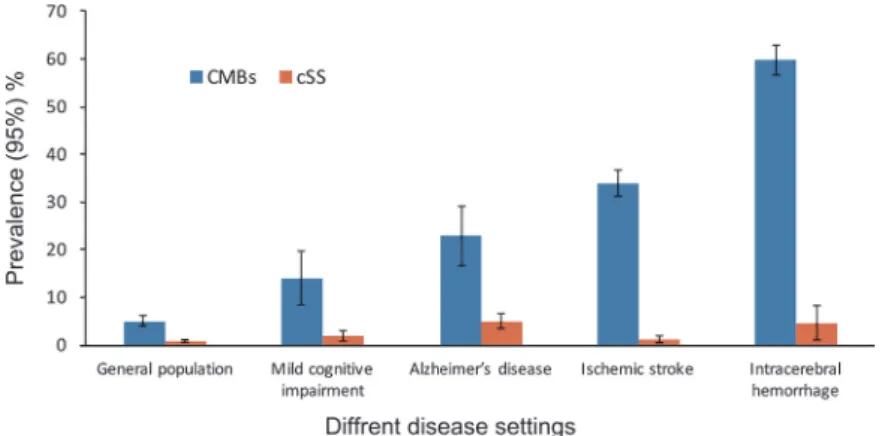
塞が,出血性変化には CMBs や脳出血がある.また白質血管 では中膜平滑筋細胞の増殖と脱落,中・外膜の膠原線維の増 生を特徴とする fibrohyalinosis が見られる.これは,血管弾 性の喪失による脳灌流圧の変化に対する血管反応性低下を介 して,白質血流低下による大脳白質病変の原因となる. 2 型 SVD は孤発性,もしくは遺伝性の脳アミロイド血管症 (cerebral amyloid angioapthy,以下 CAA と略記)で,髄膜や 大脳皮質の毛細血管,小・中サイズの動脈・細動脈の血管壁 への脳アミロイド蛋白沈着を特徴とする.CAA の病型は沈着 する脳アミロイド蛋白の種類で分類される.その種類には, アミロイド β(Aβ),シスタチン C,プリオン蛋白,ABri/ ADan,トランスサイレチン,ゲルゾリン,AL があるが,こ こでは頻度が最も高い “Aβ 沈着による CAA” を論じる.アル ツハイマー型認知症で見られる老人斑は Aβ42 が主だが,CAA での血管に沈着するものは Aβ40 が主である.CAA は侵され る血管サイズの範囲により,CAA1 型(5 μ~2 mm),CAA2 型(10 μ~2 mm)に区別される.すなわち,5 μ~10 μ の毛 細血管も障害されうるのは CAA1 型である.CAA1 型と 2 型 では,アポリポ蛋白 E の ε2 や ε4 アレルへの関連や4),血液 脳関門の透過性などが違うため5),異なる病理背景を有する と考えられている.Aβ 沈着は血管平滑筋の外側から始まり, 平滑筋細胞の変性を経て,最終的には中膜全体がアミロイド 線維で置換される.障害が高度になると,フィブリノイド壊 死,微小動脈瘤,中膜・外膜間に空隙が生じる double barrel appearance などの形態変化を生じ,出血源となる. 3 型 SVD は CAA を除いた遺伝性のもの,4 型 SVD は炎症 性・免疫介在性のもの,5 型は細・小静脈周囲の膠原線維の 増生を伴う血管硬化・閉塞を伴う venous collagenosis,6 型は その他(放射線治療関連血管症など)であるが,詳細は原著 を確認されたい3). 研究ツールとしての出血性 SVD ヒトの SVD 研究の進歩は,付随する画像所見(SVD 関連 病変)を捉える MRI 診断技術の向上によるところが大きい. SVD 関連病変の用語,定義に関しては STandards for ReportIng Vascular changes on nEuroimaging(STRIVE)として取りまと められおり6),最近の MRI を用いた SVD 研究では,この STRIVE に準拠して行うのが慣例である.これらを解釈する と,脳血管障害イベントとの時間的な関連(急性病変か否か), 発症機序(虚血,出血,その他)で分類することができる (Table 2).これらの中で,CMBs はその分布パターンで SVD 病理を推定することが可能な点で特徴的である.すなわち, 脳深部・テント下 CMBs(脳葉 CMBs との混合型も含む)は 高血圧に関連する 1 型 SVD を,脳葉限局性 CMBs は 2 型 SVD を示唆する7).このように背景にある優勢な SVD 病理を 推定できる点,並びに病理学的裏付けのある読影法・評価ス ケールがあることから8)~10),CMBs は様々な SVD 研究で神 経画像マーカーとして利用されている.但し,患者コホート に比べ,一般住民コホートでの脳葉限局性 CMBs の CAA 診 断率は低いことは知っておかなければならない11).Acute convexity subarachnoid hemorrhage(SAH)や cortical superficial siderosis(cSS)は,2 型 SVD に特異的なマーカーである.前 者は,軟膜下出血の急性期に CT や MRI fluid-attenuated inversion-recovery(FLAIR)画像で見られるが,無症候のこ とも多く,タイムリーな検出が困難である.cSS は,acute convexity SAH が洗い流された後に残存する軟膜下ヘモジデ リン沈着を見ており,より遭遇する可能性は高い.それでも CMBs に比べると12),一般住民13)14),認知症疾患15)16),脳卒 中17)のいずれの集団でも検出頻度が非常に低い(Fig. 1). cSS は CAA を抽出する研究にはマーカーとして優れるが18), Table 1 Pathological classification of SVD. Pathogenesis Notes Type 1 Arteriolosclerosis (or age-related and
vascular risk factor-related SVD) Pathological findings include fibrinoid necrosis, lipohyalinosis, microatheroma,microaneurysms, segmental arterial disorganization Type 2 CAA Sporadic or hereditary
Type 3 Inherited or genetic SVDs
(distinct from CAA) Related diseases include CADASIL, CARASIL, hereditary multi-infarct dementiaof the Swedish type, MELAS, Fabry’s disease, hereditary cerebroretinal vasculopathy, hereditary endotheliopathy with retinopathy, nephropathy and stroke, small vessel diseases caused by COL4A 1 or 2 mutations
Type 4 Inflammatory and immunologically
mediated SVD Related diseases include Wegener’s granulomatosis, Churg-Strauss syndrome,microscopic polyangiitis, etc. Type 5 Venous collagenosis
Type 6 Other SVD Related diseases include post-radiation angiopathy and non-amyloid microvessel degeneration in Alzheimer’s disease
SVD = cerebral small vessel disease; CAA = cerebral amyloid angiopathy; CADASIL = cerebral autosomal dominant arteriopathy with subcortical ischemic strokes and leukoencephalopathy; CARASIL = cerebral autosomal recessive arteriopathy with subcortical ischemic strokes and leukoencephalopathy; MELAS = mitochondrial encephalopathy with lactic acidosis and stroke-like episodes; COL4A = type IV collagenα. The table was generated from the data of Pantoni2).
1 型・2 型 SVD を比較する研究用のマーカーとしては不適切 であろう. 1 型 SVD の人種差 2012 年,筆者らは本邦健常人の横断的研究結果として,脳 深部・テント下 CMBs が認知機能低下に関与することを報告 したが19),同年発表のオランダの一般住民対象研究では,脳 葉限局性 CMBs が認知機能低下に関連していた20).この二つ の論文を深読みすると,興味深いことに,CMBs 保有者内の 脳深部・テント下 CMBs の割合は,日本人では 63%で,オラ ンダ人では 32%であった.そこで筆者らは,「SVD 病理の分 布には人種差があると仮説を立て,国際共同研究を企画した. 11 件のコホート研究(東アジア住民群:日本 6 件,韓国 1 件,中国 1 件;西洋住民群:米国 1 件,アイスランド 1 件, オーストラリア 1 件)より 13,985 名の個別データを収集し, individual participant data meta-analysis を行った21).結果は東 アジア住民群で脳深部・テント下 CMBs 保有率が 2 倍以上高 く(OR 2.78, 95% CI 1.77~4.35),その数も 2.8 倍多かった (Prevalence ratio 2.83, 95% CI 1.27~6.31).脳葉限局性 CMBs に関しては,保有率,数ともに二群間で差はなかった.すな わち,東アジア人では 1 型 SVD が特に多いことが示された. 細動脈硬化による血管破綻の代表疾患である脳出血の頻度が 「日本人では欧米の 2 倍である」22)ことを加味すると,1 型 SVD は東アジア人に多いのはほぼ事実であろう. Table 2 STRIVE classification of SVD-related lesions on brain MRI.
Acute lesions Chronic lesions
Ischemic nature Recent small subcortical infarct Lacune of presumed vascular origin WMH of presumed vascular origin Microinfarcts
Hemorrhagic nature Cerebral hemorrhage Cerebral microbleed Acute convexity SAH cSS
Other — Brain atrophy
— Perivascular space
STRIVE = STandards for ReportIng Vascular changes on nEuroimaging; SAH = subarachnoid hemorrhage; SVD = cerebral small vessel disease; WMH = white matter hyperintensity; cSS = cortical superficial siderosis.
Fig. 1 The prevalence of CMBs and cSS in different disease settings.
The graph show the prevalence (%) of CMBs and cSS in the general population and in patients with mild cognitive impairment, Alzheimer’s disease, ischemic stroke, and intracerebral hemorrhage. Bars indicate the 95% confidence interval (CI). Regarding the prevalence of CMBs (data were extracted from Yakushiji12)), the values (95% CI) are 5.0% (3.9–6.2%), 13.8% (8.2–19.4%), 23.0% (17.0–30.0%), 33.5% (30.7–36.4%),
and 60.4% (57.2–63.6%), respectively. Regarding the prevalence of cSS (data were extracted from the following references: general population13)14); mild cognitive impairment and Alzheimer’s disease15)16); ischemic stroke and intracerebral hemorrhage17)), the values (95% CI)
are 0.9% (0.5–1.3%), 2.0% (0.7–3.2%), 4.9% (3.2–6.6%), 1.0% (0.1–1.9%), and 4.7% (1.0–8.4%), respectively. CMBs = cerebral microbleeds; cSS = cortical superficial siderosis.
東アジア人と 1 型 SVD このように我々の仮説は,1 型 SVD の人種別頻度差という 形で立証されたが,東アジア人で 1 型 SVD が多い理由は不明 である.国別,人種別で差が生じ得る事実から,1 型 SVD 進 行の促進因子候補を,①医療環境,②食文化,③遺伝子,④ その他,に分けて考察してみたい(Fig. 2).①に関して,1990 年代の西洋諸国と東アジアでの高血圧医療状況を比較した研 究があるが,高血圧の認知・介入の時期,コントロール状況 において西洋諸国と日本では大きく変わらなかった23).②に 関しては,日本・韓国での塩分摂取量は 12.7 g/日と,西ヨー ロッパの 8.8 g と比べ多く,一定の影響はあるだろう.③に ついては,日本人では,塩分感受性高血圧遺伝子(α アデュ シン,アルドステロン合成酵素,G 蛋白 β3 サブユニットな ど)の遺伝子多型の頻度が白人よりも高く,日本人は食塩感 受性の高い高血圧人種であると考えられている24).これらの 知見からは,高血圧は日本に多いと考えてよいはずだが,前 述の我々のメタアナリシス研究では,高血圧の頻度は白人で 高かった.このことは,高血圧への血管内皮感受性の遺伝的 差異,すなわち同程度の高血圧暴露であっても,続発する血 管内皮障害の程度には人種的な差異があることを示唆するの かもしれない.事実,内皮障害の指標とされる循環炎症性内 皮細胞(circulating inflammatory endothelial cells),並びに CMBs の炎症性バイオマーカーと期待されている可溶性 E-selection は,同程度の高血圧において,白色人種よりも黒色 人種で多いなど25)26),炎症介在性内皮障害には人種差がある
と考えられている.④については,筆者は口腔内衛生の人種 差が 1 型 SVD に関与する可能性に注目している.齲歯の主因 である Streptococcus mutans(S. mutans)は,Cnm タンパクを 菌体表層に発現しているか否かで Cnm 陽性と陰性に分けら れ,前者はコラーゲン結合能を有する27).2015 年に,CMBs 検出者では,未検出者に比べ Cnm 陽性 S. mutans 保菌者や Cnm コラーゲン結合活性上昇者が 4~5 倍多く,Cnm コラー ゲン結合活性上昇は CMBs 検出の強力な関連因子である (オッズ 14.4,95%信頼区間 5.46~38.1)ことが示された28). この関係は 1 型 SVD の表現型である脳深部・テント下 CMBs で強い29).この背景には,細動脈硬化により脳小血管から露 出するコラーゲンに結合した Cnm 陽性 S. mutans が,局所炎 症を惹起し,血管破綻に至らしめ,最終的に出血を生じると いうプロセスが考えられている.1980~90 年代の小児から採 取された唾液中で調べられた Cnm 陽性 S. mutans 保菌率は, フィンランド人に比べ,日本人で多かった30).北米の小児で も,日本人より比較的少ない保菌率であること31),S. mutans の定着は幼少期にほぼ完成してしまうことを鑑みると,成人 日本人でも保菌率は欧米人に比べ高いのかもしれない.すな わち, “国・人種別の 1 型 SVD 保有率には Cnm 陽性 S. mutans 保有率が関連する” という仮説も生まれる. 1 型 SVD と脳卒中・認知症 CMBs の分布様式が,脳卒中や認知症の発症病型に関連す ることがわかってきた(Table 3).一般住民を対象としたオ Fig. 2 Candidates for factors that facilitate CMBs with type 1 SVD pathology.
The candidates for factors that facilitate CMBs with type 1 SVD pathology (e.g., medical environment, diet culture, genetics, and other) are shown. These candidates cause uncontrolled mid-life hypertension or inflammation, and ultimately BBB disruption, which represent the underlying pathogenesis of deep or infratentorial CMBs. The right figure (brain) shows representative locations for CMBs with type 1 SVD pathology, including territories of white matter perforating arteries, basal ganglia perforating arteries, and penetrating arterioles originating from pontine arteries. BBB = blood brain barrier; CMBs = cerebral microbleeds; SVD = cerebral small vessel disease.
ランダの縦断研究では,全脳卒中,脳梗塞,脳出血のいずれ にも一貫して関連したのは脳深部・テント下型 CMBs(脳葉 型も有する混合型を含む)であった32).一般住民における CMBs 分布パターンと認知症発症病型との関連を調べた縦断 的研究は 2 件ある33)34).これら結果からは,脳深部・テント 下型 CMBs(脳葉型も有する混合型を含む)が全認知症や血 管性認知症のマーカーのようである.このように一般住民で は 1 型 SVD 病理が脳卒中・認知症発症の鍵となっている.人 種別の 1 型 SVD の頻度を考えると,一般住民における 1 型 SVD 進行予防介入は東アジア人でより効果的だろう. SVD の包括的評価法 各々の SVD 付随病変は共存することが多く,完全に独立し た所見ではない12).2013 年にそれら病変の数・程度を評価し 包括する概念として total SVD score が提唱され35),その翌年 に現行版が発表された36).Total SVD score 評価には,STRIVE で規定された頭部 MRI 上の推定血管原性ラクナ(lacune of presumed vascular origin),CMBs,推定血管原性白質高信号 (WMH of presumed vascular origin),血管周囲腔の所見が用い られる(Fig. 3)6).後二者は重症度判定であり,各々 Fazekas 分類37)やエジンバラ大学が提唱する重症度分類38)が採用さ れている.Fig. 3 に示す規定を満たす所見があれば,各々 1 点が加算され,4 点が最重症となる.Staals らが発表した現行 版では36),本スコア上昇が,年齢,男性,高血圧,喫煙,ラ クナ梗塞に関連した.順位回帰分析で示されたこの結果は, 本スコア 1 点上昇に伴う各々の血管リスクと関連性(オッ ズ)の上昇は常に等しいことを証明している.すなわち,ス コア構成要素の各 1 点の重みは概ね等しいと解釈して良い. 本邦の健常人においても,本スコア上昇には加齢,高血圧の 有無,血圧値高値,および糖尿病が関連した39).つまり,本 スコアは加齢や生活習慣病により生じる細動脈硬化を基盤と した 1 型 SVD の重症度指標といえよう. SVD 疾患スペクトラム内の閾値 神経画像的な観点を例にとっても,ラクナ病変 1 個のみの 場合から,ラクナ・CMBs 多発と重症白質高信号を伴うビン スワンガー病に至るまでと40),SVD は広い疾患スペクラムを 有することがわかる.Total SVD score というコンセプトの登 場は「どこからが注意すべき SVD なのか?」という新たな疑 問を突きつけた.すなわち,本スコアの何点が実用的な将来 の疾患発症の閾値になるのかという課題が生じ,その答え が様々な縦断的コホート研究で示されつつある.虚血性脳卒 中患者を対象とした研究では,脳卒中再発に関しては 3 点以 上がリスクであり(score 3, hazard ratio(HR)2.02, 95% confidence interval(CI)1.29~3.18; score 4, HR 3.20, 95% CI 1.83~5.59)41),全死亡のリスクは 4 点であった(HR 2.11, 95% CI, 1.36~3.25)42).一般住民 1,651 名を平均 7.2 年間の追跡を したロッテルダム研究では,2 点以上が脳卒中発症のリスク (score 2, HR 2.92, 95% CI 1.32~6.49; score 3~4, HR 3.55, 95% CI 1.29~9.78)であった43).我々が行っている脳ドック 受診者を対象とした Kashima Scan study では,脳卒中ハイリ スク状態や頭蓋内主幹動脈病変のない 1,349 名(平均年齢: 57.7 歳,男性:47%)を対象に平均 6.7 年の追跡を行った44). ベースラインのスコアと脳心血管イベント(脳卒中,一過性 脳虚血発作,虚血性心疾患,急性心不全,および大動脈解離) の発症率の関連を調べた結果,スコア 0 点群では 1.5%(2.3/ Table 3 Population-based study that investigated the association distribution of CMBs and subsequent stroke or dementia.
All cause stroke Ischemic stroke Intracerebral hemorrhage Rotterdam Study32)
Any CMBs HR 1.71 (1.08–2.73) NS HR 5.68 (1.68–19.27) Strictly lobar CMBs NS NS HR 5.32 (1.39–20.37) DI CMBs* HR 2.89 (1.61–5.20) HR 2.45 (1.25–4.81) HR 5.98 (1.08–33.16) All cause dementia Alzheimer’s disease Vascular dementia Rotterdam Study33) Any CMBs HR 1.73 (1.03–2.90) HR 1.83 (1.00–3.33) — Strictly lobar CMBs NS NS — DI CMBs* HR 2.42 (1.18–4.96) NS — AGES–Reykjavik Study34) Any CMBs NS NS NS Strictly lobar CMBs NS NS NS DI CMBs* NS NS p = 0.010
CMBs = cerebral microbleeds; DI = deep or infratentorial; HR = hazard ratio; NS = not significant. * with or without lobar CMBs. The table was generated from the data of Akoudad32)33) and Ding34).
1,000 人・年),1 点群では 3.3%(4.8/1,000 人・年),2 点以 上群では 11.5%(17.7/1,000 人・年)と,total SVD score が高 いほどイベント発生率が高かった(Fig. 4).本スコア別グルー プ単位が 1 上昇する毎にイベント発症例のハザード比は 2.17 上昇した(95%信頼区間:1.36~3.46).つまり,スコア 0 点 群に比べ,2 点以上群では脳・心血管イベント発生のオッズ は 4.71(=2.172)であった.認知症発症に関しては,前述の ロッテルダム研究では,ベースラインの本スコアとの関連は Fig. 4 Kaplan–Meier curves for the cerebro-cardiovascular event-free survival rate stratified by total SVD score (0, 1, and 2 or more points).
SVD = cerebral small vessel disease. Permission to reuse the figure has been granted by the publisher (SAGE).
なかった43).中国の一般住民 456 名を 4.6 年間追跡した研究 でも,本スコア 4 点と MMSE 低下幅との関連しか見出され ていない45).このように,対象とするコホートとアウトカム 毎に total SVD score の至適カットオフ値は異なる.現時点で は,本スコアは脳・心血管イベント・死亡の指標であるが, 認知症についてはまだ十分な根拠がない.また,脳・心血管 イベントを予見する本スコアの閾値は,健常人で低く(2 点 以上),脳卒中患者では高い(3 点以上).この閾値の違いは, 健常人に比べ,脳卒中患者ではベースライン時の SVD 重症度 が進行しているためだろう. SVD の自動評価 Total SVD score という概念の誕生は,SVD(特に 1 型 SVD)の研究領域に一石を投じた.その後,2 型 SVD のマー カーである CAA 版 total SVD score も登場している46).これ らのコンセプトはシンプルであり,将来の脳卒中・認知症発 症のリスク層別化への有用性の探索を中心に,研究は今しば らく活発に行われるであろう.しかし,目視による各所見の 評価は未だ煩雑で,現状では臨床現場への普及は困難である. これを打開するには自動定量・定性化が現実的な解決法であ ろうが,現時点ではまだ開発途上の段階である47).SVD 付随 病変のコピューター解析に関しては,ラクナや血管周囲腔に ついては再現性の証明された解析法はまだない.脳萎縮,白 質高信号,及び CMBs に対しては,再現性のある自動解析法 Fig. 3 Definitions of total SVD score components.
The figure, generated from references 6)36) and 37), shows visual assessments, definitions, assigned scores, and MRI positive examples* of components for the total SVD score. A) A FLAIR image shows a lacunae with a hyperintense rim adjacent to the left lateral ventricle. B) A gradient echo T2*-weighted MRI shows 5 small, round, and hypointense lesions in the deep regions of the brain. C) A T2-weighted MRI shows
dot-like hyperintense lesions (11 or more) in the bilateral basal ganglia. D) A FLAIR image shows patchy confluent lesions with hyperintensity on deep white matter regions (Fazekas grade 3). * The presented MRIs are our original images. FLAIR = fluid-attenuated inversion recovery; PVS = perivascular spaces; SVD = cerebral small vessel disease; WMH = white matter hyperintensity.
が報告されているが,それらの施設間一致率は,磁場強度, シークエンスタイプ,装置,ヘッドコイルなどの違いにより ばらつきが多くなる.将来的にはこれら MRI パラメーターの 違いの補正を可能にする大規模研究が必要である. 1 型 SVD 進展修飾薬の展望 1 型 SVD が加齢の影響を受ける以上,その根治薬は望めな い.よって,1 型 SVD の現実的治療は,生活習慣病による影 響を排除し,年齢相応の脳小血管構造を維持する進展修飾に なる.1 型 SVD の予防的介入効果のエビデンスは,高血圧治 療による脳出血予防効果のエビデンス,抗血小板薬長期多剤 併用による脳出血増加のエビデンスなど,参考になる支持所 見が揃っている.すなわち,安全かつ適切な降圧治療,抗血 栓薬治療が 1 型 SVD 進展予防の大前提である.SVD 進展修 飾効果を期待して行われている既存薬の臨床試験としては, シロスタゾール(ホスホジエステラーゼ 3 阻害薬),タダラ フィル(ホスホジエステラーゼ 5 阻害薬),アロプリノール (尿酸生成阻害薬),セロリに含まれるブチルフタリドがあ る48).この中でもシロスタゾールは,脳卒中二次予防効果に 関する大規模臨床試験で,ラクナ梗塞患者で再発を抑制し49), かつ脳出血も増やさなかったため50)51),1 型 SVD 予防の有望 薬剤であろう.同薬剤は,2 型 SVD である CAA のモデルマ ウスの血管への Aβ 沈着を予防することも証明されている52). これらはシロスタゾールによる内皮機能保護や平滑筋細胞増 殖抑制を通じた,血栓形成予防,血管保護51),及び血管周囲 腔の Aβ 排泄能維持によると考えられている53). おわりに 我々が臨床でよく遭遇する SVD は,加齢と高血圧などに よって惹起される 1 型 SVD である.1 型 SVD は東アジア人 に多く,我々日本人こそが,本領域の予防法開発にリードし ていくべきである.SVD 研究の現実的目標は,高齢者での脳 卒中・認知症という老年期の 2 大神経疾患への進行過程修飾 である. 謝辞:Cnm 陽性 Streptococcus mutans に関する最新知見をご教授いた だいた英国 University of Southampton の Clinical and Experimental Sciences 教室 齊藤聡先生に深謝します.
※著者に本論文に関連し,開示すべき COI 状態にある企業,組織, 団体はいずれも有りません.
文 献
1)Bullmore E, Sporns O. The economy of brain network organization. Nat Rev Neurosci 2012;13:336-349.
2)Pantoni L, Sarti C, Alafuzoff I, et al. Postmortem examination of vascular lesions in cognitive impairment: a survey among neuropathological services. Stroke 2006;37:1005-1009. 3)Pantoni L. Cerebral small vessel disease: from pathogenesis
and clinical characteristics to therapeutic challenges. Lancet Neurol 2010;9:689-701.
4)Thal DR, Ghebremedhin E, Rub U, et al. Two types of sporadic cerebral amyloid angiopathy. J Neuropathol Exp Neurol 2002; 61:282-293.
5)Magaki S, Tang Z, Tung S, et al. The effects of cerebral amyloid angiopathy on integrity of the blood-brain barrier. Neurobiol Aging 2018;70:70-77.
6)Wardlaw JM, Smith EE, Biessels GJ, et al. Neuroimaging standards for research into small vessel disease and its contribution to ageing and neurodegeneration. Lancet Neurol 2013;12:822-838.
7)Yakushiji Y, Werring DJ. Cerebrovascular disease: lobar cerebral microbleeds signal early cognitive impairment. Nat Rev Neurol 2016;12:680-682.
8)Gregoire SM, Chaudhary UJ, Brown MM, et al. The Microbleed Anatomical Rating Scale (MARS): reliability of a tool to map brain microbleeds. Neurology 2009;73:1759-1766.
9)Cordonnier C, Potter GM, Jackson CA, et al. Improving interrater agreement about brain microbleeds: development of the Brain Observer MicroBleed Scale (BOMBS). Stroke 2009; 40:94-99.
10)Knudsen KA, Rosand J, Karluk D, et al. Clinical diagnosis of cerebral amyloid angiopathy: validation of the Boston criteria. Neurology 2001;56:537-539.
11)Martinez-Ramirez S, Romero JR, Shoamanesh A, et al. Diagnostic value of lobar microbleeds in individuals without intracerebral hemorrhage. Alzheimers Dement 2015;11: 1480-1488.
12)Yakushiji Y. Cerebral microbleeds: detection, associations and clinical implications. Front Neurol Neurosci 2015;37:78-92. 13)Vernooij MW, Ikram MA, Hofman A, et al. Superficial siderosis
in the general population. Neurology 2009;73:202-205. 14)Pichler M, Vemuri P, Rabinstein AA, et al. Prevalence and
natural history of superficial siderosis: a population-based study. Stroke 2017;48:3210-3214.
15)Zonneveld HI, Goos JD, Wattjes MP, et al. Prevalence of cortical superficial siderosis in a memory clinic population. Neurology 2014;82:698-704.
16)Shams S, Martola J, Charidimou A, et al. Cortical superficial siderosis: prevalence and biomarker profile in a memory clinic population. Neurology 2016;87:1110-1117.
17)Suda S, Shimoyama T, Suzuki S, et al. Prevalence and clinical characteristics of cortical superficial siderosis in patients with acute stroke. J Neurol 2017;264:2413-2419.
18)Linn J, Halpin A, Demaerel P, et al. Prevalence of superficial siderosis in patients with cerebral amyloid angiopathy. Neurology 2010;74:1346-1350.
19)Yakushiji Y, Noguchi T, Hara M, et al. Distributional impact of brain microbleeds on global cognitive function in adults without neurological disorder. Stroke 2012;43:1800-1805.
20)Poels MM, Ikram MA, van der Lugt A, et al. Cerebral microbleeds are associated with worse cognitive function: the Rotterdam Scan Study. Neurology 2012;78:326-333.
21)Yakushiji Y, Wilson D, Ambler G, et al. Distribution of cerebral microbleeds in the East and West: individual participant meta-analysis. Neurology 2019;92:e1086-e1097.
and functional outcome of intracerebral haemorrhage over time, according to age, sex, and ethnic origin: a systematic review and meta-analysis. Lancet Neurol 2010;9:167-176.
23)Jones DW. Hypertension in East Asia. Am J Hypertens 1995; 8:111s-114s.
24)Katsuya T, Ishikawa K, Sugimoto K, et al. Salt sensitivity of Japanese from the viewpoint of gene polymorphism. Hypertens Res 2003;26:521-525.
25)Eirin A, Zhu XY, Woollard JR, et al. Increased circulating inflammatory endothelial cells in blacks with essential hypertension. Hypertension 2013;62:585-591.
26)Rouhl RP, Damoiseaux JG, Lodder J, et al. Vascular inflammation in cerebral small vessel disease. Neurobiol Aging 2012;33:1800-1806.
27)Nakano K, Hokamura K, Taniguchi N, et al. The collagen-binding protein of Streptococcus mutans is involved in haemorrhagic stroke. Nat Commun 2011;2:485.
28)Miyatani F, Kuriyama N, Watanabe I, et al. Relationship between cnm-positive streptococcus mutans and cerebral microbleeds in humans. Oral Dis 2015;21:886-893.
29)Watanabe I, Kuriyama N, Miyatani F, et al. Oral cnm-positive Streptococcus mutans expressing collagen binding activity is a risk factor for cerebral microbleeds and cognitive impairment. Sci Rep 2016;6:38561.
30)Nomura R, Nakano K, Taniguchi N, et al. Molecular and clinical analyses of the gene encoding the collagen-binding adhesin of Streptococcus mutans. J Med Microbiol 2009;58:469-475. 31)Momeni SS, Ghazal T, Grenett H, et al. Streptococcus mutans
serotypes and collagen-binding proteins Cnm/Cbm in children with caries analysed by PCR. Mol Oral Microbiol 2019;34: 64-73.
32)Akoudad S, Portegies ML, Koudstaal PJ, et al. Cerebral microbleeds are associated with an increased risk of stroke: the rotterdam study. Circulation 2015;132:509-516.
33)Akoudad S, Wolters FJ, Viswanathan A, et al. Association of cerebral microbleeds with cognitive decline and dementia. JAMA Neurology 2016;73:934-943.
34)Ding J, Sigurethsson S, Jonsson PV, et al. Space and location of cerebral microbleeds, cognitive decline, and dementia in the community. Neurology 2017;88:2089-2097.
35)Klarenbeek P, van Oostenbrugge RJ, Rouhl RP, et al. Ambulatory blood pressure in patients with lacunar stroke: association with total MRI burden of cerebral small vessel disease. Stroke 2013;44:2995-2999.
36)Staals J, Makin SD, Doubal FN, et al. Stroke subtype, vascular risk factors, and total MRI brain small-vessel disease burden. Neurology 2014;83:1228-1234.
37)Fazekas F, Chawluk JB, Alavi A, et al. MR signal abnormalities at 1.5 T in Alzheimer’s dementia and normal aging. AJR Am J Roentgenol 1987;149:351-356.
38)Doubal FN, MacLullich AM, Ferguson KJ, et al. Enlarged perivascular spaces on MRI are a feature of cerebral small vessel disease. Stroke 2010;41:450-454.
39)Yakushiji Y, Charidimou A, Noguchi T, et al. Total small vessel
disease score in neurologically healthy Japanese adults in the Kashima scan study. Intern Med 2018;57:189-196.
40)Hanyu H, Tanaka Y, Shimizu S, et al. Cerebral microbleeds in Binswanger’s disease: a gradient-echo T2*-weighted magnetic
resonance imaging study. Neurosci Lett 2003;340:213-216. 41)Lau KK, Li L, Schulz U, et al. Total small vessel disease score
and risk of recurrent stroke: validation in 2 large cohorts. Neurology 2017;88:2260-2267.
42)Song TJ, Kim J, Song D, et al. Total cerebral small-vessel disease score is associated with mortality during follow-up after acute ischemic stroke. J Clin Neurol 2017;13:187-195. 43)Yilmaz P, Ikram MK, Niessen WJ, et al. Practical small vessel
disease score relates to stroke, dementia, and death. Stroke 2018;49:2857-2865.
44)Suzuyama K, Yakushiji Y, Ogata A, et al. Total small vessel disease score and cerebro-cardiovascular events in healthy adults: The Kashima scan study. Int J Stroke Advance Publication, 2020: 1747493020908144. doi: 10.1177/ 1747493020908144.
45)Jiang Y, Wang Y, Yuan Z, et al. Total cerebral small vessel disease burden is related to worse performance on the mini-mental state examination and incident dementia: a prospective 5-year follow-up. J Alzheimers Dis 2019;69:253-262.
46)Charidimou A, Martinez-Ramirez S, Reijmer YD, et al. Total magnetic resonance imaging burden of small vessel disease in cerebral amyloid angiopathy: an imaging-pathologic study of concept validation. JAMA neurology 2016;73:994-1001. 47)Blair GW, Hernandez MV, Thrippleton MJ, et al. Advanced
neuroimaging of cerebral small vessel disease. Curr Treat Options Cardiovasc Med 2017;19:56.
48)Smith EE, Markus HS. New treatment approaches to modify the course of cerebral small vessel diseases. Stroke 2020; 51:38-46.
49)Gotoh F, Tohgi H, Hirai S, et al. Cilostazol stroke prevention study: a placebo-controlled double-blind trial for secondary prevention of cerebral infarction. J Stroke Cerebrovasc Dis 2000;9:147-157.
50)Shinohara Y, Katayama Y, Uchiyama S, et al. Cilostazol for prevention of secondary stroke (CSPS 2): an aspirin-controlled, double-blind, randomised non-inferiority trial. Lancet Neurol 2010;9:959-968.
51)Toyoda K, Uchiyama S, Yamaguchi T, et al. Dual antiplatelet therapy using cilostazol for secondary prevention in patients with high-risk ischaemic stroke in Japan: a multicentre, open-label, randomised controlled trial. Lancet Neurol 2019;18: 539-548.
52)Yakushiji Y, Kawamoto K, Uchihashi K, et al. Low-dose phosphodiesterase III inhibitor reduces the vascular amyloid burden in amyloid-beta protein precursor transgenic mice. Int J Mol Sci 2020;21.
53)Maki T, Okamoto Y, Carare RO, et al. Phosphodiesterase III inhibitor promotes drainage of cerebrovascular beta-amyloid. Ann Clin Transl Neurol 2014;1:519-533.
Abstract
Trends and challenges in clinical research on cerebral small vessel disease,
with a particular emphasis on type-1 small vessel disease
Yusuke Yakushiji, M.D., Ph.D.
1)1) Department of Neurology, Kansai Medical University