第 93 回総会教育講演
結核・非結核性抗酸菌症の教育
― 肺結核と肺非結核性抗酸菌症を対比して ―
藤田 次郎
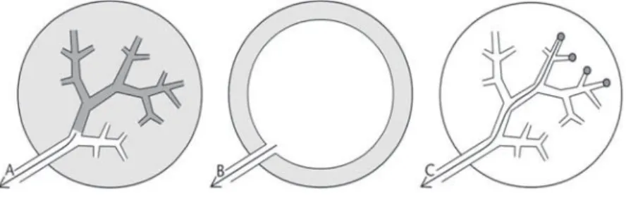
は じ め に 琉球大学医学部附属病院は,全国の大学病院では稀な, 結核病床( 4 床)と感染症病床(第一種 2 床,第二種 4 床)の両者を有する病院である。車で 5 分の近隣に,国 立病院機構沖縄病院があり,内科医の多くは医局から派 遣であることから両病院間の連携をとりやすい環境にあ る。このような背景から,琉球大学医学部附属病院の結 核病床では,重症例,妊婦,透析患者,精神疾患の合併, 重症基礎疾患(糖尿病,心不全など)を有する症例を主 として加療している。またエイズ中核拠点病院でもある ことから,免疫抑制患者(特に HIV 感染症)における多 彩な抗酸菌感染症を経験することも可能である。症例を 通して学ぶことで抗酸菌感染症の臨床像を身近に捉える ことが可能となる。 近年,呼吸器疾患の臨床現場において非結核性抗酸菌 症(特に肺Mycobacterium avium-intracellulare complex,以 下肺 MAC 症)の重要性が高まりつつある。肺 MAC 症の 病型として,i) 線維空洞型,ii) 結節・気管支拡張型,お よび iii) 免疫抑制患者に認められる播種型など,きわめ て多彩である。臨床的には,それぞれの病型の好発年 齢,性別,基礎疾患,臨床像,画像所見,臨床経過,お よび予後などを理解しておく必要がある。一方,病理像 からは,画像所見では得られない生体反応を解釈するこ とが可能となる。単純に肉芽腫の組織像,菌量,菌の分 布からも生体の免疫応答は理解可能である。さらに肺結 核と肺非結核性抗酸菌症を対比して学ぶことで両者の理 解が深まることも事実である。 1. 肺結核症 ( 1 )肺結核の疫学 肺結核は,社会面,および経済面に大きな影響を与え る疾患であり,全世界の 3 分の 1 の人が結核菌に感染し ているという推計がある。この数字の大きさは結核が慢 性の感染症であることを示すものである。このため疫学 的な観点から,感染のみの場合と,臨床的に発病するか 琉球大学大学院感染症・呼吸器・消化器内科学(第一内科), 琉球大学医学部附属病院 連絡先 : 藤田次郎,琉球大学大学院感染症・呼吸器・消化器内 科学(第一内科),〒 903 _ 0215 沖縄県中頭郡西原町字上原 207 (E-mail : [email protected]) (Received 28 Aug. 2018) 要旨:抗酸菌感染症の診断は,病歴,身体所見,微生物検査,および画像診断などを用いて総合的 に行うべきものであることはいうまでもない。もちろん最終診断は微生物検査,または病理所見で あるものの,これを補助するものが画像診断であり,近年胸部高分解能 CT(HRCT)の詳細な解析に より,pathologic-radiologic correlation が確立したことにより,HRCT 所見から病理所見を推定するこ とが可能になってきている。この考え方が確立したのは,肺結核の画像診断と病理所見との対比に よるものであった。すなわち肺結核の画像診断は胸部画像診断の基本とも言うべき内容を含んでい る。このため肺結核の画像診断を理解することで,胸部画像診断全体に対する理解が深まることに なる。さて抗酸菌感染症に関して学ぶ方法として,まず一般臨床で経験することの多い非結核性抗 酸菌症の臨床的特徴を理解し,加えて結核診療を主体となって担っている国立病院機構などで集中 的に肺結核症例を経験することが有用であると考える。国立病院機構病院に研修病院としての機能 を強化することで,結核を理解することができ,抗酸菌感染症全体に対する理解が深まると考える。 キーワーズ:結核,非結核性抗酸菌症,教育,画像診断,鑑別診断Fig. 1 Schemata of pulmonary tuberculosis lesions: airspace consolidation (A), cavitation (B), and centrilobular opacities (C)1).
A, In air space consolidation, alveolar air is replaced by fluid and the necrotic material drains through the airway. B, Cavitation occurs when the caseous necrotic material liquefies and is extruded through the connecting airway. The cavity therefore essentially connects to a relatively large airway. C, Centrilobular opacities reflect peribronchiolar intralobular nodules, which are localized around bronchioles. Note that the distance from the affected area to the central airway is longer in the case of centrilobular opacities than in that of air space consolidation and cavitation.
次性結核症は小児に多いことから小児型結核症ともいわ れる。成人の結核症の大部分が二次性結核症に属するの で成人型結核症といわれる。両者の病像は大きく異なる ので別々に記載する。 ( 4 )一次性結核症2) 3) 一次性結核症は,結核菌を含んだ飛沫核を吸入するこ とにより発症する。結核菌が定着する肺胞は各肺葉の胸 膜直下(大部分は胸膜下 1 cm 以内,換気がこの部分で 最もよく行われている)であることが多い(Fig. 2)2) 3)。 最初の実質内の感染巣は,Ghon focus(初感染巣)と 名づけられている。結核菌は多くは所属リンパ節まで到 達し,前述した Ghon focus と所属リンパ節の病変を合わ せて,Ranke complex(初期変化群)と呼ぶ。リンパ節の 病変は,肺内病変と同様に,肉芽腫を形成し,壊死に至 り,周囲の線維化,および石灰化をきたす。初感染巣の 多くは自然に治癒する。ただし結核菌は被膜に被われた 壊死組織の中で生きており,これが後に再活性化される 部位となる。 初期変化群リンパ節病変に関連し,わずかな菌が肺下 流のリンパ節を経て静脈角リンパ節に達し,血中に入 る。血中に入った結核菌は,主として肺に転移して軽微 な病巣を作る。いったん治癒するものの,宿主が免疫抑 制状態に陥った際に,既存の病巣から発病すると考えら れる。血中移行の菌数が多ければ粟粒結核になり,少な 感染症である。肺結核はヒトからヒトに感染する伝染性 疾患であり,主たる感染経路は経気道感染であり,感染 様式は,飛沫核感染(空気感染)である。このため感染 対策として,空気感染対策(医療従事者は N95 マスクの 使用,患者はサージカルマスクの着用)が必要となる。 2007 年 4 月からは結核予防法が廃止され,結核対策は改 正感染症法により第 2 類感染症として取り扱われている。 感染の危険性は,感染者の伝染力の程度,曝露者の抵 抗力,および接触の程度による。病変の程度がひどいほ ど,菌量が多いほど,および咳がひどいほど伝染しやす い。特に空洞形成の有無が病気の伝染力と関連する。空 洞病変の本態は,結核病巣の中心部壊死と,壊死物質の 気管支からの排出によるものであるから,空洞形成と排 菌は密接に関連する(Fig. 1)1)。 ( 3 )発病 M. tuberculosis に感染した人のわずか 10% が発病する と推定されている。いったん感染した人が発病する可能 性は一生涯あるものの,多くは感染後 2 年以内に発病す る。肺結核の発症率は年齢によって大きく異なる。発症 率は幼少期には高く,青年期では減少し,中・高年で著 しく高くなる。 様々な疾患と肺結核の発病との関連が示唆されている。 糖尿病の患者,透析患者においても肺結核を発症しやす い。免疫抑制患者,中でも最も重要なのが HIV 感染によ
Fig. 2 The clinical course of pulmonary tuberculosis2) 3)
Fig. 3 Tuberculosis observed in bats4)
Hypothesis for explaining the upper lung-fi eld predominance in secondary tuberculosis. In bats, tuberculosis in the lung is observed in the lower lung-fi elds as well as the lower part of both kidneys. The authors have suggested that the host s position strongly affects the distribution of tuberculosis.
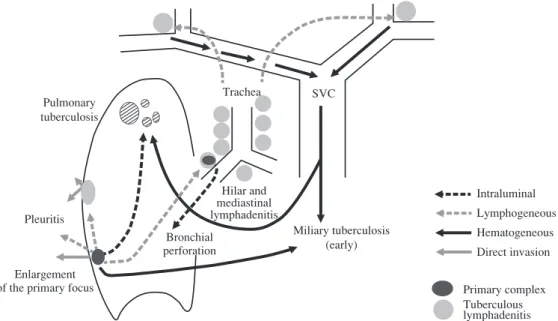
SVC Primary complex Intraluminal Lymphogeneous Hematogeneous Direct invasion Miliary tuberculosis (early) Pulmonary tuberculosis Pleuritis Enlargement of the primary focus
Bronchial perforation Trachea Hilar and mediastinal lymphadenitis Tuberculous lymphadenitis ければ主として肺尖領域に定着する(Fig. 2)。 一次性結核症では,肺胞性浸潤影は約 70% に認められ ており,しばしば右肺優位である。一次性結核症におい てはしばしば肺門リンパ節腫大を認める。無気肺は小児 においては,10∼30% に認められ,右上葉に多い。この 原因は腫大したリンパ節により気管支が圧迫されたこと による。成人においては無気肺の合併は稀である。一次 性結核症を有する多くの患者は無症状であるものの,咳, および発熱を認めることがある。 ( 5 )二次性結核症2) 3) 二次性結核症は,発病初期には,上葉の肺尖部,背側 に限局する傾向がある。その理由として,肺尖部背側に は比較的高い O2分圧を有すること,換気 ・ 血流比が高い こと,肺血流量が乏しいためリンパ流によるドレナージ が十分でないこと,などによる。また肺結核の発症部位 は体位と関連(すなわち血流の多少に関連)するという 説を提唱している論文内には,コウモリの肺結核の病理 像が示されており,コウモリの肺結核は下葉に病変の強 いことが示されている(Fig. 3)4)。約 5 ∼10% の患者に, 肺門 ・ 縦隔リンパ節腫大を認める。 二次性結核症のおよそ 50∼70% において,限局性の 浸潤影を認める。多くの例において,浸潤影は 1 つの区 域に限られているか,1 葉のいくつかの区域に存在する。 典型的には,上葉の肺尖部,背側に多い。時に病気は葉 全体を占めるようになる(結核性大葉性肺炎,Fig. 4)。浸 潤影の部分は,境界が不明瞭であり,融合傾向を示し, 周囲に娘病巣を有する。しばしば同側肺の肺門に向かう 血管 ・ 気管支束陰影の増強を認める。 二次性結核症の画像診断として重要な所見が,細葉単 位で病変が進展することにある。特に重要な画像所見は Aschoff が 1924 年に結核に特徴的な病理所見として記載 した acinar nodule と呼ばれる小結節性陰影である。ただ し実際の症例でこの細葉単位を認めることは稀であり, むしろi) tree-in-bud lesion,ii) 細葉性結節性病変(同じく Aschoff による),および iii) 小葉中心性粒状病変,が画 像所見として重要である。
ここで用語を整理しておきたい。胸部高分解能 CT が 導入され,画像所見と病理所見の対比が可能となった。 知見が集積されたことで,i) tree-in-bud lesion,ii) 細葉性 結節性病変,および iii) 小葉中心性粒状病変を厳密に定 義することが可能となった。これらはしばしば混同され
Fig. 5 Comparison of sizes of acinar lesions (A) and centrilobular lesions(B)
The size of acinar lesions is about 1.5_2 mm ( tree-in-bud appearance), and the size of centrilobular lesions is about 5 mm.
Reid’s lobulus
Reid’s lobulus Reid’s lobulus Miller’s secondary lobule
A B Respiratory bronchiole Terminal bronchiole Respiratory bronchiole Terminal bronchiole
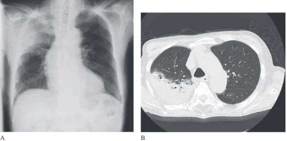
Fig. 4 Radiological fi ndings of caseous pneumonia caused by pulmonary tuberculosis
Plain chest X-ray (A) and chest computed tomography (B) shows consolidation in the right upper lobe. Air bronchograms are also observed (B). It is very diffi cult to distinguish caseous pneumonia caused by Mycobacterium tuberculosis from lobar bacterial pneumonia.
B A るものの,伊藤は,病変のサイズに着目し,tree-in-bud lesion(病変の幅が 1 mm を超えない微細分岐状影)と小 葉中心性粒状病変(サイズが大きく分布が疎)の違いを 明確に示した。小葉内における両者のサイズの対比を Fig. 5に示した。また細葉性結節性病変は,小葉内でtree-in-bud lesion が集簇したものであることを報告した5) 6)。 次に重要なことは,前述した病変の分布である。大部 分の肺結核症は肺尖領域ないしは背部上肺野(Fig. 6) および下葉 S6を好発部位として始まり,経気道的な菌の 転移で上背部に進展する特徴をもっている。二次性結核 症において病変は進展しやすく,炎症,および壊死は拡 大し,より広範囲に広がる傾向がある。この経過中,気 道との交通をしばしば認め,壊死物質の排出に伴って 20 ∼45% の患者に空洞形成を認める(Fig. 6,Fig. 7)。 空洞形成の意味として,外の環境と交通することによ り病原体を排出することにある。この交通には 2 つの大 きな意味がある。 1 つは十分に酸素化された空気を継続 的に空洞内に取り込むことであり,結果的に細胞外にお ける結核菌の増殖を促すことになる。もう 1 つは結核菌 を肺の他の部位に広げること,あるいは別の人への感染 源となることである。空洞から排出された液化した壊死 物質が経気道的に進展することにより,同一葉のみなら ず,他の葉にまで広がりうる(Fig. 7)。そのような機序 においては細気管支レベルに進展し,典型的な肉芽腫を 形成することにより,多発性の実質性の結節影を形成す る(Fig. 7)。 時に,小∼中等度のサイズの肺動脈が空洞壁周囲の線 維化したカプセル内に接線方向に存在し,この血管が拡
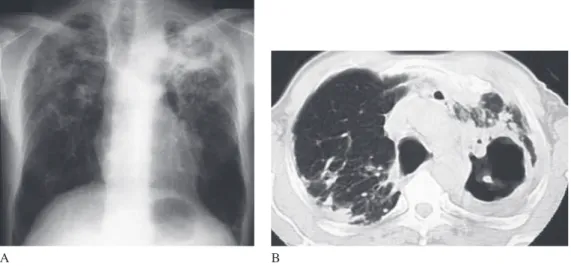
Fig. 6 Radiological fi ndings of the typical secondary pulmonary tuberculosis
Chest computed tomography shows cavitary lesions in both upper lobes. Multiple small nodular shadows (these correspond to acinar nodules) around cavities are also observed.
Fig. 7 Cavity formation by pulmonary tuberculosis (A) and its peribronchial spread (B)
Cavity formation in the apical portion of the left upper lobe is demonstrated (A). Small acinar nodules (mixture of tree-in-bud lesions and centrilobular lesions) caused by peribronchial spread are observed in both lower lung fi elds (B).
Fig. 8 Presence or absence of genes in mycobacterial strains7)
Although M.tuberculosis and other mycobacteria share many antigens in common, this is particularly true of the closely related species within the tuberculosis complex such as M.tuberculosis, M.bovis, and the derived BCG vaccine strains. These strains share several so-called tuberculosis complex specifi c genes, which distinguish them from environmental mycobacteria. Deleted regions in the genome of BCG that fall within tuberculosis-complex-specifi c genes are thus of particular interest as they encode antigens that can potentially discriminate patients with tuberculosis both from donors sensitised by non-tuberculous mycobacteria or vaccinated by BCG.
B A
Common mycobacterial genes Tuberculosis complex-specifi c genes Deleted region
M. bovis BCG
ESAT-6 ESAT-6 ESAT-6
M. tuberculosis M. avium 張することがある(Rassmussen の動脈瘤)。この動脈瘤 が破裂することにより喀血を来たし,死に至ることもあ る。慢性の空洞を形成した際には,Aspergillus 種による 真菌球を形成することがある。 ( 6 )身体所見,検査所見,および診断 身体所見上の特徴として,肉芽腫を形成する結核病巣 の際には聴診所見の弱いことがあげられる。胸部 X 線で 広汎な陰影が認められるにもかかわらず,聴診所見が弱 い際には,肺結核を考える根拠となりうる。 全血からのインターフェロン-γの産生能を測定する ことによりツベルクリンに対する細胞性免疫の程度をみ る方法も用いられている。この方法は BCG 接種による 影響を受けず,肺結核の高危険群内(結核蔓延地域から の移民,あるいは活動性結核患者と接触したもの)での スクリーニング検査に有用な手法である(Fig. 8)7)。
36% 27% 10% 10% 6% 6% 5% M.abscessus M.intracellulare M.fortuitum M.chelonae M. avium M.gordonae Others
Fig. 9 Epidemiology of NTM lung disease (N=114)8)
NTM lung disease cases, defi ned according to the American Thoracic Society criteria, were extracted from NTM detected cases. Others category includes: Mycobacterium kansasii (0.9 %), M.scrofulaceum (0.9%), M.szulgai (0.9%), M.terrae (0.9 %), and M.xenopi (0.9%).
染症の多くは 2 ∼ 3 の病原体によるものであり,特に Mycobacterium kansasii,Mycobacterium chelonae complex, およびMycobacterium avium-intracellulare complex(MAC) によることが多い。 肺結核と同様,非結核性抗酸菌による感染症は世界中 に広がっている。AIDS の流行に伴って,非結核性抗酸 菌,なかでも MAC による感染症は最近の 20 年間で有意 に増加している。またわが国においては,近年,中年女 性に発症する MAC 症の重要性が高まりつつある。ただ しこの頻度は地域差が大きく,日本で唯一,亜熱帯地方 に位置する沖縄県においては,他県と異なりM. abscessus の頻度が最も高い(Fig. 9)8)。 ( 2 )感染経路 MAC は,自然界の土壌,水系,塵埃に広く存在するこ とから,菌を含んだ塵埃や霧などの小水滴を吸入するこ とにより,経気道的に体内に入ると考えられている。土 や水などの環境に生息する MAC は容易にエアロゾル化 することが知られている。このためヒトや動物は,環境 中から病原体を経口摂取,あるいは菌体を含むエアロゾ ルとして吸入することにより感染する。ただし健常人に おいては,生体内に侵入した菌は排除されるか,限局さ 能の低下をもつ宿主にのみ発症するとこれまで推測され ているものの,その詳細は不明である。 結核では,初感染結核をはじめ,経気道的に菌が散布 された場合,病変は肺胞道から始まることが多い。しか しながら肺 MAC 症における初期感染病巣は必ずしも明 らかではない。CT 画像上,小粒状陰影は,末梢気道, すなわち細気管支レベルを中心に観察される。症例によ っては末梢領域の浸潤影を呈する。こうした病巣を病理 学的に調べると,病巣は細気管支から気管支レベルまで 広汎に局在する。画像上,浸潤影を呈した症例では,肺 胞腔内を肉芽腫が占拠し,肺胞壁の炎症性肥厚を認める。 器官培養による研究から,MAC は気道の粘液細胞や 線毛上皮には感染せず,線毛を有さない細気管支上皮細 胞に感染することが明らかにされている。肺胞上皮細胞 に対する感染性を調べた研究では,ヒト肺胞 II 型上皮細 胞である A549 細胞に対して感染および増殖することが 示されている。このような基礎的検討を加味すると MAC の経気道感染初期の病態の主座は,線毛を有する終末細 気管支より末梢の,線毛を有さない呼吸細気管支,また は肺胞上皮にあると考えられる。 ( 3 )肺 MAC 症の各種病型に関して
肺 MAC 症の病型として,i) 線維空洞型,ii) 結節・気 管支拡張型,および iii) 過敏性肺炎の 3 つの型があるこ とに留意しておく。iii) の病型はきわめて稀なので,臨 床的には i) および ii) が重要である。 ①線維空洞型について(Fig. 10)9) 主として上肺野を主体に空洞形成を示すもので,高齢 者,喫煙者,アルコール依存症,あるいは塵肺など既存 の肺疾患を有するものに多く認められる。この病型にお いては肺結核との鑑別が困難である。従来,肺 MAC 症 は,この病型が主体であったが,最近ではこの病型は減 少しつつある。本病型の特徴的病態は空洞形成にある。 本病型においては臨床症状を伴うことが多いものの, 基礎疾患による臨床症状との鑑別が求められる。一般的 には咳,および喀痰などの症状を伴うことが多い。息切 れ,血痰,および全身 怠感,体重減少,発熱,および 寝汗などの全身症状を伴うこともありその際には,病変
Fig. 10 Mycobacterium intracellulare pulmonary disease associated with pneumoconiosis (example case of fi brocavitary disease)9)
Chest X-ray (A) shows large cavities in both upper lung fi elds and upper lung fi eld predominance. Chest computed tomography (B) also shows large cavities of both upper lung fi elds.
B A
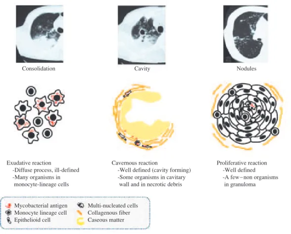
Fig. 11 Nodular/bronchiectatic disease caused by Mycobacterium avium-intracellulare complex In chest computed tomography (A, B), bronchiectasis (A, B) and small nodular shadows (A) are recognized in addition to volume reduction of the right middle lobe. B A が進行していることが多い。 ②結節・気管支拡張型について(Fig. 11) わが国においては,近年この病型が急増している。こ の病型では既存の肺疾患を有しておらず,中・高年の女 性に多い。画像所見として,気管支拡張症と,空洞を有 さない班状の結節を呈する。臨床症状として慢性の咳, および膿性痰を認めるものの,全身症状,および血痰を 認めることは稀である。進行はきわめて緩徐であるこ と,画像所見は結節性陰影が主体であり空洞形成は稀で あること,特に中葉,および舌区の小粒状影と気管支拡 張を特徴とする。 ( 4 )肺非結核性抗酸菌症の病理 岩崎龍郎著の「結核の病理」10)においては,結核の病 理像を滲出型と増殖型に分類している。われわれは同様 の分類を非結核性抗酸菌症にもあてはめ,これら 2 つの 病型と病理像の対比を実施した。 まず線維空洞型の病理所見の検討結果を示す。乾酪性 肺炎を示した肺 MAC 症における病理学的な検討では滲 出性の肉芽腫病巣であり,また抗結核菌モノクローナル 抗体(MAC とも cross react)を用いた検討では,線維空 洞型の特徴的な画像所見である浸潤影と空洞病変におい ては抗酸菌の菌量が多いことを示した11)。 一方,結節・気管支拡張型においては,増殖性肉芽腫 形成が主要な病理学的変化であり,菌量は少ないことが 示された11)。また孤発性結節病巣での抗酸菌抗原の存在 を検討したところ,中心部に壊死を認めない結節影にお いて,抗酸菌抗原を認めないか,ごくわずかであり,中 心部に壊死を認める結節影の内部には少数の抗酸菌抗原 が検出された。さらに気管支粘膜下の肉芽腫においては, 抗酸菌抗原は検出されなかった。 以上の結果をまとめたものが Fig. 12 である。すなわち 画像所見と病理所見との対比により,線維空洞型の特徴
Fig. 12 Characteristics of different granulomatous lesions11)
Most consolidation or ground-glass opacity on the chest computed tomography (CT) indicates histologically exudative reaction. Monocyte-lineage cells infi ltrated into alveoli containing the rod-shaped bacteria as well as the amorphous bacterial antigens, and were possibly phagocytized and processed antigens. Cavitary lesion on CT image indicates cavernous reaction. Histologically, caseous granuloma is shown, but most of mycobacterial antigens are seen in the epithelioid cells or multi-nucleated cells on the margin of the cavitary wall. Most nodules on CT image indicate proliferative reaction with or without central caseous necrosis. Histologically, mycobacterial antigens are rarely observed in the proliferative granuloma. Phagocytized mycobacterial antigens might be observed in the cytoplasm of multi-nucleated giant cells.
Exudative reaction
-Diffuse process, ill-defined -Many organisms in monocyte-lineage cells
Cavernous reaction
-Well defined (cavity forming) -Some organisms in cavitary wall and in necrotic debris
Proliferative reaction -Well defined -A few_non organisms in granuloma
Mycobacterial antigen Monocyte lineage cell Epithelioid cell Multi-nucleated cells Collagenous fiber Caseous matter 的な画像所見である浸潤影と空洞病変においては抗酸菌 の菌量が多いことが示され,結節・気管支拡張型におい ては肉芽腫形成がしっかりしており,抗酸菌量が少ない ことが示された。この 2 つの病型においては,病変の分 布も対照的であることから,著者らは,これらの 2 つの 病型の病態を,感染型,および宿主反応型と表現するこ とが適切ではないかと考えている(Fig. 12)11)。 3. 肺結核と肺 MAC 症との鑑別 画像所見で非結核性抗酸菌症を疑っておらず,喀痰の 抗酸菌染色にて抗酸菌陽性であるとの結果が届いた場合 には,肺結核との鑑別が必要である。その際には,以下 のポイントが重要である。 a)まず肺 MAC 症を念頭におくこと b)ガフキー陽性でもただちに結核専門病院に送るので はなく,PCR の結果を待てる状況かを判断すること c )患者の性別,基礎疾患の有無を考慮すること(女性で, かつ糖尿病などの基礎疾患がないときには肺 MAC 症 を考慮) d)病変の場所は上肺野優位か,中葉・舌区主体かを判 断すること(中葉 ・ 舌区主体であれば肺 MAC 症を考 慮) e )気管支拡張所見を認めるか,否かを判断すること(気 管支拡張所見を認めれば肺 MAC 症を考慮) f )小粒状影を認めるか,否かを判断すること(小粒状 影が中葉・舌区主体であれば肺 MAC 症を考慮) 著者の COI(confl icts of interest)開示:本論文発表内 容に関して特になし。
文 献
1 ) Kosaka N, Sakai T, Uematsu H, et al.: Specifi c high-resolution computed tomography fi ndings associated with sputum smear-positive pulmonary tuberculosis. J Comput Assist Tomogr. 2005 ; 29 : 801 804.
冨岡洋海編, 医学書院, 東京, 2006, 26 40.
3 ) 藤田次郎:患者さんの人生を理解した肺結核の画像診 断. 結核. 2013 ; 88 : 763 773.
4 ) Rothlin E, Undritz E: Beitrag zur Lokalisationsregel der Tuberkulose. Schweiz Z allg Path Bakt. 1952 ; 15 : 690 700. 5 ) 伊藤春海:肺結核の画像 ― 呼吸器画像診断学の貴重な
教育資源. 結核. 2010 ; 85 : 869 879.
6 ) 伊藤春海:肺結核の画像診断 ― Radiologic-Anatomic-Pathologic Correlation. 結核. 2016 ; 91 : 667 676. 7 ) Andersen P, Munk ME, Pollock JM, et al.: Specifi c
immune-based diagnosis of tuberculosis. Lancet. 2000 ; 356 (9235) : 1099 1104.
8 ) Nagano H, Kinjo T, Nei Y, et al.: Causative species of nontuberculous mycobacterial lung disease and compara-tive investigation on clinical features of Mycobacterium
ab-scessus complex disease: A retrospective analysis for two major hospitals in a subtropical region of Japan. PLoS One. 2017 ; 12 : e0186826.
9 ) Fujita J, Kishimoto T, Ohtsuki Y, et al.: Clinical features of eleven cases of Mycobacterium avium-intracellulare com-plex pulmonary disease associated with pneumoconiosis. Respir Med. 2004 ; 98 : 721 725.
10) 岩崎龍郎:「改訂 結核の病理」, 財団法人結核予防会, 東京, 1997.
11) Hibiya K, Shigeto E, Iida K, et al.: Distribution of myco-bacterial antigen based on differences of histological char-acteristics in pulmonary Mycobacterium avium infectious diseases―consideration of the extent of surgical resection from the pathological standpoint. Pathol Res Pract. 2012 ; 208 : 53 58.
Abstract The diagnosis of an acid-fast bacterial infection should be performed comprehensively, using medical history, physical fi ndings, microbiological examination, and radio-logical fi ndings. However, the fi nal diagnosis of mycobac-terial diseases is traditionally based on a microbiological examination or pathological fi nding, but radiological fi ndings can assist this. In recent years, a pathologic-radiologic cor-relation has been established through the detailed analysis of chest high-resolution CT (HRCT); specifi cally, the radio-logical fi ndings of pulmonary tuberculosis patients compared with their pathological fi ndings. Now, it is possible to estimate pathological fi ndings from HRCT images. In other words, the radiological fi ndings of pulmonary tuberculosis contain fun-damentals of chest radiological diagnosis. Potentially, under-standing the radiological fi ndings of pulmonary tuberculosis will, in turn, deepen the entire understanding of chest imag-ing diagnoses. Indeed, to learn more about mycobacterial lung diseases, we should fi rst understand the clinical features of the non-tuberculous mycobacteria often experienced in general clinical practice. Then, the clinical experiences of the National Hospital Organization will be useful to examine
pulmonary tuberculosis cases, intensively. By strengthening the National Hospital Organization Hospitals function as training hospitals, our knowledge of tuberculosis and other mycobacterial lung diseases will deepen.
Key words : Pulomanry tuberculosis, Non-tuberculous my-cobacteria, Education, Radiological fi ndings, Differential diagnosis
Department of Infectious, Respiratory, and Digestive Medi-cine, Control and Prevention of Infectious Diseases, Graduate School of Medicine, University of the Ryukyus; University of the Ryukyus Hospital
Correspondence to: Jiro Fujita, Department of Infectious, Respiratory, and Digestive Medicine, Control and Prevention of Infectious Diseases, Graduate School of Medicine, University of the Ryukyus, 207 Uehara, Nishihara-cho, Nakagami-gun, Okinawa 903_0215 Japan.
(E-mail: [email protected]) −−−−−−−−Review Article−−−−−−−−
EDUCATION FOR TUBERCULOSIS AND NON-TUBERCULOUS
MYCOBACTERIAL DISEASES
―