緒 言
近 年, 頚 動 脈 ス テ ン ト 留 置 術(Carotid artery stenting;CAS)は広く行われるようになった.その合 併症としては血管拡張時に生じるデブリスによる脳塞栓 が最も多く,それを予防するためにフィルターデバイス を用いて行われている2,14).われわれは術後にフィルタ ーを Hematoxylin-Eosin(HE)染色し,回収されたデブ リスを観察している4,5).本研究では,デブリスを頚動
脈内膜剥離術(carotid endarterectomy;CEA)で得ら れた病理標本と比較し,デブリスの性状の同定を試みた.
対象と方法
2007年12月から2010年12月に当科にて頚動脈狭窄 症に対してフィルターデバイスを用いて CAS を施行し た27例(男性24例,女性3例)28病変,CEA を施行 した49例(男性46例,女性3例)49病変を対象とした.
デブリスの観察は,CAS 施行後にフィルターを生理食
頚動脈ステント留置術中に回収されたデブリスの同定:
頚動脈内膜剥離術病理標本と対比して
林 健太郎1) 堀江信貴1) 森川 実2) 宗 剛平1) 竹下朝規1) 陶山一彦1) 永田 泉1)
Identification of the debris collected during carotid artery stenting, comparing with carotid endarterectomy specimen
Kentaro HAYASHI
1)Nobutaka HORIE
1)Minoru MORIKAWA
2)Gohei SO
1)Tomonori TAKESHITA
1)Kazuhiko SUYAMA
1)Izumi NAGATA
1)1) Department of Neurosurgery, Nagasaki University School of Medicine 2) Department of Radiology, Nagasaki University School of Medicine
●Abstract●
Objective: Carotid artery stenting (CAS) in high-surgical-risk patients is considered as an effective alternative to carotid endarterectomy (CEA). Since the occurrence of distal embolization with CAS is still a major concern, an embolus protection device is usually employed during the procedure. We examined the debris in the embolic protection filter and compared the characteristics of the debris with the characteristics of CEA specimens.
Materials and Methods: CAS was performed for 27 patients with carotid artery stenosis (28 lesions). After completing CAS, each filter membrane was stained with Hematoxylin-Eosin (HE) solution, removed from the filter strut, mounted onto a glass slide, and evaluated under a microscope. Forty-nine patients (49 lesions) were treated with CEA. Histopathological examination was performed with HE stain, Azan stain, and Elastica van Gieson stain. The characteristics of the debris were compared with those of the CEA specimens.
Results: HE stain facilitated the characterization of the debris. Microscopically, thrombotic debris, calcified debris, lipid-rich debris, fibrous debris, cellular debris, and strips of endothelium were observed.
Conclusion: Carotid plaque debris captured during carotid stenting with a protection filter can be visualized on the filter by HE staining. Almost all components of the carotid plaque are identifiable as debris.
●Key Words●
carotid artery stenting, debris, carotid endarterectomy, operative specimen
(Received July 4, 2011:Accepted October 21, 2011)
1)長崎大学医学部 脳神経外科
2)同 放射線科
<連絡先:林健太郎 〒852-8501 長崎市坂本1-7-1 E-mail:[email protected]-u.ac.jp>
CEA 標本では49病変中,プラーク内出血40病変,
石灰化42病変,線維性被膜48病変,コレステリン結晶 39病変,炎症細胞浸潤38病変,平滑筋細胞の増殖35 病変,血管増生26病変がみられ,割合はそれぞれ異な るものの,多くは混在していた.また,血管内腔に付着 した壁在血栓も5病変にみられた.Azan 染色は細胞核 と膠原線維の同定に有用で,EVG 染色は弾性線維の同 定に有用であった.
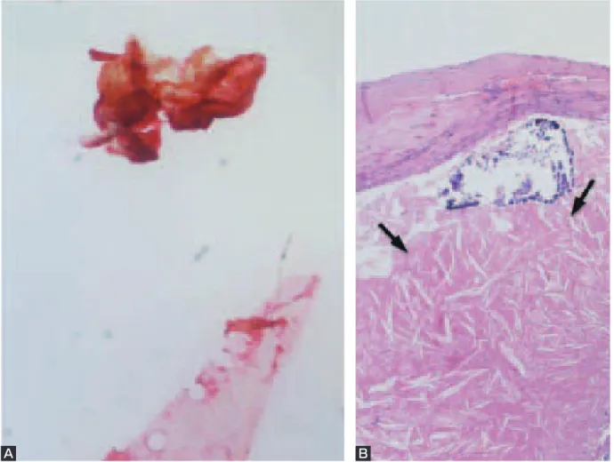
CAS で回収されたデブリスはフィルター上に HE 染 色された状態で観察された.大小の赤色調で細胞成分が 少ないデブリスは血栓性デブリスと同定され,28病変 すべてに観察された(Fig. 1A).大きな血栓性デブリス は CEA 病本におけるプラーク内出血(Fig. 1B)に由来 し,小さな血栓性デブリス(Fig. 1C)はプラーク内出 血の断片や壁在血栓
(Fig. 1D)
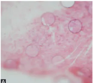
に由来すると考えられた.Hematoxylin 好性で多角形のデブリスは石灰化デブリス と考えられ,28病変中20病変に観察された(Fig. 2A). CEA 標本では,石灰化は42病変にみられており,脂質 成分と混在しているものも18病変で認めた.1病変で は石灰化が内腔に突出していた(Fig. 2B).細胞成分が 少なく黄色調のデブリスは脂質性デブリスと同定され,
28病変中13病変に観察された(Fig. 3A).プラークの コレステリン結晶といった脂質成分に由来するものと考 えられた(Fig. 3B).紡錘状の細胞が散在し,線維成分 が主体のデブリスは線維性デブリスと同定され,28病 変中13病変に観察された(Fig. 4A).CEA 標本におけ る線維性被膜の構造とよく類似していた(Fig. 4B). その他,細胞成分に富んだ細胞性デブリスは28病変中 8病変に観察された(Fig. 5A).CEA 標本では細胞成分 の多い壁在血栓も観察されており,そのような部位から のデブリスと推測された(Fig. 5B).細胞成分と器質化
これまで,われわれは CAS 中に回収された血液をろ 過したり,フィルターを HE 染色したりしてデブリスを 観察してきた4,5).また,デブリスの量と術中の血流障 害の関係やデブリスの性状と術前の画像診断と対比し て,その妥当性を評価してきた6,8,9).本研究では,デブ リスを CEA で得られた病理標本と比較し,その同定を 試みた.
最も多くみられるのは血栓性デブリスである.成分も 器質化した組織と混じったようなものもある.CEA 標 本ではプラーク内出血が血液性の成分としては最も多 く,そのような病変は不安定であり,デブリスとなりや すいと考えられる.また,Fig. 1Dに示したような壁在 血栓もバルーン血管拡張などで容易に血管壁から剥離し てデブリスになるものと考えられる.石灰化デブリスは Hematoxylin に良く染色されるため,顕微鏡の視野にお いても目立つ.石灰化は病変が器質化し,陳旧化,安定 化したものといった印象があるが,CEA 標本を観察す ると脂質成分と混在していたり,炎症細胞浸潤を伴って いたりすることもあり,必ずしも安定した病変とは言え ない.Fig. 2Bに示したように血管内腔に露出した石灰 化は,容易にデブリスとなりえると考えられる.脂質性 デブリスはコレステリン結晶といった脂質由来と考えら れる.当科でフィルターデバイスを使用し始めた頃に,
術前の画像診断で lipid-rich プラークと診断された症例 に CAS を施行したところ,術中に shower embolism を 来した7).術後の観察では脂質性デブリスが認められた.
このようなデブリスは液状化したりして,フィルターで 回収できないこともある.線維性デブリスには紡錘形細 胞と線維成分がみられる.線維性プラークは安定プラー クと考えられ,断片化しにくいと思われるが,線維性被 膜は血管内腔側に位置しており,バルーン拡張やステン トの影響を受けやすいため,デブリスになりうると思わ
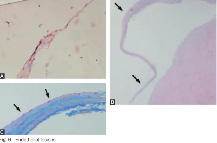
れる.プラークで細胞成分が多いのは炎症細胞浸潤のみ られる部分である.また,プラーク内に血管平滑筋細胞 が遊走し,増殖することもある.このような部位は不安 定プラークと位置づけられ,細胞性デブリスの起源と考 えられる.その他にも,Fig. 5Bに示したような壁在血 栓も細胞成分が多いものがある.血管内膜は血管の長軸 に平行に紡錘形の内皮細胞が並んでいる.それが剥がれ る際に紐状になって断片化したものが血管内皮片と考え られる.Fig. 6Bのように内膜や内膜下組織は CEA 標 本を作製する際にも剥離しやすい.Azan 染色すること で,その構造を把握することができた.血管内皮片は血 管造影検査でのガイドワイヤーやカテーテルの操作でも
生じうると報告されている3,11).
冠動脈の血管拡張術後に組織を観察した報告では,プ ラークを押しつぶすというよりも,プラークに亀裂が入 ることで血管が拡張されることが示されている12).亀 裂は中膜にも及び,動脈解離を引き起こすこともある.
その場合,プラークの内腔側ばかりでなく,深部からも デブリスが発生することになる.CAS においても拡張 機序は同様と考えられ,それを裏付けるように,本研究 でも,さまざまな種類のデブリスが観察される.
デブリスの性状に関する報告では,Malik らはデブリ スをフィルターから取りはずして HE 染色や Von Kossa 染色し,石灰化病変や無形性無細胞性のデブリスの存在 Fig. 1 Thrombotic lesions
A:Debris from a large thrombotic lesion. It is acellular and amorphous. Original magnification (OM)× 50.
B:Carotid endarterectomy (EA) specimen. Intraplaque hemorrhage (arrow). OM ×50.
C:Debris from a small thrombotic lesion. OM ×100.
D:CEA specimen. Intraluminal thrombus (arrow). OM ×50.
A B
C D
Fig. 2 Calcified lesions
A:Calcified debris. It is mainly stained by hematoxylin. OM ×50.
B:CEA specimen. Calcification, some of which is intraluminal (arrow). OM ×50.
A B
Fig. 3 Lipid-rich lesions
A:Lipid-rich debris. It is yellowish and acellular. OM ×50. B:CEA specimen. Deposit of cholesterol (arrow). OM × 50.
A
B Fig. 4 Fibrous lesions
A: Fibrous debris. Fusiform cells are seen within fibrous tissue. OM × 100.
B: CEA specimen. Fibrous cap (arrow). OM × 100.
Fig. 5 Cellular lesions A: Pure cellular debris. OM
×200.
B: CEA specimen. Intraluminal thrombus. OM ×100. C: Cellular debris with organic
lesions. OM ×100. D: CEA specimen. Inflammatory
cells infiltration (arrow).
OM ×50.
A
C D
B
を報告している10).Angelini らはフィルターごと組織切 片を作成し,Heidenhain trichrome 染色し,壊死性物質,
コレステロール,石灰化,血栓性物質,線維性物質を確 認している1).Piñero らはデブリスを電子顕微鏡下に観 察し,フィブリン,血小板,コレステロール,コラーゲ ン,カルシウム,平滑筋,毛細血管などを同定している13). われわれのデブリス観察法はフィルターに捕捉された デブリスを HE 染色し,そのままの状態で観察できるの が特長である.また,手技も単純で,特別な機器や薬品 を必要としない.ただし,デブリスの性状を正確に同定 するには組織切片を作成し,免疫染色などを行い,細胞 や構成成分を同定する必要がある.本法では切片を作成 できず,形態学的な観察にとどまるのが欠点である.
今後,デブリス所見を術前の画像所見対比し,プラー クの性状をより正確に把握することで,治療適応の判断 や周術期の管理に役立つように研究を発展させていきた い.
結 論
CAS で回収されたデブリスは CEA 病理標本と比較す ることでその性状を同定できた.プラークの構成成分の 多くが,CAS 時のデブリスとして観察された.
文 献
1) Angelini A, Reimers B, Della Barbera M, et al: Cerebral protection during carotid artery stenting: collection and histopathologic analysis of embolized debris.
33:456-461, 2002.
2) Brott TG, Hobson RW 2nd, Howard G, et al: Stenting versus endarterectomy for treatment of carotid-artery stenosis. 363:11-23, 2010.
3) Coleman CC, Posalaky IP, Robinson JD, et al:
Atheroablation with the Kensey catheter: a pathologic study. 170:391-394, 1989.
4) Hayashi K, Kitagawa N, Morikawa M: Observing the carotid debris aspirated during carotid stenting:
technical note. 27:22-26, 2005.
5) Hayashi K, Kitagawa N, Morikawa M, et al: Observation of the embolus protection filter for Carotid Artery Stenting. 72:532-537, 2009.
B
C
Fig. 6 Endothelial lesions A:Strips of endothelium. OM ×100.
B:CEA specimen. Intima artificially detached from the media (arrow). OM ×50.
C:CEA specimen. Azan stain of the intima. Location of the endothelium is apparent (arrow). OM ×100.
6) 林健太郎,北川直毅,森川 実,他:Embolus protection filter を用いたステント留置術中に filter obstruction を来
した内頚動脈狭窄症の1例. 36:1133-
1138, 2008.
7) 林健太郎,北川直毅,森川 実,他:Embolus protection filter を用いたステント留置術で shower embolism を来し た内頚動脈狭窄症の1例. 61:83-87, 2009. 8) Honda M, Kitagawa N, Tsutsumi K, et al: High-resolution
magnetic resonance imaging for detection of carotid plaques. 58:338-346, 2006.
9) Kawahara I, Morikawa M, Honda M, et al: High-resolution magnetic resonance imaging using gadolinium-based contrast agent for atherosclerotic carotid plaque.
68:60-66, 2007.
10) Malik RK, Landis GS, Sundick S, et al: Predicting embolic potential during carotid angioplasty and stenting: analysis of captured particulate debris, ultrasound characteristics, and prior carotid endarterectomy. 51:317-
322, 2010.
11) Manninen HI, Räsänen HT, Vanninen RL, et al: Stent placement versus percutaneous transluminal angioplasty of human carotid arteries in cadavers in situ: distal embolization and findings at intravascular US, MR imaging and histopathologic analysis. 212:483- 492, 1999.
12) Naruko T, Ueda M, Becker AE, et al: Angiographic- pathologic correlations after elective percutaneous transluminal coronary angioplasty. 88:1558- 1568, 1993.
13) Piñero P, González A, Martínez E, et al: Volume and composition of emboli in neuroprotected stenting of the carotid artery. 30:473-478, 2009.
14) Yadav JS, Wholey MH, Kuntz RE, et al: Protected carotid-artery stenting versus endarterectomy in high- risk patients. 351:1493-1501, 2004.
JNET 5:99-105, 2011
要 旨
【目的】頚動脈ステント留置術(carotid artery stenting;CAS)は主にフィルターデバイスを用いて行われる.
我々は術後にフィルターを Hematoxylin-Eosin(HE)染色し,回収されたデブリスを観察している.本研究では 頚動脈内膜剥離術(carotid endarterectomy;CEA)病理標本とデブリスを比較し,デブリスの性状の同定を試 みた.【対象と方法】当科にて CAS を施行した27例(28病変)と CEA を施行した49例(49病変)を対象とした.
デブリスの観察はフィルターを HE 染色し,filter membrane を strut より離断し,プレパラートを作成後に観察 した.CEA で摘出されたプラークは固定後に包埋し,切片を作成した.HE 染色および特殊染色し,観察した.【結 果】CEA 標本にはプラーク内出血,血栓,石灰化,コレステリン結晶,線維性被膜,炎症細胞浸潤,平滑筋増殖 などが観察された.CAS で回収されたデブリスは血栓性デブリス,石灰化デブリス,脂質性デブリス,線維性デ ブリス,細胞性デブリスなどと同定された.【結論】CAS で回収されたデブリスは CEA 病理標本と比較すること で,その性状を同定できた.
〈第26回日本脳神経血管内治療学会学術総会優秀演題推薦論文〉