Study&of&Zn&finger&proteins&and&the&homeodomain&protein&
Otx2&in&early&Xenopus&eye&development&
&
Zn
9
7
2
Otx2
&
&
Contents
! Abstract(...(5! Abbreviations(...(8! Gene(names(and(gene(symbols(...(9! General(introduction(...(10! Chapter(I(...(12! Analysis(of(C2H2.type(zinc(finger(proteins,(XXXX(and(YYYY,(for(eye( development(...(12! 1.1(Introduction(...(13! Chapter(II(...(30! Modulation(of(the(homeodomain(protein(Otx2(activity(in(cell(proliferation,( anteroposterior(patterning(and(eye(formation(by(phosphorylation(states(30! 2.1(Introduction(...(31! 2.2(Results(...(34! Phosphorylation-of-exogenous-Otx2(...(34! Phosphorylation-of-endogenous-Otx2(...(36! Phosphorylation-of-Otx2-depends-on-Cdk-activity(...(38! Otx2-mutants-4A-and-4E-have-distinct-functions-in-eye-formation(...(41! Otx2>4E-stimulates-cell-proliferation(...(44! Activities-of-Otx2>4E-and->4A-for-anteroposterior-patterning(...(47! Otx2>4E,-but-not->4A-interacts-with-Tle1(...(50! Otx2>4E-but-not->4A-interacts-with-XXXX(...(51! 2.3(Discussion(...(52! Repressor-activity-of-Otx2-conferred-by-post>translational-modification(...(52! Role-of-cyclin/Cdk>dependent-phosphorylation-of-TFs(...(53! Repression-activity-by-phosphorylated-Otx2-and-XXXX(...(56! How-does-Otx2-coordinate-its-activator-and-repressor-functions-for-development?(...(57!General(Discussion(...(62! Conclusion(...(66! Experimental(procedures(...(67! cDNA-cloning,-sequence-analysis,-and-constructs(...(67! Xenopus-embryo-and-microinjection(...(68! Observation-of-subcellular-localization-of-Venus>fused-XXXX-constructs-and-YYYY-by-confocal-microscopy(...(69! Whole>mount-in-situ-hybridization-(WISH)(...(70! Western-blot-analysis(...(70! Co>immunoprecipitation-assay(...(71! Luciferase-reporter-assays(...(72! In-vitro-translation-and-protein-phosphatase-treatment(...(72! Immunoprecipitation-(IP)-assays-for-putative-phosphorylated-Akt-sites-and-the-detection-of-endogenous-Otx2-protein(...(72! Quantitation-of-the-eye-size-in-tailbud-stage-embryos(...(73! Immunostaining-for-PH3-and-DAPI-nuclear-staining(...(74! Tables(and(Figures(...(76! Table.-1.-The-list-of-plasmid-constructs(...(76! Table.-2.-The-list-of-cutting-sites-and-RNA-polymerases-for-the-in-vitro-transcription-of-anti> sense-RNA-probe.(...(79! References(...(126! Acknowledgements(...(134!
Abstract
During early Xenopus development, the neuroectoderm acquires regional identities along the anteroposterior axis and develop into brain, eye, and so on. In this process, various region-specific transcription factors (TFs) have been shown to play roles in the specification and differentiation of these organs. However, whether other TFs are involved in this process, how those TFs interact and regulate each other, and how TF activity is modulated by post-transcriptional modifications remain to be elucidated. In this thesis, I addressed these three subjects by focusing on two uncharacterized zinc finger (Znf) genes and the homeobox gene otx2, which are expressed in the anterior neuroectoderm (ANE).
In Chapter I, I investigated the roles of two uncharacterized Znf proteins, XXXX and YYYY, in eye formation, in concert with Otx2. I chose XXXX and YYYY as candidate regulators of early eye development based on a systematic expression pattern database using a Xenopus ANE (ANE) cDNA library. I first showed that both proteins were localized in the nucleus, suggesting their functions as TFs. At the early neurula stage, xxxx and yyyy were expressed in the ectodermal sensory layer, and their expression domains were gradually restricted to the eye field and the neural fold as development proceeds. Gain-of- and loss-of-function experiments using Xenopus embryos suggested that XXXX and YYYY are involved in eye development by regulating the expression of eye marker genes at the early neurula stage. In addition, I found that XXXX and YYYY
physically interact with Otx2 as assayed by co-immunoprecipitation (Co-IP) analysis. Reporter analysis suggested that XXXX enhances the transrepression activity of Otx2 for posterior genes, whereas YYYY inhibits the transactivation activity of Otx2 for anterior genes. These data suggest that XXXX and YYYY function as regulators of Otx2 in a distinct manner to execute eye development.
In Chapter II, I investigated the role of modifications on Otx2, and demonstrated that both exogenous and endogenous Otx2 are phosphorylated at multiple sites in Xenopus embryos. I identified three possible cyclin-dependent kinase (Cdk) sites and one possible Akt site, and analysed biological activities of phosphomimetic (4E) and non-phosphorylatable mutants (4A) of Otx2 for those four sites. In the neuroectoderm, the 4E, but not the 4A mutant downregulated the Cdk inhibitor gene p27xic1, and promoted cell proliferation, possibly forming a positive feedback loop consisting of Cdk, Otx2, and p27xic1 for cell proliferation. In addition, the 4A mutant downregulated the expression of the hindbrain marker gene gbx2 for anteroposterior patterning. In contrast, the 4A mutant functioned as an activator on its own and upregulated the expression of eye marker genes, and later enlarged the eyes. Consistent with these results, the interaction of Otx2 with the corepressor Tle1 is suggested to be phosphorylation-dependent. Finally, I found that XXXX enhances the repression activity by Otx2 through phosphorylation-dependent interaction of Otx2 with XXXX. These data suggest that the ternary complex of Otx2, XXXX and Tle1 is formed upon Otx2 phosphorylation for repressing target genes in the ANE.
Taken together, I propose a molecular mechanism in which transcriptional activator and repressor activities of Otx2 are regulated not only by its interaction with the corepressor Tle1 as well as the partner TFs XXXX and YYYY, but also by its phosphorylation states. Thus, Otx2 could coordinate cell proliferation and differentiation during neural patterning and early eye development in Xenopus.
Abbreviations AD, activation domain ANE, anterior neuroectoderm
BTB, bric-a-brac, tramtrack, broad-complex Cdks, cyclin dependent kinases
ChIP, chromatin immunoprecipitation CNS, conserved non-coding sequence Co-IP, co-immunoprecipitation CRM, cis-regulatory module
EFTFs, eye field specific transcription factors λPP, λ protein phosphatase
MAPK, mitogen-activated protein kinase PH3, Phospho-Histone H3
RPE, retinal pigment epithelium TFs, transcription factors
NLS, nuclear localization signal
WISH, whole mount in situ hybridization Znf, zinc finger
Gene names and gene symbols
Official gene name (gene symbol): synonymous gene symbol
goosecoid homeobox (gsc) gsc, Xgsc, goosecoid, gsc-a, gsc-b Meis homeobox 3 (meis3) XMeis3, mrg2, meis3-a, meis3-b
orthodenticle homeobox 2 (otx2) Xotx2, otx-2, Xotx-2, otxA, otx2-a, otx-b cyclin-dependent kinase inhibitor xic1
(cdknx)
Xic-1, Xic1, p27XIC1, p28
paired box 2 (pax2) XPax2, XPax-2, pax-2, pax2-a, pax2-b
paired box 6 (pax6) XLPAX6, xpax6, pax-6, an2, mgda, wagr, pax6-a, pax6-b
retina and anterior neural fold homeobox (rax)
rx1, Xrx1, Xrax, Rx2A, rx, rx1a, rax-a, rax1, Xrx1A, rax-b, rx-1
SIX homeobox 3 (six3) XSix3, hpe2, six3-1, six3-b
SRY-box 2 (sox2) XLSOX-2, Sox-2, XSox2, Xsox-2, anop3, mcops3 T-box 3 (tbx3 ) xtbx3, Xltbx3, tbx3-a, tbx3-b
transducin like enhancer of split 1 (tle1) grg1, xgrg1, esg, esg1 XXXX (xxxx)
General introduction
Detailed molecular mechanisms in establishing the vertebrate basic body plan have been elucidated by analyses of various signaling molecules, intracellular signal transducers, and transcription factors (TFs) (De Robertis et al., 2000; Niehrs, 2004). Among these factors, TFs directly regulate various genes that are involved in tissue-specific gene cascade or gene regulatory networks during development, so I have been interested in how TFs design outputs of gene expressions to drive a tissue-specific program in cellular and developmental regulatory processes. Recently, comprehensive analyses such as RNA-seq and ChIP-seq provide the information about tissue-specific gene expression profiles and TF-binding genomic loci, but how TFs are coordinately regulated to exert their transcriptional activities by physical and functional interactions with other partner TFs and transcriptional cofactors (coactivators and corepressors) or by posttranslational modifications still needs to be elucidated by focusing on individual TFs.
Xenopus laevis and X. tropicalis are useful model systems for molecular embryological studies, because large numbers of easily manipulated embryos can be obtained, and gain-of- and loss-of function analyses have been established. Especially, using X. laevis embryos, many region- and tissue-specific TFs have been identified, such as the organizer genes, which are specifically expressed in the Spemann and Mangold
organizer (also called the gastrula organizer), including goosecoid (gsc), lim1 (the same as lhx1), and otx2, and the neural genes including otx2, rax, pax2, and gbx2. From the extensive analyses of these TFs, we have learned about molecular mechanisms of early vertebrate development (Andoniadou and Martinez-Barbera, 2013; Lake and Kao, 2003; Yasuoka and Taira, 2018) Compared to the study of the gastrula organizer and early neural patterning, it remains elusive how the early development of the brain and eye is regulated at the level of TFs.
In this thesis, I focused on region- and tissue-specific TFs that are expressed during neural patterning and eye formation, and addressed two subjects; (i) analysis of two uncharacterized Znf genes, xxxx and yyyy isolated from the systematic expression pattern screening of an anterior neuroectoderm (ANE) cDNA library (Takahashi et al., 2005), (ii) analysis of the regulatory mechanism of transcriptional activities of the homeodomain TF Otx2 through posttranslational modifications in early development.
Chapter I
Analysis(of(C2H2.type(zinc(finger(proteins,(XXXX(and(
YYYY,(for(eye(development(
1.1 Introduction
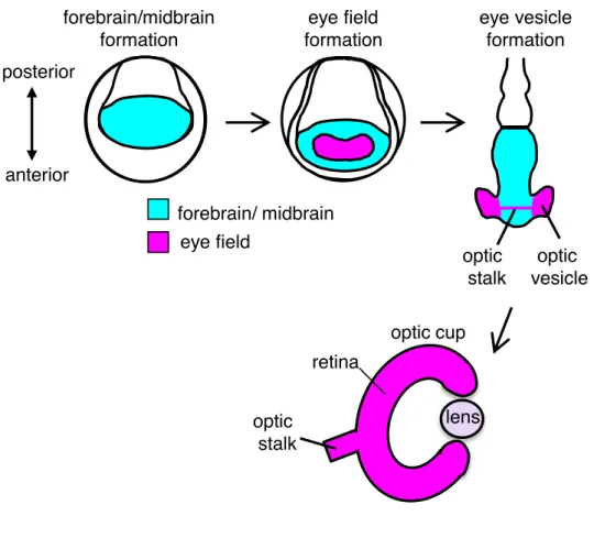
During early eye development in vertebrates, a part of the anterior neural plate is fated to the eye field that will form eye structures, such as eye vesicles including the retina and optic stalks at later stages. Subsequently, sequential processes progress for eye formation: splitting the eye field, the formation of the optic vesicle and the optic cup, and lens induction (Graw, 2010; Zuber, 2010) (Fig. 1). There are many reports for later eye development in vertebrate such as the specification of the optic vesicle and stalk, retina differentiation and lens placode formation. However, the molecular mechanism to specify the eye field, which is one of the earliest event in eye development, is not fully understood, though a rough genetic cascade has been proposed (Zuber et al., 2003).
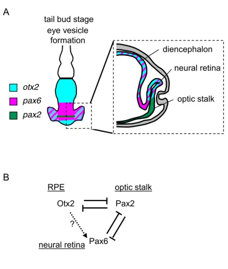
The homeobox gene, otx2 is reportedly involved in early eye formation in Xenopus. In the neuroectoderm, otx2 starts to be expressed in the anterior domain which gives rise to the forebrain and midbrain, and plays an essential role in anteroposterior patterning by preventing Meis3 and Gbx2-mediated caudalization (Agoston and Schulte, 2009; Katahira et al., 2000) (Fig. 2A). In early eye formation, Otx2 is involved in the specification of the eye field by upregulating an eye field-specific gene, rax, in the center of the otx2-expressing domain at the gastrula stage (Danno et al., 2008). In turn, Rax downregulates otx2 to discriminate the eye field from the diencephalon (Zuber, 2010). Subsequently, eye field specific transcription factors (EFTFs) including pax6, which are essential for eye field specification, are expressed in the center of the region where otx2 is expressed (Fig.2A,B). In Xenopus, a gene cascade of TFs starting from otx2 and tbx3
through rax to pax6 is reported to define the eye field, and this region starts to express other downstream eye-specific genes, six3, lhx2, tll, and six6 (Zuber et al., 2003) (Fig. 2C), suggesting that Otx2 functions at the top of the hierarchy in ocular morphogenesis. Although the genetic relationship of Otx2 with other EFTFs in the early eye gene cascade has been proposed, it remains elusive how individual TFs interact with each other to exert their functions in this cascade.
Chapter II
Modulation(of(the(homeodomain(protein(Otx2(activity(in(cell(
proliferation,(anteroposterior(patterning(and(eye(formation(
2.1 Introduction
As described in Chapter I, I found several bands of Otx2 in western blotting (Fig. 10A,B), and Dr. Sudou, a previous member in our laboratory, also noticed that multiple bands of overexpressed Otx2 in Xenopus embryos migrate more slowly than an in vitro translated product, suggesting a post-translational modification of Otx2. Similarly, a few previous studies using western blotting showed multiple bands of exogenous mouse Otx2 in transfected cultured cells (Mallamaci et al., 1996) and endogenous Otx2 in mouse embryos (Boyl et al., 2001), but post-translational modification of Otx2 has remained to be characterized.
otx2 is required for the formation of the anterior portion of the head and eye development. In early eye development, Otx2 upregulates an eye field-specific gene, rax, in the center of the otx2-expressing domain at the gastrula, and subsequently this region starts to express other downstream EFTFs including pax6 (Zuber, 2010). In later eye development, otx2 restarts to be expressed in the developing optic vesicle (Martinez-Morales et al., 2001; Viczian et al., 2003), and is involved in the specification of presumptive neural retina (pax6-positive cells) from the optic stalk (pax2-positive cells) in mice (Martinez-Morales et al., 2001) (Fig. 13). After the retinal specification, otx2 expression becomes restricted to postmitotic retinal progenitors, and determines bipolar cell fate (Brzezinski and Reh, 2015). Thus, the knowledge of the role of Otx2 for head and eye formation is being accumulated.
Otx2 has both transactivation and transrepression activities for the regulation of downstream target genes. Otx2 has a transactivation domain in the C-terminal region (Gammill and Sive, 2001), and directly upregulates a cement gland marker xcg1 in Xenopus (Gammill and Sive, 1997). Otx2 also has a repression domain with a motif of SIWSPAS, which belongs to the engrailed homology region 1 (eh1) (Heimbucher et al., 2007). This domain is conserved in vertebrate Otx family proteins, and interacts with the corepressor TLE/Groucho (Heimbucher et al., 2007). With this domain, Otx2 functions as a repressor that inhibits the posterior gene gbx2 to form the midbrain-hindbrain boundary (MHB) (Nakamura et al., 2005). Furthermore, genome wide analysis of chromatin immunoprecipitation (ChIP) sequencing using X. tropicalis gastrula embryos has shown that Otx2 cooperates with the transcriptional activator Lim1/Lhx1 to bind specific cis-regulatory modules (CRMs) to activate anterior genes in the head organizer, whereas Otx2 cooperates with the repressor Goosecoid (Gsc) to inhibit non-head organizer genes (Yasuoka et al., 2014). Thus, both the transactivation and transrepression activities of Otx2 are important for regionalization and tissue patterning and function during embryogenesis.
Recently, several reports have shown that Otx2 directly influences both cell proliferation and differentiation. In mice, aberrant expression of Otx2 in the hindbrain induces the ectopic proliferation of neural progenitor cells (Wortham et al., 2012), and overexpression of Otx2 causes proliferation of dopaminergic progenitors in the ventral mesencephalon (Omodei et al., 2008; Vernay et al., 2005). In addition, in a
medulloblastoma cell line, Otx2 upregulates cell cycle-positive regulator genes, such as cyclin D3, and downregulates cell cycle-negative regulator genes including p27 and differentiation-specific genes, such as neuroD (Bunt et al., 2012). Thus, Otx2 is likely to be involved in both proliferation and differentiation. However, the molecular mechanism of how Otx2 coordinates cell proliferation and differentiation remains unknown.
Here, I show that Otx2 is phosphorylated, which confers transrepression activity, otherwise functioning as a transactivator on its own. Functional analyses using phosphomimetic or non-phosphorylatable Otx2 mutants suggest that phosphorylated Otx2 represses the Cdk inhibitor gene p27xic1 and the posterior gene gbx2 for promoting cell proliferation and anteriorizing the neuroectoderm, respectively, whereas unphosphorylated Otx2 enhances the formation of the eye field and later the retina in the place of the optic stalk. Additionally, I have shown that XXXX or Tle1 interacts with the phosphorylated mutant of Otx2 and potentiates the repression activity of Otx2. These functional and biological data suggest that phosphorylated Otx2 interacts with partner proteins including XXXX and Tle1 to form a repressive complex, leading to the downregulation of repressed target genes of Otx2, such as cell cycle inhibitor genes and posterior genes. Thus, my data provide a new possibility that Otx2 exerts its versatile functions with cell proliferation- and differentiation-orientated activities through its post-translational regulation.
2.2 Results
Phosphorylation of exogenous Otx2
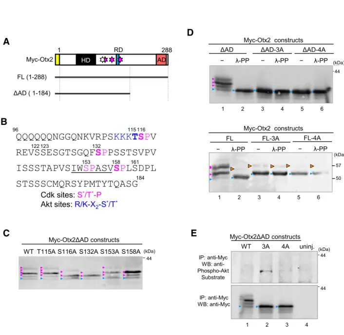
Using western blotting analysis of N-terminally Myc-tagged full length (Myc-Otx2FL) and several deletion mutants of Otx2 constructed by previous members of our laboratory (Ms. Hosono, Mr. Minami, and Mr. Okada), we showed that modification mainly occurs in the region of amino acid positions 96-184 (Fig. 14A). The amino acid sequence from position 96 to 184 indicated possible phosphorylation sites for Cdk (cyclin-dependent kinase; S*/T*-P or S*/T*-P-X-R/K; *, phosphorylatable residues), MAPK (mitogen-activated kinase), casein kinase (S*/T*-X0-2-D/E/Sp1-3; Sp, primary phosphorylatable
serine residue), and Akt kinase (R/K-X2-S*/T*) (Fig. 14B). Notably, mouse Otx2
reportedly possesses an Akt phosphorylation site at S115 (the corresponding site of Xenopus laevis Otx2 is T115; see Fig. 15), according to the database for the prediction of phosphorylation sites (see Materials and Methods). I first took over this work from previous members, and tried to identify modification sites of Otx2 by the examination of four serine residues (S116, S132, S153, S158) and one threonine (T115) by introducing point mutations. Because we found the suitable conditions in SDS-PAGE to separate the shifted bands of modified Otx2 by using Myc-Otx2ΔAD constructs, and that modification of Otx2 mainly occurs around the repression domain, we used Myc-Otx2ΔAD constructs in the following experiments. Replacement of a single serine with alanine revealed that S116A, S132A and S158A constructs eliminated the uppermost
band and increased the lower bands, compared with the wild type (WT), T115A and S153A construct (Fig. 14C). Furthermore, a triple alanine mutant of Myc-Otx2ΔAD for S116, S132 and S158 (named ΔAD-3A) showed no slowly migrating bands (Fig. 14D; lane 3). These data suggest that S116, S132 and S158, which have the S-P consensus motif for Cdk and MAPK, are responsible for multiple shifted bands (up to seven bands by combinations of three sites) of Myc-Otx2. To examine this hypothesis, embryonic lysates expressing Myc-Otx2 constructs were treated with λ-protein phosphatase (λ-PP). As shown in Fig. 14D (upper panel), λ-PP treatment removed all of the modified bands of Myc-Otx2ΔAD (lanes 1, 2) and did not change the band of ΔAD-3A itself (lanes 3, 4), suggesting that slowly migrating bands of Myc-Otx2ΔAD were caused by phosphorylation at S116, S132 and S158. The same experiment was performed with full-length Otx2 (FL), showing a similar result, in which all modified bands of Myc-Otx2FL were removed by λ-PP treatment except for a weak shifted band (Fig. 14D, lower panel). Consistently, Myc-Otx2FL-3A had only a single weak shifted band, and this band was not removed with λ-PP (Fig. 14D, lower panel; orange arrowheads), suggesting another distinct modification site around the activation domain.
I next asked whether T115 of Xenopus Otx2 is an Akt phosphorylation site, although I did not detect a shifted band in Otx2 mutant 3A (ΔAD-3A) in western blotting (see Fig. 14D, upper panel; lanes 3, 4). In addition, there was no difference in band positions between ΔAD-3A (remaining T115) and ΔAD-4A (alanine mutation at T115 in addition to 3A) regardless of λ-PP (Fig. 14D, upper panel; lanes 3-6). However, it was still
possible that phosphorylation at T115 may not have caused band shift in ΔAD-3A in western blotting, or may have been inhibited by alanine mutations at the three sites including S116 next to T115. Therefore, I performed IP-western assays using anti-Phospho-Akt Substrate antibody, which recognizes the R-X-X-Sp/Tp motif (Sp/Tp, phosphorylated residues). Myc-Otx2ΔAD in embryonic lysates was immunoprecipitated with anti-Myc antibody and then was subjected to western blotting with anti-Phospho-Akt Substrate antibody. The result showed that a band was detected with mutant ΔAD-3A but not with ΔAD-4A (Fig.14E, upper panel, lanes 2, 3), suggesting T115 as an Akt site. Reasons why no clear band was detected in WT (lane 1) might be (i) phospho-Akt signals were dispersed in several shifted bands caused by phosphorylation at other sites, and (ii) phosphorylation at T115 was inhibited by phosphorylation at S116 or the other sites. Thus, I identified four possible phosphorylation sites for Akt (T115) and Cdk/MAPK (S116, S132 and S158) in Otx2, and these sites are well conserved in vertebrate Otx2 as well as an Otx2 paralog, Otx5/Crx (Fig. 15).
Phosphorylation of endogenous Otx2
It is important to show whether endogenous Otx2 is phosphorylated, but no antibodies
against phosphorylated sites of Otx2 are available. As far as I know, there is no report
Xenopus embryos, like Otx2, as evidenced by band shift in western blotting, possibly due to their limited amounts in the whole embryo. Therefore, I first estimated sensitivity of
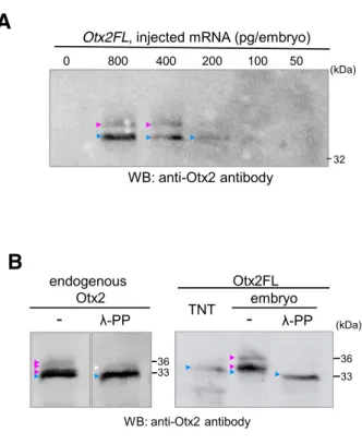
detection using a diluted series of otx2 mRNA injected into Xenopus embryos. As
shown in Fig. 16A, modified bands of full-length Otx2 without Myc-tag (Otx2FL) was
clearly detected when otx2FL mRNAs were exogenously injected with greater than 200
pg per embryo. Based on an RNA-seq data from X. tropicalis (Owens et al., 2016), the
amount of otx2 mRNA was estimated to be the order of 1 pg per embryo at the neurula
stage. I therefore predicted that endogenous Otx2 can be detected by western blotting
with more than 200 X. tropicalis embryos. As expected, I successfully detected multiple
bands of the endogenous Otx2 protein immunoprecipitated from lysates of approximately
800 X. laevis neurula embryos in total, and the upper bands were reduced by λ-PP
treatment (Fig. 16B, left). Similarly, shifted bands of exogenous Otx2FL were detected,
and those were abolished by λ-PP treatment (Fig. 16B, right). Thus, endogenous Otx2
is actually phosphorylated, though the intensities of phosphorylated bands of endogenous
Phosphorylation of Otx2 depends on Cdk activity
Because S116, S132 and S158 sites have the S-P motif (Fig. 14B), I speculated that
MAPK or cyclin/Cdk complexes may phosphorylate Otx2. To examine this, I tested the
effect of MAPK on Otx2 modifications. Overexpression of the constitutively active
form of MAPKK, MAPKK* (Kosako et al., 1993), did not strongly affect the modified
band patterns of Myc-Otx2ΔAD even with the high-dose injection of MAPKK* (Fig.
17A). In addition, Mr. Minami, a previous member, tested the blastocoel injection of a
chemical inhibitor for MAPK, U0126, and reduced the activation form of MAPK, in the
embryonic lysate, but this did not affect the modified band patterns of Myc-Otx2ΔAD
(data not shown). These data suggest that Otx2 does not respond to MAPKK and its
downstream kinases.
I next tried to hyperactivate endogenous Cdk activity in Xenopus embryos by overexpression of stable mutants of cyclin B1 (cyclin B1* for Cdk1) and cyclin A1 (cyclin A1* for Cdk1 and Cdk2), both of which lack the destruction box (Geley et al., 2001; Yamano et al., 2004). Hyperactivation of Cdks by mRNA injection for HA-cyclin B1* or HA-cyclin A1* in the Xenopus embryo was verified by cell-cycle arrest of mRNA-injected blastomeres (Fig. 17B, white asterisks). Under these conditions, modifications of Myc-Otx2ΔAD were dramatically enhanced (Fig. 17C,D), and these modified bands
were almost removed by λ-PP treatment (Fig. 17C,D). These data suggest that Cdk is involved in phosphorylation of Otx2.
If Otx2 is phosphorylated by cyclin/Cdks, a preference of cyclin B/Cdk and cyclin A/Cdks for Otx2 phosphorylation might emerge. To examine this, I constructed double alanine mutants (2A) for S116, S132 and S158 in ΔAD, in which only one site is responsible for Otx2 phosphorylation (Fig. 18A). The intensity of uppermost bands of S132 (S116A/S158A) and S116 (S132A/S158A) constructs, but not that of S158 (S116A/S132A), was increased by HA-cyclin B1* expression (Fig. 18B), whereas all modified bands were enhanced by HA-cyclin A1* expression (Fig, 18C), with an additional modified band (white arrowheads). These data suggest that S116 and S132 have a preference for both cyclin B/Cdk and cyclin A/Cdks, and that S158 has it for cyclin A/Cdks, if Otx2 is directly phosphorylated by cyclin/Cdks.
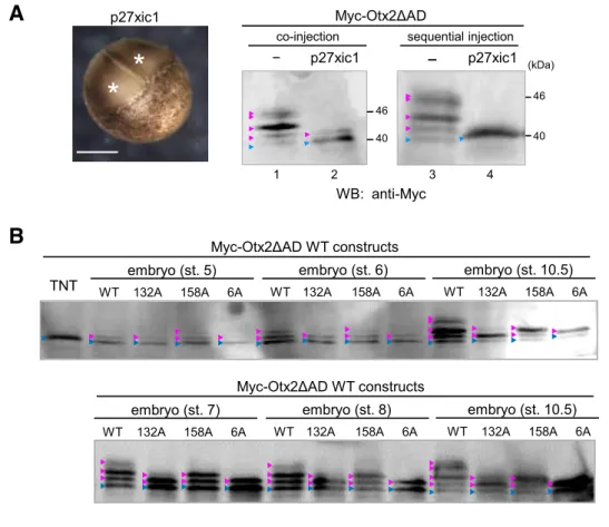
To further examine Cdk-dependent phosphorylation of Otx2, I overexpressed the Cdk inhibitor p27xic1 in the embryo (Vernon, 2003). I first verified cleavage arrest by p27xic1 mRNA injection (Fig. 19A left; white asterisks) as reported (Ohnuma et al., 1999). When a mixture of p27xic1 and Myc-Otx2ΔAD mRNAs were injected at the 2-cell stage, modified bands of Myc-Otx2ΔAD were reduced, but a weak band still remained, which might be due to phosphorylation occurring prior to the accumulation of p27xic1 protein translated from injected mRNA (Fig. 19A, right; lanes 1, 2). Therefore, I first injected p27xic1 mRNA alone at the 2-cell stage, then Myc-Otx2ΔAD mRNA was injected into the cleavage-arrested blastomeres at the 32-cell stage equivalent. As
shown in Fig. 19A (right; lanes 3, 4), p27xic1 completely abolished modified bands of Myc-Otx2ΔAD, suggesting that phosphorylation of Otx2 depends on Cdk activity. These data also imply that the reduction of modified Otx2 by p27xic1 expression was probably not caused by dephosphorylation, because, once Otx2 was phosphorylated at the 2- to 32-cell stages (lane 2), it appeared to be stable until the late blastula stage when western blotting was performed (compare lanes 2 and 4). Because phosphorylation of Otx2 appears to start very early, I examined developmental changes of phosphorylation levels using Myc-Otx2ΔAD (Fig. 19B). The wild type (WT) and alanine mutants of a single serine at S132 (S132A) and S158 (S158A), and of 6 serines at S122, S123, S132, S153, S158, and S161 (6A) in Myc-Otx2ΔAD constructs were analyzed from stages 5 to 10.5, as indicated. Note that the 6A construct has S116, which is a phosphorylation site. The data showed that modification of Otx2ΔAD was already detected at the 16-cell stage (stage 5), gradually increased until the early gastrula stage (stage 10.5), and reached almost a plateau at stage 10.5 onwards (Fig. 19B). In addition, mutation at S132 (S132A and 6A constructs) reduced phosphorylation levels compared to S158 mutation, indicating that S132 and S116 are more efficiently phosphorylated than S158 during cleavage stages. Increase of phosphorylation levels until stage 9 correlates well with the cleavage cycle, supporting again the possibility that Otx2 is a substrate of cyclin/Cdks.
Taken together, Otx2 has four possible phosphorylatable sites, and I referred to T115 as A-site (putative Akt site) and S116, S132 and S158 as C-sites (putative Cdk sites) in the following experiments.
Otx2 mutants 4A and 4E have distinct functions in eye formation
To ask whether known Otx2 activities are affected by phosphorylation states, I
overexpressed phosphomimetic or non-phosphorylatable mutants and analysed eye
phenotypes, which have been well-documented for Otx2 functions (Martinez-Morales et
al., 2001; Nishihara et al., 2012). A phosphomimetic mutant, Otx2-4E, was made by
replacing A-site (T115) and three C-sites (S116, S132 and S158) with glutamate in
Otx2FL. Non-phosphorylatable mutants, named Otx2-T115A, -3A and -4A, were
similarly made by replacing A-site, C-sites or all four sites with alanine, respectively.
Each Otx2 construct was expressed unilaterally in the anterior neuroectoderm (ANE),
which includes the eye field, and the phenotypes were scored at stages 38-42. I
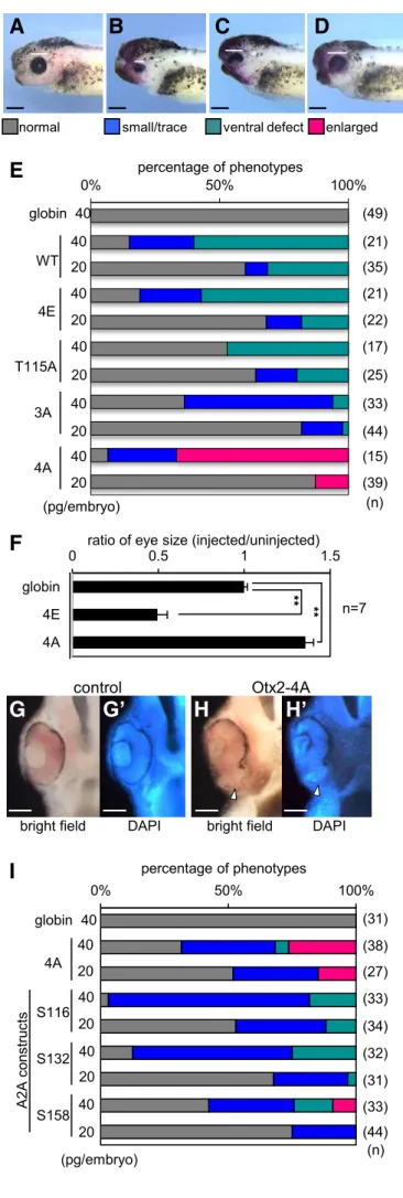
categorized abnormal eye phenotypes into three types compared with the normal looking
phenotype (Fig. 20A-D). Expression of Otx2-WT, -4E -T115A, or -3A caused
small/trace eye and ventral defect phenotypes in a dose-dependent manner (Fig. 20E), in
which stronger phenotypes appeared to be obtained by expression of Otx2-WT, -4E, -3A,
have not previously been reported by expressing any other Otx2 mutants in Xenopus
(Andreazzoli et al., 1997; Isaacs et al., 1999). Notably, either Otx2-T115A or -3A did
not enlarge, but rather reduced eye sizes (Fig. 20E), indicating that mutations at both
A-site and C-A-sites are necessary for the activity of Otx2-4A. Quantitative analysis for the
area of the eye vesicle showed that the eye size in Otx2-4A expressing embryos was
enlarged by approximately 1.3 times, whereas that in Otx2-4E expressing embryos was
reduced by approximately 0.5 times, compared with control (Fig. 20F). Section
examination for the enlarged eye phenotype by Otx2-4A showed that the retinal tissue
was expanded ventrally with the reduction of the retinal pigment epithelium (Fig. 20H,H’;
arrowheads) compared with the control (Fig. 20G,G’). These results clearly
demonstrated that Otx2-4A has different activities in eye formation compared to the other
mutants, and imply that exogenous Otx2-WT functions as phosphorylated forms, because
WT exhibits similar activities to 4E (Fig. 20E).
To further examine whether all C-site mutations are required for the activity of Otx2-4A, I constructed triple alanine mutants of Otx2 (named A2A constructs), in which only one of C-sites (S116, S132 or S158) is intact, referred to as A2A-S116, -S132, or -S158, respectively. As shown in Fig. 20I, A2A-S116 and A2A-S132 both caused small/trace
and ventral defect eye phenotypes. By contrast, A2A-S158 caused enlarged eye phenotype in a dose dependent manner, though the fraction of enlarged eye phenotypes by A2A-S158 was lower than that of Otx2-4A. This data indicates that mutations at T115, S116, and S132 are essential for enlarged eye phenotypes.
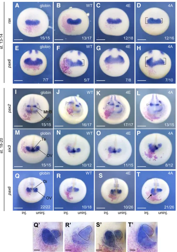
I next examined the early effect of Otx2-4A and -4E by focusing on the eye field marker genes, rax and pax6. Overexpression of Otx2-4A increased the expression of rax compared with the uninjected side (Fig. 21D), whereas Otx2-WT and -4E as well as the globin control did not affect (Fig. 21A-C). The expansion of rax expression by Otx2-4A may be a reason for enlarged phenotypes. Similarly, Otx2-4A enhanced the expression of pax6 compared with the uninjected side (Fig. 21H) and globin injected control (Fig. 21E), whereas 4E showed no effect (Fig. 21G). The effect of Otx2-WT on pax6 was complicated; Otx2-Otx2-WT expanded pax6 expression but partially reduced it (Fig. 21F), possibly due to a combination of various phosphorylation states. These data suggest the possibility that unphosphorylated Otx2, as mimicked by Otx2-4A, promotes eye field formation. The enlarged eye phenotypes with aberrant ventral eye structures (see Fig. 20H,H’) raised another possibility that Otx2-4A converts the presumptive optic stalk to the retina. Therefore, I examined the expression of pax2, six3 and pax6 in the late neurula, in which pax2 is expressed in the optic stalk (OS) and MHB (Fig. 21I), whereas six3 is expressed in the optic vesicle (OV) and telencephalon (Te) (Fig. 21M), and pax6 is expressed in the OV and diencephalon (Di) side by side with different WISH intensities (Fig. 21Q). Overexpression of Otx2-WT, -4E, and -4A all
inhibited pax2 expression in the OS (Fig. 21J-L), while Otx2-WT or -4E, but not -4A caused posterior shift of pax2 expression in the MHB (open arrowheads). The reduction of pax2 in the OS by Otx2-WT, -4E, and -4A (Fig. 21J-L) are well consistent with the aberrant eye morphology at the tailbud stage on the ventral side where the OS is formed (see Fig. 20C,E). Regarding six3 expression, Otx2-WT, -4E, and -4A expanded in the OV (Fig. 21N-P), and Otx2-4A appeared to be most effective for this expansion (Fig. 21P). As for pax6 expression, Otx2-WT, -4E, and -4A differently affected. WT reduced OV but expanded Di pax6 expression (Fig. 21R,R’; black dashed lines, Di; white dashed lines, OV); and Otx2-4E slightly reduced the OV expression similar to WT (Fig. 21S,S’), consistent with their small/trace eye phenotypes (see Fig. 20B,E). Opposite to them, Otx2-4A expanded the OV pax6 expression (Fig. 21T,T’), consistent with its enlarged eye phenotypes (see Fig. 20D,E). These data demonstrated that Otx2-4E and -4A have different activities in eye formation. This raised the next question whether Otx2-4E and -4A have different activities in cell proliferation and anteroposterior patterning, which have been reported for Otx2 functions (Pilo et al., 2001; Acampora et al., 2000).
Otx2-4E stimulates cell proliferation
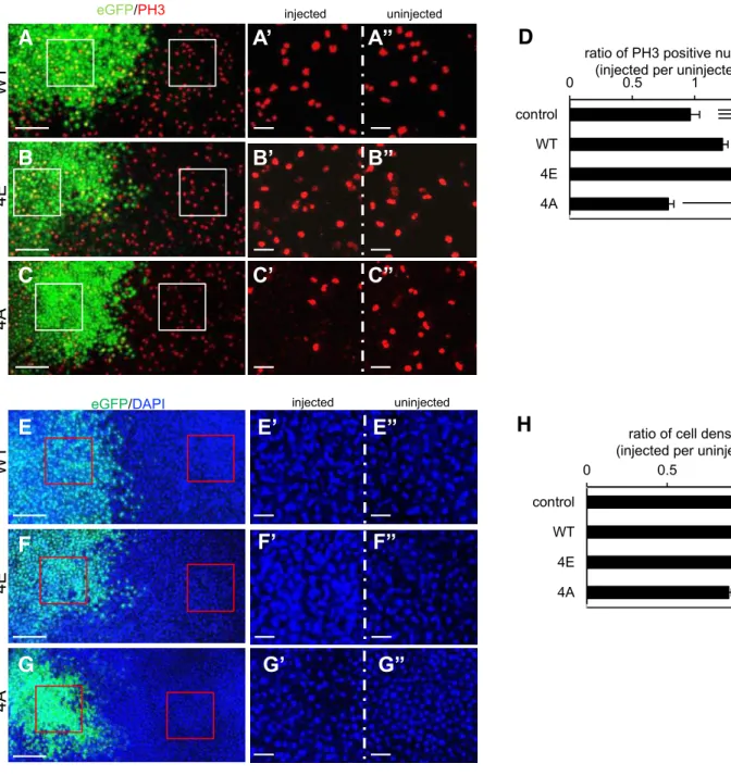
To investigate the effect of Otx2 on cell proliferation, Otx2-WT, -4E or -4A was
performed with anti-PH3 (phospho-Histone H3) antibody as a mitosis marker. The
numbers of PH3-positive nuclei were counted in eGFP-positive and negative areas for
quantitative analysis as shown in Fig. 22A-C. The data showed that the ratio of the
number of PH3-positive nuclei in injected versus uninjected areas was 1.27 ± 0.07 (mean
± s.e.m.) and 1.38 ± 0.05 in Otx2-WT and -4E expressing embryos, respectively, whereas
that was 0.727 ± 0.062 in Otx2-4A expressing embryos. These ratios significantly
differed from the control (0.944 ± 0.014) (Fig. 22D), suggesting that Otx2-WT and -4E
both stimulate cell proliferation whereas Otx2-4A reduces it. To confirm this, I counted
the number of nuclei stained by DAPI (Fig. 22E-G). Quantitative analysis showed that
the ratio of cell number in injected versus uninjected areas was 1.08 ± 0.061 and 1.13 ±
0.02 in Otx2-WT and -4E expressing embryos, respectively, whereas that was 0.894 ±
0.019 in Otx2-4A expressing embryos. The ratios in 4E and 4A embryos differed from
the control (1.02 ± 0.04) (Fig. 22H). Thus, these data suggest that Otx2-4E and -4A
exhibited opposite effects on cell proliferation: Otx2-4E stimulates but -4A reduces
To gain in vivo relevance of the Otx2 function in cell proliferation, I next performed loss-of-function experiments using antisense morpholino oligos (MOs) for otx2 and its paralog otx5. Previous study using MO-injection in X. tropicalis embryos has shown that Otx2 and its paralogue Otx5, which are expressed in the ANE, have functional redundancy in head development (Yasuoka et al., 2014). X. tropicalis is a diploid species closely related to the tetraploid species X. laevis and suitable for MO knockdown experiments, and all three C-sites of Otx2 are conserved in Otx5. MOs together with FITC dextran as a tracer were injected into one blastomere at the 4-cell stage of X. tropicalis embryos; followed by PH3 immunostaining and DAPI staining at early neural stage. The numbers of stained nuclei were counted and compared between injected and uninjected areas. As shown in Fig. 23, the number of PH3-positive nuclei was almost unchanged in the ANE injected with standard control MO (A,A’,A”,C), whereas it significantly decreased in the region injected with otx2/otx5-MOs (B,B’,B”,C). Consistent with this, the number of nuclei decreased in the otx2/otx5-MOs injected area (Fig. 23E,E’,E”,F), but not affected in the control MO-injected area (Fig. 23D,D’,D’’,F). These data indicate that Otx2 and Otx5 are required for cell proliferation in the ANE, and this in vivo data is consistent with the enhancement of cell proliferation by exogenous Otx2-WT and -4E (see Fig. 22D,H). This may also suggest that the reduction of cell proliferation by Otx2-4A is caused by dominant-negative action of Otx2-4A against endogenous phosphorylated Otx2 that stimulates cell proliferation, and the unphosphorylated form of Otx2 cannot take its place.
To ask how Otx2 stimulates proliferation, I focused on p27xic1 because Otx2 reportedly inhibits the p27 gene (Bunt et al., 2012). Analysis of our ChIP-seq data using X. tropicalis gastrula embryos (Yasuoka et al., 2014) showed co-occupancy of Otx2 and Tle1 to several potential CRMs near p27xic1 (Fig. 24A), suggesting that p27xic1 is a direct target gene of Otx2. Using mRNA injection, Otx2-WT, -4E, or -4A with nβ-gal as a tracer was unilaterally expressed, and the expression of p27xic1 was examined by WISH. Expression of Otx2-WT and -4E, but not -4A, downregulated p27xic1, compared with globin-injected negative control and on the uninjected side (Fig. 24B-E), consistent with the stimulation of cell proliferation by Otx2-4E (see Fig. 22D,H).
To assess the gain-of- and loss-of-function data shown above, the expression domains of otx2 and p27xic1 and the distribution of PH3-positive cells in the early neurula were examined using sagittal hemisections. The data showed that the otx2 expressing region did not overlap with that of p27xic1 (Fig. 24F,G), and has more mitotic cells than the posterior region (Fig. 24H), consistent with the involvement of otx2 in cell proliferation in the ANE. These data suggest the existence of a positive feedback loop involving Otx2, Cdk, and p27xic in the regulation of cell proliferation (Fig. 24I).
Activities of Otx2-4E and -4A for anteroposterior patterning
In the neural plate, Otx2 is involved in anteroposterior patterning by directly inhibiting
and by directly activating anterior genes such as xcg1 (Gammill and Sive, 1997) and rax
(Danno et al., 2008). I first examined gbx2 by ectopically expressing Otx2-WT, -4E or
-4A, in the posterior neuroectoderm. Both Otx2-WT and -4E downregulated the
expression of gbx2 (Fig. 25B,C,C’; blue arrowheads) compared with the uninjected side
and globin-injected control (Fig. 25A,A’). This activity explains the posterior shift of
pax2 expression in the MHB by WT and -4E (see Fig. 21J,K). Conversely, Otx2-4A did not downregulate gbx2 expression (Fig. 25D,D’; white arrowheads), suggesting
the possibility that only phosphorylated Otx2 has the ability to inhibit posteriorization.
xcg1 is expressed in the cement gland, which is the anterior most organ (Fig. 25E). Ectopic expression of Otx2-WT, -4E and -4A all upregulated xcg1 (Fig. 25F,G,H; magenta arrowheads), and this activation was observed in the ventrolateral ectoderm, but not in the neuroectoderm as reported (Gammill and Sive, 1997). To assess whether the ectopic upregulation of xcg1 by the Otx2 mutants requires their transactivation activity, I made transactivation domain-deleted constructs of 4E (ΔAD-4E) and 4A (ΔAD-4A). Neither ΔAD-4E nor ΔAD-4A upregulated xcg1 (Fig. 25I,J), indicating that the transactivation domain of Otx2-4E and -4A is required for their activation of xcg1. These data suggest that Otx2 has transactivation activity regardless of its phosphorylation states.
The above data suggest that the difference in function between Otx2-4E and -4A is repression activity. To further examine whether phosphorylation states affect the repression and activation activity of Otx2, I performed luciferase reporter assays. I first examined the response of the silencer of meis3, a posterior gene, which is expected to be repressed by Otx2. The meis3-D2 silencer (Yasuoka et al., 2014) was inserted upstream of the SV40 promoter in the reporter construct, and this reporter alone showed a high level of luciferase activity in the animal pole region. Under this condition, reporter activity was significantly repressed by Otx2-WT and -4E, but not by -4A (Fig. 25K). I next tested combinatorial repressive activity of Otx2 with the interacting partner Gsc and the corepressor Tle1, which form the repression complex for negative target genes of Otx2 (Yasuoka et al., 2014). Repression of the meis3-D2 reporter by Gsc and Tle1 was enhanced by co-expression of Otx2-WT, and this enhancement was also observed with Otx2-4A (Fig. 25L), suggesting that neither phosphorylation of Otx2 is required for nor unphosphorylated Otx2 interferes with the formation of a ternary repression complex. Because Otx2-4A and Tle1 did not repress the reporter (Fig. 25L), repression activity by a combination of Otx2-4A, Gsc, and Tle1 is probably due to recruitment of Tle1 to Gsc. Although Otx2-4A does not interfere with complex formation, Otx2-4A may weaken repression activity of the complex as implied in Fig. 25M (compare 4A with Otx2-4E).
I next examined an activated target gene of Otx2, rax, using the reporter gene SOP-FLASH, which has 8 tandemly repeated rax enhancer elements binding for Otx2 and
Sox2 (Danno et al., 2008). The data showed that both Otx2-4E and -4A exerted transactivation activity (Fig. 25N), similarly to activation of the xcg1 gene by Otx2-4E and -4A (see Fig. 25G,H). Because Sox2 is expressed in the neuroectoderm (Mizuseki et al., 1998) and has a transactivation domain (Nowling et al., 2000), a combination of Sox2 and Otx2 may function as activator regardless of phosphorylation states of Otx2. Because Otx2-4E did not upregulate the endogenous rax gene (see Fig. 21C), activation of the rax reporter by Otx2-4E may be due to the “boosted CRM” by 8 tandem repeats. Taken together, it is suggested that phosphorylation of Otx2 is required for its repression activity, but does not affect transactivation activity itself.
Otx2-4E, but not -4A interacts with Tle1
To investigate how phosphorylated Otx2 functions as a gene repressor, I tested the association with other repressor factors. The eh1 repression domain of Otx2 reportedly interacts with TLE/Groucho (Heimbucher et al., 2007). Therefore, I tested the physical interaction between Otx2 mutants and HA-tagged Tle1 by Co-IP assays (Fig. 25O). A co-immunoprecipitated band of HA-Tle1 was detected with Myc-Otx2-4E as well as Myc-Otx2-WT (magenta arrowheads), but barely with Myc-Otx2-4A, consistent with the data that neither Otx2-4A alone nor co-expression with Tle1 exerted repressive activity for meis3-D2 luciferase reporter (see Fig. 25K,L). The reason why the intensity of the co-immunoprecipitated Tle1 band with WT was weaker than that of 4E (Fig. 25O) is possibly due to partial phosphorylation of WT. These data suggest that phosphorylation
of Otx2 is required for the interaction with Tle1 to exert its own repression activity. The phosphorylation-dependent interaction of eh1 domains with TLE has not previously been reported.
Otx2-4E but not -4A interacts with XXXX
2.3 Discussion
Repressor activity of Otx2 conferred by post-translational modification
In this study, I have shown that Otx2 exists in two distinct states, phosphorylated and
unphosphorylated states. Using phosphomimetic and non-phosphorylatable constructs
of Otx2, I demonstrated that phosphorylated Otx2 can function as both repressor and
activator, whereas non-phosphorylated Otx2 can function solely as an activator. To date,
only a few TFs have been reported to be converted between being an activator and a
repressor by post-translational modification. The SoxE group of TFs including Sox9
are converted by SUMOylation from an activator to a repressor by displacing CBP/p300
and recruiting Tle4/Grg4 (Lee et al., 2012). Another case is Pax2, transactivation of
which is enhanced through JNK kinase-mediated phosphorylation in the activation
domain by blocking TLE/Groucho (Cai et al., 2003). Thus, I have shown the third case,
in which phosphorylation of Otx2 enables it to function as a repressor through the
interaction with Tle1 and XXXX (Fig. 27), opposite to the case of Pax2.
It is noted that Otx2 has a novel eh1 domain with a Cdk site (SIWSPASISP; underlined is a possible Cdk site), which is only present in the Otx family proteins (Otx1,
Otx2, and Otx5/Crx) as well as a few other TFs (Copley, 2005). The eh1 domain is present in many metazoan TFs (Copley, 2005) and known to interact with TLE family proteins through the conserved C-terminal WD40 repeat domain (Heimbucher et al., 2007; Pickles et al., 2002). Because some WD40 domains of ubiquitin ligases and phosphatases, such as β-Trcp and Cdc4, are known to interact with a phosphorylated serine residue in their substrates (Yaffe and Elia, 2001), it is possible that one of the WD40 repeats of TLE may interact with the eh1 domain with a phosphorylated site in Otx2.
Role of cyclin/Cdk-dependent phosphorylation of TFs
From my observations, I propose a positive-feedback-loop model for Otx2 functions in
cell proliferation (Fig. 24I). In this model, in a proliferative state, Otx2 is
phosphorylated by elevated Cdk activity, and phosphorylated Otx2 becomes to repress
p27xic1, which, in turn, derepresses cyclin/Cdk activity to enhance phosphorylation of
Otx2, thereby forming a positive feedback loop (Fig. 24I, left). When the cell cycle is
arrested, cyclin/Cdk activity decreases and hence phosphorylated Otx2 decreases, causing
derepression of p27 expression to repress Cdk, keeping unphosphorylation state of Otx2
toggled by the cell cycle. However, it is not known how the phosphorylated state of
Otx2 is changed to the unphosphorylated state. There are two possibilities: (i)
dephosphorylation of Otx2 by phosphatase, and (ii) degradation of phosphorylated Otx2.
I did not observe any difference in instability of the Otx2-4E mutant compared to -4A by
western blotting, which does not support the second possibility, and denies a
phosphorylation dependent degradation. A putative PEST sequence, an indicator of
rapidly degraded protein, is present in the Xenopus Otx2 protein (Mori et al., 1994;
Williams and Holland, 1998), and Otx2 may have a short half-life. Therefore,
unphosphorylated Otx2 could be generated through the rapid turn-over of Otx2 regardless
of its phosphorylation state.
I have shown that endogenous Otx2 is phosphorylated, but its phosphorylation levels were much lower than those of exogenous Otx2 (Fig. 16B). This difference might be caused by the timing of protein production and the number of proliferating cells where phosphorylation of Otx2 supposedly occurs. For exogenous Otx2, mRNA was injected into the animal pole region at 2- or 4-cell stages, and translated in the presumptive ectoderm from early cleavage stages, and the translation product is phosphorylated until the blastula stage (Fig. 19B) by Cdk1 activity, which oscillates with a period of ~30 minutes during the cleavage cycle (Hörmanseder et al., 2013). By contrast, in the in
vivo situation, otx2 starts to be transcribed in the dorsal endoderm and mesoderm at the late blastula stage and in the dorsal ectoderm at the early gastrula stage (Sudou et al., 2012), and then the endogenous Otx2 protein accumulates in the head organizer and the ANE, and the proliferation rates in these regions dramatically decrease especially in the head organizer (Fig. 24H). Therefore, even if phosphorylation levels of endogenous Otx2 in proliferating cells are high, the average phosphorylation level of major non-proliferating and minor non-proliferating cell populations in a gastrula or neurula embryo is much lower than those of exogenous Otx2.
I have proposed a positive feedback loop involving Cdk-dependent phosphorylation of Otx2 for cell proliferation (Fig. 24I). Similar feedback loops involving other TFs are also reported, but the regulatory mode is different from Otx2. That is, TFs are switched “on” or “off” by Cdk-dependent phosphorylation to promote cell cycle progression. An example for regulation by an “on” switch is FOXM1. FOXM1 is activated by the phosphorylation of the N-terminal inhibitory domain by cyclin/Cdk to upregulate cyclin B and cdc25B, thereby forming a positive feedback loop for cell proliferation (Park et al., 2008; Major et al., 2004). An example for an “off” switch is FOXO. FOXO upregulates the Cdk inhibitor genes p27kip1 and p21WAF1, but, upon phosphorylation by cyclin/Cdk, undergoes nuclear export, which virtually switches off the function of FOXO, leading to cell proliferation (Schuff et al., 2010; Liu et al., 2008; Stahl et al., 2002). In the case of Otx2, Cdk-dependent phosphorylation confers repressor activity to Otx2
for repressing p27xic, otherwise it functions as an activator on its own as mentioned above, differing from simple switch on or off regulations.
Regarding tissue specification and differentiation, Cdk-dependent phosphorylation generally negatively regulates activity of TFs that directly activate differentiation genes. For example, the phosphorylation of the myogenic TF MyoD by cyclin/Cdk leads to its degradation to prevent muscle differentiation (Kitzmann et al., 1999). During neurogenesis, the Cdk-dependent phosphorylation of Ngn2 (neurogenin 2) inhibits its neural differentiation activity (Ali et al., 2011; Hindley et al., 2012). In these cases, Cdk-dependent phosphorylation of the TFs switches off their functions. Taken together, phosphorylation of TFs by cyclin/Cdks either promotes cell cycle progression (e.g., FOXM1 and FOXO) or inhibits tissue differentiation (e.g., MyoD and Ngn2). In contrast, Cdk-dependent phosphorylation does not simply inhibit the function of Otx2 in differentiation, but at the same time converts it to function in proliferation. Thus, this study has shown a new category of TFs, in which post-translational modification of a single TF converts from a differentiation- to proliferation-orientated role.
Repression activity by phosphorylated Otx2 and XXXX
It is not well understood how Otx2 properly functions as an activator or a repressor for its target genes. An answer to this question was reported (Yasuoka et al., 2014), showing that Otx2 functions as an activator together with Lim1 and also as a repressor
together with Gsc, suggesting that the combination with partner proteins determines transcriptional activity of Otx2. This combinatorial regulation may dominate over phosphorylation states because the Otx2-4A construct, which does not have repression activity by itself (Fig. 25K), still exert repressive activity together with Gsc for repressing the meis3-D2-luc reporter (Fig. 25L,M). However, it should be noted that gsc is not expressed, in the neural plate, and it is not known yet what is a partner protein of Otx2 for repressing meis3 and gbx2. In this study, I demonstrate a possible partner TF of Otx2 in the neural plate, XXXX.
How does Otx2 coordinate its activator and repressor functions for development? Based on these lines of evidence together with other observations, I propose a model for
Otx2 functions (Fig. 27). In this model, during cell cycle progression (in proliferation),
Otx2 is phosphorylated by cyclin/Cdk and Akt upon growth stimulation, and
phosphorylated Otx2 is able to interact with Tle1 and XXXX to repress p27xic1, and, in
turn, derepresses cyclin/Cdk activity, thereby forming a positive feedback loop (Fig. 27,
upper). In the ectoderm, phosphorylated Otx2 also represses posterior genes (gbx2 and
meis3) and activates anterior genes (xcg1, rax) to establish anteroposterior patterning. When growth stimuli are reduced, and the cell cycle is arrested, unphosphorylated Otx2
functions as an activator for anterior development (cement gland formation and retinal
differentiation) (Fig. 27, lower). Thus, Otx2 has ability to orchestrate cell proliferation
and differentiation through changing its phosphorylation states. This model is
consistent with the expression patterns of related genes. The ANE (future forebrain and
midbrain), which expresses otx2 as well as cyclin and cdk genes (Fig. 24F) (Vernon and
Philpott, 2003), exhibits high mitotic rate with no expression of p27xic1 (Fig. 24G,H).
By sharp contrast, the posterior neuroectoderm, which expresses no otx2 but cyclin and
cdk to a lesser extent (Fig. 24F) (Vernon and Philpott, 2003), exhibits low mitotic rate with the expression of p27xic1 (Fig. 24G,H). However, at the tailbud stages (stages
20-40), the developing retina expresses both otx2 (Wang and Harris, 2005 Viczian et al.,
2003) and p27xic1 (Ohnuma et al., 1999). It was reported that, in developing retinal
cells, otx2 is transcribed, but not translated during the early to mid-retinal neurogenesis
(stages 33, 37), and Otx2 protein was only detectable from late retinal neurogenesis (stage
38) in bipolar cells (Decembrini et al., 2006). Therefore, the absence of the Otx2 protein
at the early to mid-retinal neurogenesis in the developing retina may explain
Notably, both Otx2-4E and -4A upregulate xcg1 (Fig. 25G,H), and this activity needs the activation domain (Fig. 25I,J), implying that phosphorylated Otx2 retains transactivation activity, consistent with reporter analysis (Fig. 25N), and that Otx2 can directly upregulate xcg1 as has been shown (Gammill and Sive, 1997). During cement gland formation, xcg1 appears to be expressed in both proliferating and non-proliferating cells because the expression of xcg1 starts at the gastrula stage in the anterior-most ectoderm at the dorsoventral border (Gammill and Sive, 1997 Gammill and Sive, 2001), and continues during neurula stages beyond stage 18, at which cell proliferation is undetectable (Saka and Smith, 2001). This observation is consistent with the data that Otx2-4E and 4A both activate xcg1 expression, supporting the model that activator activity of Otx2 is unchanged by phosphorylation.
In eye formation, it was reported that expression of a repressor form of Otx2 (Otx2-EnR) by mRNA injection in Xenopus embryos results in small eye or eyeless phenotype (Isaacs et al., 1999), and that expression of another type of repressor form, EnR-Otx2, in chick eyes by electroporation caused pigmentation defects of the retinal pigment epithelium and the reduction of pax6 (Nishihara et al., 2012). These activities of repressor forms of Otx2 are similar to that of Otx2-4E in Xenopus embryos (Fig. 20E and Fig. 21R,S), consistent with the repressor activity of Otx2-4E (Fig. 25K). In contrast with Otx2-4E, Otx2-4A expanded the expression of rax at early neurula stage (Fig. 21D) without stimulating cell proliferation (Fig. 22D,H); at later stages, Otx2-4A expanded the expression of pax6 (retina) but inhibited pax2 (optic stalk) on the ventral side, leading to
ventral expansion of the retina. These data suggest that Otx2-4A can expand the eye field, and subsequently alter patterning of the neural retina and optic stalk. In other words, enlarged eye phenotypes by Otx2-4A is caused by changes in patterning, not by proliferation. Our data is reminiscent of the previous report on the function of Otx2 for Xenopus retina formation, in which an activator type of Otx2 (Otx2-VP16) promotes the bipolar cell fate without stimulating retinal proliferation (Viczian et al., 2003), supporting the possibility that unphosphorylated Otx2 acts mainly as an activator in retina formation.
Several heterozygous Otx2 mutations in human were reportedly linked with severe ocular malformation. For example, point mutations of P133T, P134A, and P134R, which occur near one of the four putative phosphorylation sites, S132, found in our study (see Fig. 15), are associated with microphthalmia, anophthalmia, sclerocornea and retinal detachment (Beby and Lamonerie, 2013). It is not certain whether these mutations actually affect phosphorylation of Otx2, and if it affects the interaction with putative binding partners of Otx2, but it is possible to speculate that ocular defects in human are caused by the alteration of phosphorylation states of Otx2.
In summary, I demonstrated that Otx2 undergoes phosphorylation in vivo, and that phosphomimetic and non-phosphorylatable mutants of Otx2 exhibit distinct activities in cell proliferation, patterning and differentiation in Xenopus embryos. In vivo analysis
of the phosphorylation sites, such as a mutated gene knock-in approach, is now awaited to explore the role of phosphorylated Otx2 in embryonic development.
General Discussion
To understand the molecular mechanisms of gene regulation during development, it is important to elucidate not only genome-wide binding profiles of individual transcription factors (TFs) to cis-regulatory modules (CRMs) near target genes together with their loss-of- and gain-loss-of-function data, but also the regulation of individual TFs at the protein level, such as the combinatorial interplay among TFs and their binding partners or cofactors as well as the post-translational modification of TFs. In this thesis, I investigated the molecular mechanism of how transcriptional activities of Otx2 are regulated by the combination with XXXX and YYYY, and by its phosphorylation states in developmental processes, such as the patterning of neuroectoderm and early eye formation in Xenopus.
I started this study by investing two uncharacterized genes, xxxx and yyyy, which are expressed in the anterior neuroectoderm (ANE), and found that these two genes are involved in the gene cascade for eye development at positions downstream of Otx2. This finding led me to hypothesize functional and physical interactions of XXXX and YYYY with Otx2. In Chapter I, I found that XXXX enhances the gene repression activity of Otx2, whereas YYYY inhibits the gene activation activity of Otx2. From the data of WISH and RNA-seq, xxxx and yyyy are maternally expressed in the animal pole (Fig. 6), in which otx2 is not expressed (Sudou et al., 2012), suggesting the possibility that XXXX and YYYY associate with other TFs rather than Otx2. Such TF could be a maternally expressed gene, otx1, because the possible phosphorylation sites are conserved
between Otx1 and Otx2 (Fig. 15). In chapter II, I focused on post-translational modifications of Otx2, and found that the modification is phosphorylation. Then, I investigated the role of phosphorylation at the three possible Cdk sites and one possible Akt site by using phosphomimetic and non-phosphorylatable mutants of Otx2. Although these data in this thesis obtained from overexpression approach, the differences of the activities between phosphomimetic or non-phosphorylatable Otx2 were clearly shown, thereby reasonably speculating in vivo functions of phosphorylated and unphosphorylated Otx2 proteins. It would now be necessary to carry out knock-in experiments with a phosphomimetic or non-phosphorylatable otx2 gene for obtaining in vivo relevance data, but those are not feasible at this moment, in the Xenopus system. In near future, it may be possible to examine this by CRISPR-Cas9-mediated generation of knock-in and knock-out approaches.
My data suggest that phosphorylation of Otx2 confers repression activities of Otx2, otherwise functioning as a transactivator on its own. Importantly, the interaction of Otx2 with Tle1 and XXXX is dependent on the phosphorylation of Otx2 (Fig. 25O and 26A). By contrast, Otx2 expressed in the head organizer does not require phosphorylation for interacting with Goosecoid to repress target genes (Fig. 25L,M). Thus, the data in my thesis suggest various regulatory modes of Otx2, which are summarized in Fig. 28. To repress Otx2-target genes (Fig. 28A), Otx2 takes two modes, phosphorylation-dependent and -independent transrepression activity: (i) In the anterior neuroectoderm, where cell proliferation rate is high (see Fig. 24H), Otx2 requires
Cdk/cyclin-dependent phosphorylation to interact with Tle1 and XXXX, thereby linking cell proliferation and Otx2-mediated repression of target genes including p27 (see Fig. 22D,H and 24D); and (ii) in the head organizer, where cell proliferation rate is low (see Fig. 24H), Otx2 does not require phosphorylation to repress its repressed target genes. Thus, phosphorylation-dependent regulation of Otx2 takes place in proliferative cells in the ANE, and the possible four phosphorylation sites (A-site and 3 C-sites) are evolutionarily conserved in vertebrates but only A-site and S132 are conserved in the echinoderm sea urchin, only S132 is conserved in the amphioxus, and no site seems to be conserved in the sea anemone Nematostella (Fig. 29), implying that this regulatory system might have started to be evolved in the common ancestor of deuterostomes and have been established in the common ancestor of vertebrates, whereas the amphioxus lost A-site. This evolutionary scenario of the Otx2 regulatory system might be related to the evolutionary development of the ANE in the chordate lineage.
For activated target genes (Fig. 28B), Otx2 could upregulate them regardless of its phosphorylation stage in the case of the cement gland (Fig. 25G,H), but, in the neuroectoderm, phosphorylated Otx2 may tend to be recruited in a repression complex by interacting with XXXX, thereby reducing the contribution of Otx2 to activation of target genes. In the head organizer, Otx2 requires a partner TF like Lim1 to activate target genes, because Otx2 alone does not confer the organizer activity when expressed alone in the ventral region (Yasuoka et al., 2014). The third regulatory mode of Otx2 is shown in Fig. 28C, in which YYYY inhibit Otx2 transactivation activity, but whether
YYYY inhibits binding of Otx2 to a coactivator (p300) or CRMs of target genes remains to be elucidated.
Thus, post-translational regulation of Otx2 repressor activity (Chapter II) in addition to combinatorial regulations with partner TFs (Chapter I) may allow Otx2 to play versatile roles during development. As exemplified by Otx2, other TFs could be modulated by post-translational modifications including phosphorylation, ubiquitination, methylation, and so on, and by combinations of partner TFs as well as coactivators or repressors, thereby generating the diversity of multiple outputs in gene regulation for cellular and developmental processes. Further elucidation of how TFs are regulated at various levels is necessary to understand the general principal of gene regulatory networks during development including cell proliferation, patterning, and differentiation.
Conclusion
The thesis study has shown that XXXX and YYYY are involved in eye development in Xenopus, and both interact with Otx2 to modulate transcriptional activities of Otx2. I also have shown that Otx2 undergoes phosphorylation in vivo, and that phosphomimetic and non-phosphorylatable mutants of Otx2 exhibit distinct activities in cell proliferation, patterning, and differentiation in Xenopus embryos. Based on these findings, the combinatorial regulation with XXXX and YYYY, and post-translational regulation of Otx2 bring a proper gene expression of Otx2 for cell proliferation, the specification of the eye field and the patterning of neuroectoderm.
Experimental procedures
cDNA cloning, sequence analysis, and constructs
EST clones of xxxx (clone name XXXX) and yyyy (YYYY) in X. laevis were previously reported (Takahashi et al., 2005). Dr. Mamada PCR-amplified the full-length cDNA for yyyy and a 3’-portion of the xxxx and cloned them into the pCSf107-mT vector, which contains SP6 terminator sequences downstream of the SV40 polyadenylation signal (Mii and Taira, 2009). The 5’-portion of xxxx (2078 bp) was purchased from Open Biosystems (IMAGE 5065565) and cloned into the 3’-portion of the xxxx at BamHI/AflII sites of pCSf107-mT to reconstruct the full-length cDNA xxxx.S (accession number XM_018249724). The coding sequences (CDSs) of XXXX [amino acid numbers, 1-1136] and YYYY [1-545] were PCR-cloned into the pCSf107mT, pCSf107_Venus_mT (for Venus constructs), pCSf107_MTmT (Myc constructs) and pCSf107_4HAmT (HA constructs) vectors (Shibano et al., 2015). PCR fragments of deleted CDSs of XXXX (BTB domain [1-581]), ZF domain [582-1136]) were cloned into the pCSf107-Venus_mT. Predicted domain search was done using Pfam (http://www.sanger.ac.uk/Software/Pfam/). Point mutants and deletion constructs of Otx2 were made by using PCR-mediated methods. PCR fragments of Otx2 mutants were cloned into the pCSf107mT, pCSf107_MTmT and pCSf107_4HAmT vectors. To construct the stable mutant of cyclin B1, pGEX-GST-ΔN106cyclin B1 (Iwabuchi et al.,
2002) was re-cloned into pCSf107_HAmT. To construct the stable mutant of cyclin A1, pGEM-Δcyclin A (deletion of 55 amino acids from the N terminus of the protein) gifted by Dr. Furuno (Hiroshima University) was re-cloned into pCSf107_HAmT. All constructs made in this study are listed in Table. 1.
Xenopus embryo and microinjection
Xenopus laevis and Xenopus tropicalis embryos were obtained by artificial fertilization, dejellied, and incubated in 0.1x Steinberg’s solution (Peng, 1991). Embryos were staged according to the normal table of Nieuwkoop and Faber (Nieuwkoop and Faber, 1967). Microinjection of mRNA or antisense morpholino oligos (MOs) were done with a fine glass capillary and a pneumatic pressure injector IM300 (Narishige) in 3% Ficoll in 1x Modified Bath’s solution (Peng, 1991). Injected embryos were kept in 3% Ficoll in 1x Modified Bath’s solution for 2–3 h, transferred into 0.1x Steinberg’s solution containing 50 µg/ml gentamicin sulfate, and incubated until embryos reached the appropriate stages. For mRNA synthesis, pCSf107mT constructs, which possess 4x SP6 terminators, were transcribed with SP6 polymerase (mMESSAGE mMACHINE SP6 kit, Ambion). mRNAs were injected into one or two dorsal blastomeres of 2- or 4-cell stage embryos. Nuclear β-galactosidase (nβ-gal) mRNA (50-100 pg/embryo) or eGFP mRNA (250 pg/embryo) was co-injected for lineage tracing. Antisense and standard control MOs were dissolved in water and injected into a dorsal blastomere at 4-cell stages in X. tropicalis embryos. FITC-dextran (5 ng/embryo) was coinjected for lineage