東北医科薬科大学 審査学位論文(博士)
氏名(本籍) ダン ゼンウェイ
段 程偉(中国)
学位の種類 博士(薬科学)
学位記番号 博薬科第20号
学位授与の日付 令和2年3月10日
学位授与の要件 学位規則第4条1項該当
学位論文題名 Importance of core fucosylation in FLT3-mediated cellular signaling
論文審査委員
主査 教 授 山 口 芳 樹 副査 教 授 東 秀 好 副査 教 授 顧 建 国
Importance of core fucosylation in FLT3-mediated cellular signaling
令和
2
年3
月東北医科薬科大学大学院薬学研究科 段 程 徫
Contents
1. Introduction ... 1
2. Materials and Methods ... 5
3. Results ... 13
4. Discussion ... 25
5. References ... 30
6. Abbreviations ... 45
7. Acknowledgments ... 46
1
1. Introduction
Acute myeloid leukemia (AML) is a heterogeneous aggressive disease that accounts for approximately 12% of all hematologic malignancies (1). AML is characterized by the expansion of an abnormal stem cell clone and accumulation of immature blasts in the bone marrow (2). Over the past decade, accumulating evidence has suggested that leukemogenesis is a process in which multiple events involving independent genetic alterations in proto- oncogene or suppressor genes, together with epigenetic or environmental factors, contribute to the development of a full malignant phenotype (3-7).
Fms-like tyrosine kinase 3 (FLT3) is a proto-oncogene expressed in both normal hematopoietic cells and AML cells. FLT3 plays a pivotal role in hematopoiesis through regulating hematopoietic cell proliferation, survival, and differentiation (8-12). Internal tandem duplication (ITD) of the juxtamembrane domain of FLT3 is the most frequent kinase mutation in human AML (affecting 20–30% of adult AML), and is significantly associated with leukocytosis and a poor prognosis (11, 13, 14). The predominant point mutation within the tyrosine kinase domain (TKD) is D835Y mutation, which is found in 5-10% of AML patients (15, 16), as shown in Fig1 (17).
Figure1. Structure presentation of the FLT3 receptor.
2
FLT3-ITD and FLT3-TKD are known to induce ligand-independent cell proliferation in cytokine-dependent Ba/F3 and 32D cells via the display of different signaling properties (18- 21). The most significant difference in these signaling properties is STAT5, which FLT3- ITD strongly and constitutively activates (18, 20). FLT3-ITD mutations lead to an endoplasmic reticulum (ER)-retained intracellular localization, and a change in the maturation of surface glycosylation and autophosphorylation of the receptor results in efficient activation of STAT5 (22, 23), as shown in Fig2 (24).
Figure2. Signaling pathway activation of FLT3 or FLT3-ITD mutant.
Mass spectrometry analysis exhibited 9 potential N-glycosylation sites in the extracellular domain of FLT3 (25). These facts strongly suggest that surface glycosylation changes in the FLT3 receptor may affect the function and downstream signaling of FLT3.
N-Glycosylation plays critical roles in folding, stability, and a vast degree of biological functions of glycoproteins (26-28). These different effects on glycoproteins mainly result
3
from the different N-linked glycan structures determined by various glycosyltransferases.
Among these, α1,6-fucosyltransferase (Fut8) is the only enzyme that catalyzes the transfer of a fucose from GDP-fucose to the innermost GlcNAc residue via α1,6-linkage to form α1,6- fucosylation in mammals (29), as shown in Fig3.
Figure3. Reaction of α1,6-fucosyltransferase (Fut8).
The resulting core fucosylated N-glycans are widely distributed in a variety of glycoproteins (30). Accumulating data suggests that Fut8 and its products may play important roles in various physiological and pathological processes, such as tumor formation (31, 32), inflammation and immune response (33-35), as well as in central nervous system diseases (36-38). Core fucosylation is known to be crucial for the ligand-binding affinity of transforming growth factor (TGF)-β1 receptor (39), epidermal growth factor (EGF) receptor (40), and integrin α3β1 (41) for their downstream signaling. Conversely, a deficiency in core fucose leads to a marked enhancement of complex formation such as in activin receptors (42) and α-amino-3-hydroxy-5-methyl-4-isoxazolepropionate receptors (AMPAR) (37), which constitutively activates intracellular signaling. Depletion of the core fucosylation of IgG1 is reported to be the most critical role in the enhancement of antibody-dependent cellular
4
cytotoxicity (43, 44). Recently, the blocking of core fucosylation in programmed cell death protein-1 (PD-1) was shown to reduce the cell-surface expression of PD-1 and enhance T cell activation, which led to more efficient tumor eradication (45). Those studies clearly suggest that core fucosylation plays important roles in cell-signal transduction.
In the present study, to clarify the impact of core fucosylation on FLT3, we employed a CRISPR/Cas9 system to knockout (KO) Fut8 gene in the Ba/F3 cell line, an interleukin-3 (IL-3) dependent hematopoietic progenitor cell (46). We found that a deficiency of Fut8 increased the FLT3 dimeric formation and intracellular signaling, which led to IL-3 independent cell growth of the FLT3-WT cells, but greatly chemosensitized the cells to PKC412, a tyrosine kinase inhibitor. These novel findings suggest that core fucosylation plays a pivotal role in both normal and malignant FLT3 signaling.
5 2. Materials and Methods
2.1 Antibodies and reagents
Experiments were performed with the following antibodies: p-STAT5 (#4322S), ERK1/2 (#9102), p-ERK1/2 (#4370), AKT (#9272), p-AKT (#4060) and FLT3 (#3462S), all of which were acquired from Cell Signaling Technology (Danvers, MA, USA); Total STAT5 (#13- 3600) was from Thermo Fisher, and monoclonal antibody against α-tubulin was acquired from Sigma (St. Louis, MO, USA). Rabbit polyclonal antibody against glyceraldehyde-3- phosphate dehydrogenase (GAPDH) (#sc-25778) and Mouse monoclonal antibodies against FLT3 (#sc-19635), p-Tyr (sc-7020) and p-Tyr agarose (sc-508 AC), all were from Santa Cruz (Santa Cruz, CA, USA). Biotinylated aleuria aurantia lectin (AAL) and wheat germ agglutinin (WGA) were obtained from the Seikagaku Corp (Tokyo, Japan). Biotinylated pholiota squarrosa lectin (PhosL), which specifically recognizes core fucosylated N-glycans, was a generous gift from Dr. Yuka Kobayashi (J-oil Mills, Tokyo, Japan). TO-PRO-3 (#T3605) was obtained from Molecular Probes, Inc. (Thermo Fisher, Waltham, MA, USA);
The peroxidase-conjugated goat antibodies against mouse and rabbit IgG were from Cell Signaling Technology; Goat anti-mouse/rabbit IgG Alexa Fluor 568, goat anti-rabbit/mouse IgG Alexa Fluor 488, streptavidin-conjugate Alexa Fluor 647 and 488 were purchased from Invitrogen (Carlsbad, CA, USA). Recombinant murine IL-3 was obtained from PeproTech (London, United Kingdom). Neomycin (G418) was purchased from Nacalai Tesque (Kyoto, Japan). BS3 linker (A39266, 2 mg×10) was obtained from Thermo scientific; PKC412 (#M1323) and DMSO were (#276855) from Sigma; and 2-fluoro-L-fucose (2FF) was from Synchem, Inc., IL, USA.
6 2.2 Cell line and cell culture
Parental Ba/F3 cells (CTR) were maintained in RPMI with 10% heat-inactivated fetal bovine serum, 1 ng/ml recombinant murine IL-3, and 50 µmol/L of 2-mercaptoethanol. The stably transduced Ba/F3-FLT3-WT cell lines were maintained in RPMI containing 10% fetal bovine serum (FBS), 1 ng/ml recombinant murine IL-3, 400 µg/ml G418 and 50 µmol/L 2- mercaptoethanol (47). Ba/F3-FLT3-ITD and Ba/F3-FLT3-TKD cells were grown with 400 µg/ml G418 in RPMI containing 10% heat-inactivated FBS and 50 µmol/L 2- mercaptoethanol (47).
The 293T (Human embryonic kidney cells) cell line was obtained from the RIKEN cell bank (Tsukuba, Japan). The 293T cells were grown in Dulbecco modified Eagle medium containing 10% FBS. Cells were cultured at 37 °C in a humidified 5% CO2 atmosphere.
2.3 Expression plasmids and transfection
To confirm the glycosylation status of FLT3 proteins, we transfected them with expression pcDNA3.1 vectors containing human FLT3-WT(48), FLT3-ITD (49) or FLT3- TKD (47) in 293T cells using PEI MAX (molecular weight, 40 kDa; Polysciences Inc., PA) and followed the dictates of the U.S. patent document (US20110020927A1) with minor modifications. Briefly, 12 h prior to transfection, 5x10⁶ cells were seeded on a 10 cm dish.
Each expression vector (12 µg dissolved in 1 ml of dilution buffer (20 mM CH3COONa, pH=4.0, 150 mM NaCl) and PEI MAX (36 µg dissolved in 36 µl of 0.2 M HCl and then diluted with the dilution buffer to 1 ml) were mixed and incubated at room temperature (RT) for 20 min, and then 2 ml of the mixture was gently transferred to the cell cultured dish for
7
transfection. After incubation for 6 h, the conditioned medium was replaced with a normal culture medium for further incubation for 48 h (50). At 48 h post transfection, the cell lysates were treated with or without peptide-N-glycosidase F (PNGase F) (New England BioLabs, Ipswich, MA, USA) according to the manufacturer’s instructions. Then, the digested cell lysates were analyzed by Western blotting. For immunoprecipitation, cell lysates (1 mg) were immunoprecipitated with anti-FLT3 antibody (1.25 µg) and 5 µl of Ab-Capcher MagTM beads (P-050-1, Protenova). The immunoprecipitates were analyzed by Western blotting.
2.4 Plasmid constructions and establishment of Fut8KO cells
The pSpCas9 (BB)-2A-GFP plasmid was purchased from Addgene (PX458: Addgene
#48138), which was deposited by Dr. Feng Zhang (51). The target vector of mouse Fut8 gene was constructed as previously described (52). The plasmid was transfected into cells according to the manufacturer's instructions (Amaxa Cell Line Nucleofector kit V). After 2 days of transfection, GFP-positive cells were sorted using a FACSAria II (BD Bioscience).
Following 3 weeks of culture, Fut8KO cells were sorted by PhosL lectin, and confirmed by FACS analysis and lectin blotting as described above. Fut8 expression was rescued by electroporating the Fut8KO cells with the pcDNA3.1 vector containing the Fut8 gene as previously described (42).
To establish a stable rescue cell line, CSIV-TRE-RfA-CMV-KT-Fut8-lentivirus production and infection were performed as described previously (53). In brief, the CSIV- TRE-RfA-CMV-KT-Fut8 vectors were co-transfected with pCAG-HIVgp and pCMV-VSV- G-RSV-Rev into 293T cells via a calcium phosphate transfection method. After transfection
8
for 48 h, the lentivirus supernatants were collected. The Fut8KO FLT3-WT Ba/F3 cells were infected with the CSIV-TRE-RfA-CMV-KT-Fut8-lentivirus. First, the infected cells were selected via Kusabira Orange marker and then were sorted twice with PhosL lectin using FACS Aria II. The stable cell lines were used in the subsequent studies.
2.5 Flow cytometry analysis of cells
Flow cytometric analysis was performed as previously described (52). Briefly, cells that indicated a semi-confluency were collected from the 10 cm culture dishes and centrifuged at 90×g, for 10 min. Subsequently, collected cells were washed with chilled phosphate-buffered saline (PBS) and stained with biotinylated PhosL (1:1,000) for 1 h on ice, followed by incubation with streptavidin-conjugate Alexa Fluor 647 for 1 h. During incubation, the cells were mixed gently every 10 min by flicking. After incubation, cells were washed 3 times with ice-cold PBS and then analyzed using a FACS Calibur flow cytometer (BD Biosciences, San Jose, CA, USA).
2.6 RT-PCR for detection of mRNA expression levels
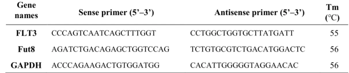
Total RNAs of Ba/F3 cell lines were extracted by Trizol reagent (Invitrogen), and then reverse-transcribed using a Prime Script RT Reagent kit with gDNA Eraser (Takara, Japan) according to the manufacturer’s instructions. The specific primers used for the PCR amplification are listed in Table 1. The GAPDH mRNA served as a control. The obtained reaction products were then subjected to 1.2% agarose gels containing ethidium bromide for electrophoresis.
9
Table 1. Primer sequences and annealing temperatures (Tm) for RT-PCR Gene
names Sense primer (5’–3’) Antisense primer (5’–3’) Tm (℃) FLT3 CCCAGTCAATCAGCTTTGGT CCTGGCTGGTGCTTATGATT 55
Fut8 AGATCTGACAGAGCTGGTCCAG TCTGTGCGTCTGACATGGACTC 56 GAPDH ACCCAGAAGACTGTGGATGG CACATTGGGGGTAGGAACAC 56
2.7 Western blotting and lectin blotting
Western blot analysis was performed as described previously (52). To prepare the cell lysate, cells were washed with chilled PBS three times and then lysed with cold lysis buffer (20 mM Tris-HCl (pH=7.4), 150 mM NaCl, 1% TritonX-100) containing proteases and phosphatase inhibitor cocktail (Nacalai Tesque). After being subjected to a rotor shaker for 40 min at 4 °C, cell lysates were cleared by centrifugation at 15,000 rpm, for 15 min at 4 ˚C, and the supernatant was transferred to new marked tubes. The protein concentration was determined using a PierceTM BCA protein assay kit (Thermo Fisher Scientific, Munich, Germany). Equal amounts of protein lysate were resolved by reducing SDS-PAGE. After electrophoresis, separated proteins were transferred to PVDF membranes (Millipore, Billerica, MA, USA) and incubated with indicated primary and secondary antibodies or with biotinylated lectins as indicated, and immunoreactive bands were detected using either an Immobilon Western Chemiluminescent HRP Substrate (Millipore) or a Vectastain ABC kit (Vector Laboratories, Burlingame, CA, USA), according to the manufacturer's instructions.
10 2.8 Cell proliferation assay
After washing three times with pre-warmed PBS, Ba/F3 parent cells then resuspended with the RPMI 1640 medium containing 10% FBS, 1 ng/ml recombinant murine IL-3, and 50 µmol/L of 2-mercaptoethanol. Ba/F3-FLT3-WT cells were then resuspended with the RPMI 1640 medium containing 10% FBS, 1 ng/ml recombinant murine IL-3, 400 µg/ml G418 and 50 µmol/L of 2-mercaptoethanol. Ba/F3-FLT3-ITD and Ba/F3-FLT3-TKD cells were maintained with RPMI 1640 medium containing 10% FBS, 400 µg/ml G418 and 50 µmol/L of 2-mercaptoethanol. At daily intervals, following staining with trypan blue, the viable cell number was calculated by counting the unstained cells with a hemocytometer.
Experiments were performed in biological triplicates.
2.9 Confocal microscopy analysis
Cells were processed for immunofluorescence microscopy as previously described (54).
Briefly, cells were washed and suspended in cold PBS containing WGA lectin for 30 min at 4 °C to stain the cell membrane. For intracellular staining, cells were fixed with 4%
paraformaldehyde for 20 min at RT, and washed twice with wash buffer (0.3% BSA in PBS).
Cells were plated on poly-L-lysine coated slides for 20 min at RT. To block non-specific staining, 400 µl of blocking buffer (10% normal donkey serum, 0.3% TritonX-100) was added with incubation for 45 min at RT. Cells were then stained with FLT3 antibody in dilution buffer (PBS, 1% bovine serum albumin, 1% normal donkey serum, 0.3% TritonX- 100, and 0.01% sodium azide) overnight at 4 °C followed by washing with wash buffer and incubation in secondary antibody and with TO-PRO-3. Slides were rinsed with PBS and
11
mounted using coverslips, and fluorescence was detected via sequential excitation using an Olympus FV1000 laser-scanning confocal microscope with an UPlanSApo×60/1.35 oil objective and high-sensitivity gallium arsenide phosphide detector units operated by F10- ASW version 4.02 software.
2.10 MTT assay
To evaluate the cytotoxic effects of PKC412, an MTT (#341-01823, Dojindo) assay was performed. The WT Ba/F3 cells expressing FLT3 were pre-treated with or without 2FF (100 μM) for 3 days. After 3 days, the WT Ba/F3 cells were plated in quintuplicate at 2×10⁴ cells per well in 96-well plates under normal media containing 2FF and PKC412 at indicated concentrations for another 48 hours. Then, a 10 μl MTT solution (5 mg/ml in PBS) was added to each well and let stand for 4 h at 37 °C. The plate was then centrifuged at 2,000 rpm, for 10 min. Subsequently, the supernatant was removed and 100 μl DMSO was added to each well at 37 °C, which was let stand for 10 min. Finally, absorption at 570 nm was measured using a microplate reader (Infinite® M1000, TECAN, Japan).
2.11 Chemical cross-linking of FLT3
To assay the dimerization of FLT3, a cross-linking experiment was performed as previously described (55). Briefly, 12 h prior to transfections 1x10⁶ cells were seeded on a 6 cm dish, and were then transfected with the FLT3-WT expression vector (1 μg) in 293T or Fut8KO 293T cells (42) using PEI MAX as described above. Then, the 293T cells were serum-starved for 8 h before incubation with or without human FLT3 ligand (FL) at 100 ng/ml for 30 min at 4 °C. Cross-linking was performed via incubation with 1 mM BS3 for 15
12
min at 4 °C. The reaction was stopped by adding glycine-HCl (pH=7.5) to a final concentration at 150 mM and incubating for 5 min. After washing three times with chilled PBS, these cells were harvested with RIPA buffer (Tris-HCl (pH=7.5), 150 mM NaCl, 1%
TritonX-100, 0.1% SDS). The cell lysates were subjected to 7.5% SDS-PAGE and analyzed by Western blotting with anti-FLT3 antibody.
2.12 Statistical analysis
Results are reported as the Means ± S.E.M. Statistical analyses were performed using an unpaired Student’s t test with Welch’s correction (one-tailed) using GraphPad Prism 5.0 software (GraphPad Software Inc.). p< 0.05 was regarded as statistically significant.
13 3. Results
3.1 Comparing of the glycosylation status of WT with that of FLT3 mutants
Glycosylation of proteins often results in a heterogeneous pattern on Western blotting.
Two forms of human FLT3 are known to exist. One is a mature form at around 150 kDa, which is thought to be completely glycosylated at the N-linked glycosylation sites of the extracellular domain of the Golgi apparatus to form complex types of N-glycans, and is then expressed on the cell surface. The other is an immature form around 130 kDa, which is incompletely glycosylated to form a high mannose type of N-glycans, and is mainly localized in the ER (23). To confirm the differences in the glycosylation status of FLT3 wild type and mutants, we transfected 293T cells with expression plasmids encoded with FLT3-WT (48), FLT3-ITD (49) or FLT3-TKD (47), and extracted cell lysates after 48 hours for Western blotting. The bands of FLT3-WT that migrated on SDS-PAGE were totally different from the other two mutants (Fig.4A), which could be neutralized by the removal of N-glycans via treatment with PNGase F. All bands of FLT3 were shifted to around 120 kDa upon PNGase F treatment. On the other hand, we also were curious about the effects of FLT3 on glycosylation. The expression levels of core fucosylation confirmed by AAL lectin, which preferentially recognizes core fucosylated N-glycans (42, 52), were enhanced by expression of FLT3 either WT or mutants (Fig.4B), whereas the reactive abilities with WGA lectin, which recognizes GlcNAc-containing total glycans were similar among those cells (Fig.4C).
To determine if the FLT3 was modified by core fucosylation, we performed immunoprecipitation with anti-FLT3 antibody in Ba/F3 cells. Unfortunately, the detection of FLT3 protein expression failed in the Ba/F3 cells. Therefore, we used the 293T cells instead
14
to improve the protein expression levels, and found FLT3 was core fucosylation, which was confirmed by AAL lectin blotting, as shown in Fig. 1D. To explore whether the increased core fucosylation was due to a promotion of Fut8 expression by FLT3, semi-quantitative RT- PCR was performed and showed that FLT3 was not expressed in the CTR cells (Fig.4E).
Overexpression of either WT- or mutant-FLT3 greatly induced Fut8 mRNA expression (Fig.
4F). On the other hand, the increased levels of core fucosylation in those FLT3 expressing cells were inhibited by PKC412 (Fig. 4G), a FLT3 kinase inhibitor, suggesting the FLT3- mediated signaling may induce Fut8 expression. That result suggested that core fucosylation is specifically increased by the expression of either wild type or mutant FLT3.
15
Figure 4. Glycosylation patterns of FLT3 proteins and the effects on fucosylation expression. A) The 293T cells were cultured at 70% confluency, and then transfected with plasmids containing human FLT3-WT, FLT3-ITD or FLT3-TKD. At 48 h post transfection, the cell lysates were treated with or without PNGase F, and analyzed by Western blotting with anti-FLT3 antibody. The effects of FLT3 WT or mutants on core fucosylation in Ba/F3 cells were examined by AAL lectin blot B) or WGA lectin blot C). The same amounts of cell lysates were separated on 7.5% SDS-PAGE, and the membranes were probed with AAL (top panel), and then re-probed with anti-GAPDH antibody (bottom panel), which was used as a loading control. D) The same amounts of cell lysates from those indicated transfected 293T cells were immunoprecipitated with anti-FLT3 antibody, and then the immunoprecipitates were probed with AAL lectin (upper panel) and re-probed with anti-FLT3 antibody (lower panel) as a loading control. E) RT-PCR using total RNA extracted from different Ba/F3 cells were carried out to examine the mRNA levels of FLT3. GAPDH was used as control. F) RT- PCR was carried out to examine the mRNA levels of Fut8 in different Ba/F3 cells as indicated.
GAPDH was used as control. G) The same amounts of cell lysates from the different Ba/F3 cells treated with or without PKC412 (1 nM) for 48 h, were separated on 7.5% SDS-PAGE, and the membranes were probed with AAL (top panel), and then re-probed with anti-GAPDH antibody (bottom panel), which was used as a loading control.
3.2 Establishment of FLT3-expressed Fut8KO Ba/F3 cells
To understand the mechanisms of induction for core fucosylation by FLT3, we established Fut8KO cell lines using a murine Ba/F3 cell line, an IL-3-dependent cell line that exhibits IL-3 independence in the presence of oncogenic signals such as FLT3-ITD and -
16
TKD (18). The pSpCas9 (BB)-2A-GFP vector containing target sequences were transduced into those Ba/F3 cell lines as indicated by electroporation, and stable cell lines were established by positive and negative sorting with GFP and PhosL lectin as described in the
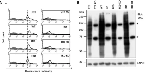
“Methods and Materials” section. Finally, we successfully confirmed stable Fut8KO cell lines by flow cytometric analysis using biotinylated PhosL lectin (Fig. 5A) and lectin blotting with AAL (Fig.5B), which specifically recognizes core fucosylated N-glycans (52, 56).
Figure 5. Established Fut8 deficient Ba/F3 cells. A) The parent (CTR), FLT3 WT and mutants of Ba/F3 cells, and Fut8KO cells were collected and incubated with (bold line) or without (gray shadow) the PhosL lectin, which preferentially recognizes core fucosylated N- glycans, followed by incubation with Biotinylated Alexa Fluor 647 streptavidin and flow cytometric analysis. The vertical dashed lines indicate the peak reacted with PhosL lectin expression in CTR cells. B) Equal amounts of cell lysates from CTR and stably expressed FLT3-WT or mutants were analyzed by immunoblot with AAL lectin; GAPDH served as a loading control. Asterisks represent non-specific bands.
17
3.3 IL-3 independent proliferation in Fut8 knockout- FLT3-WT Ba/F3 cells
FLT3 plays a crucial role in normal hematopoietic processes such as proliferation, differentiation, and survival (8, 12). To test whether Fut8KO regulates cell proliferation, we compared the viable cell number in different Ba/F3 cell lines. Fut8KO slightly increased cell proliferation in the presence of IL-3 in the CTR (Fig. 6A). Quite interestingly, Fut8KO induced cell proliferation in FLT3-WT cells even in the absence of IL-3 (Fig. 6B), whereas the Fut8KO CTR showed no IL-3-independent proliferative activity (Fig. 6A). Furthermore, the restoration with Fut8 gene in the Fut8KO WT cells (Rescue cell) resulted in IL-3- dependent cell growth (Fig. 6B). The cell proliferation of the rescue cells was completely blocked in the absence of IL-3. These results suggest that FLT3 expression is necessary for Fut8KO-induced IL-3-independent cell proliferation. In addition, ablation of Fut8 partially blocked ITD (Fig. 6C) and TKD (Fig. 6D) cell proliferation. These results suggest that the lack of core fucosylation dramatically activates FLT3-WT. To clarify FLT3 receptor activation mechanisms related to core fucosylation depletion, we mainly focused on the FLT3-WT in subsequent studies.
18
Figure 6. Influences of core fucosylation on Ba/F3 cell proliferation. A) The parent Ba/F3 cells (CTR) and CTR Fut8KO cells were cultured under normal culture media with or without IL-3 at 1 ng/ml final concentration, and the cell numbers of living cells were measured by trypan blue exclusion assay at indicated times. CTR KO with IL-3 versus CTR KO without IL-3. B) The parent cells expressing FLT3-WT (WT) or Fut8KO cells expressing FLT3-WT (WT KO), and the WT KO cells restored with Fut8 gene (WT Rescue) were cultured. Cell numbers were counted as described above. WT KO and WT Rescue with IL-3 versus WT KO and WT Rescue without IL-3, respectively. The parent or Fut8KO cells expressing FLT3- ITD (C) or FLT3-TKD (D) were also cultured under normal culture media without IL-3 for 24, 48 and 72 hours. The numbers of living cells were measured by trypan blue exclusion assay. Data represent the average of three independent experiments. All values are Means ± S.E.M (n=3). *p< 0.05. **p< 0.01.
19
3.4 Effects of Fut8KO on cellular signaling in the FLT3-WT Ba/F3 cells
Next, we performed Western blotting to analyze the activation status of STAT5, AKT, and ERK signaling, which are important pathways for FLT3 WT and mutant downstream signaling (18, 20, 57). Consistently, FLT3-WT could not induce phosphorylation of STAT5 in the absence of IL-3. However, Fut8KO greatly induced phosphorylation of STAT5 even without IL-3 (Fig. 7A). The activation of AKT and ERK was also observed in the Fut8KO cells (Fig. 7A). Importantly, these inductions were blocked by the re-expression of Fut8 in the KO cells (Fig. 7A). The core fucosylation was confirmed by AAL lectin blotting (Fig.
7B).
Figure 7. Effects of core fucosylation on intracellular signaling. The wild type (WT), Fut8KO (KO), or restoration of Fut8 in the KO cells (Res) of Ba/F3 cells expressing FLT3 were cultured under normal culture media with (+) or without IL-3 (-) for 24 h. A) The same amounts of cell lysates were separated on 7.5% SDS-PAGE, and Western blotting with the indicated antibodies including both total and phospho-STAT5, ERK and AKT. B) The expression levels of core fucosylation in these cells were blotted with AAL (top panel) and re-probed with anti-GAPDH (low panel). GAPDH served as a loading control.
20
3.5 Deficiency of core fucosylation did not affect FLT3 intracellular localization Changes in FLT3 localization are known to influence downstream signaling. Mutations of FLT3 receptor result in localization in the endoplasmic reticulum, which affects surface glycoprotein maturation and activates STAT5, while ligand-activated wild type FLT3 is mainly localized on the cell surface and activates MAP kinase (22, 58, 59). Therefore, we next performed immunostaining with FLT3 antibody to examine the effects of core fucosylation on FLT3 intracellular localization. The FLT3-WT proteins were mainly localized on the cell surface, and co-localized with WGA staining (Fig. 8, upper panel). In Fut8KO cells, the FLT3-WT proteins were also localized on the cell surface (Fig. 8, middle panel). The localization pattern was quite different from that of FLT3-ITD, in which FLT3- ITD proteins were mainly localized in the intracellular domain (Fig. 8, lower panel), presumably in the ER, as previously described (22, 60). These results suggest that the underlying molecular mechanism for the induction of FLT3-WT by Fut8KO may be different from activation in FLT3-ITD mutation.
21
Figure 8. Effects of core fucosylation on FLT3 intracellular localization. The indicated cells were stained for plasma membrane using WGA lectin (Green), fixation and then adhered to slides coated with poly-L-lysine for further staining with FLT3 (Red) and TO-PRO-3 (Blue). Slides were visualized by immunofluorescence microscopy using an Olympus FV1000 laser-scanning confocal microscope as described in Methods and Materials. Bar shows 30 µm.
3.6 Deficiency of core fucosylation increased cellular tyrosine phosphorylation levels and dimerization of FLT3
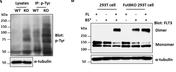
Following the revelation that Fut8KO resulted in the activation of FLT3-WT downstream signaling, we next examined the tyrosine phosphorylation levels of FLT3. The intracellular domain of FLT3 has 10 potential tyrosine phosphorylation sites (61, 62). The cell lysates and immunoprecipitates with p-Tyr agarose (PY20) from indicated cells were examined. As a
22
result, ablation of core fucosylation potently increased total tyrosine phosphorylation levels (Fig. 9A). There is evidence that the dimerization of FLT3-WT is an initial and essential event in ligand-induced signal transduction (63). Therefore, we compared FLT3 dimerization in 293T and Fut8KO 293T cells with or without FLT3 ligand (FL). As shown in Fig. 9B, the dimer formation of FLT3 in wild type or Fut8KO 293T cells could be induced by FL.
Interestingly, dimerization was observed in the Fut8KO 293T cells even without FL, but not in the wild type 293T cells. In addition, the upper band of FLT3 (around 150 kDa, so-called mature form) rather than the lower band (around 130 kDa, so-called immature form) participated in the dimer formation. Collectively, these data suggest that the deficiency of core fucosylation promotes the dimerization of FLT3 and results in the aberrant activation of FLT3-WT-mediated signaling such as p-STAT5, p-ERK and p-AKT, which induces an IL- 3-independent cell proliferation pathway in the Ba/F3 cell system.
Figure 9. Deficiency of core fucosylation increased cellular tyrosine phosphorylation levels and dimerization of FLT3. The wild type (WT) and Fut8KO (KO) of Ba/F3 cells
23
expressing FLT3 were cultured under normal culture media with or without IL-3 for 48 h, respectively. A) The same amounts of cell lysates and immunoprecipitates with p-Tyr agarose (PY20) were separated on 7.5% SDS-PAGE, and the membranes were blotted with anti-p-Tyr (PY99) and re-probed with anti-α-tubulin, as a loading control. B) The FLT3-WT plasmid was transfected into 293T or Fut8KO 293T cells. After 48 hours, cells were treated for chemical cross-linking as described under “Materials and methods”. Cell lysates were separated on 7.5% SDS-PAGE, and blotted with anti-FLT3 antibody to detect FLT3 monomer and dimer. α-Tubulin was used as a loading control.
3.7 PKC412 efficiently inhibited cell proliferation of Fut8KO cells in a relatively lower dose
Since Fut8KO greatly induced tyrosine phosphorylation as described above, we examined the effects of PKC412, a tyrosine kinase inhibitor, on cell proliferation. In fact, it was reported that the inhibitory effects of PKC412 on cell proliferation could be observed with usage at 500 nM, but even at 100 nM in Ba/F3 cells expressed FLT3 (64). However, it was interesting, that PKC412 significantly inhibited cell proliferation even at 1 nM, but did not affect cell proliferation of the WT cells (Fig.10A). A fluorinated analog of fucose, 2FF, functions as a metabolic fucosylation inhibitor, which is taken up by cells and converted to GDP-2FF through endogenous salvage pathways (65, 66). Therefore, 2FF could be considered a specific inhibitor for fucosylation. In fact, a treatment with 2FF at 100 µM suppressed core fucosylation in HepG2 cells (32). We also confirmed the inhibitory effect on Ba/F3 cells. Similar to the Fut8KO cells, the sensitivities for the inhibitory effects of PKC412 on cell proliferation were also increased in the cells pretreated with 2FF (Fig. 10B). These
24
results suggested that a deficiency of core fucosylation induces a novel signal pathway, but not like the IL-3-dependent signal pathway, which is more sensitive to treatment with a tyrosine kinase inhibitor.
Figure 10. Loss of core fucosylation chemosensitized the cells to PKC412, a tyrosine kinase inhibitor. A) The wild type (WT) and the Fut8KO (KO) of Ba/F3 cells expressing FLT3-WT were cultured in the presence of PKC412 at indicated concentrations, and then cell numbers were examined after 48 hours. The WT and KO cells were cultured with or without IL-3, respectively. The inhibitory ratio was normalized to that of each group without the inhibitor as 1. Data are Means ± S.E.M (n=3). *p<0.05. B) The WT Ba/F3 cells expressing FLT3 were pre-treated with or without 2FF (100 μM) for 3 days, and then cultured under normal media containing 2FF and PKC412 at indicated concentrations for another 48 hours.
The cell numbers were measured using MTT assay. The inhibitory ratio was normalized to that of each group without the inhibitor as 1. Data are Means ± S.E.M (n=3). *p<0.05.
25 4. Discussion
FLT3 signaling is important for normal and oncogenic hematopoiesis, but the downstream effect from the modification of FLT3 surface glycoprotein remains to be elucidated. In the current study, we used the CRISPR/Cas9 system to establish Fut8KO Ba/F3 cell lines, and found that a deficiency of Fut8 resulted in cell proliferation in an IL-3 independent manner in FLT3-expressing cells, but not in FLT3-negative cells, which suggests that this cell proliferation is dependent on FLT3 expression (Fig. 11). Of course, we could not exclude the influences of Fut8KO from other receptors including the IL-3 receptor.
We revealed a novel effect for a deficiency of core fucosylation for FLT3 homodimerization and activation of several downstream signaling pathways in the absence of a ligand.
Furthermore, blockage of core fucosylation by Fut8KO or 2FF, a fucosylation inhibitor, greatly increased sensitivities for the suppression of cell proliferation when using PKC412, a tyrosine kinase inhibitor. Therefore, the manipulation of core fucosylation could provide valuable direction for the development of drugs that could be effective in treating AML.
As mentioned above, core fucosylation is an important regulator for receptor-mediated signaling. Our group recently reported that Fut8 deficiency increases sensitivities to inflammatory stimulators such as IFN-γ or IL-6 in glial cell lines (52), and sensitivities for postsynaptic depolarization by enhancing the heteromerization of AMPARs (37), suggesting that without core fucosylation these receptors might exist in an active state. In contrast, core fucosylation represses several receptor functions as well. Deletion of core fucosylation down- regulated the EGF-induced phosphorylation of EGF receptor (40) and TGF-β1 receptor- mediated Smad activation (39). Sialylation and fucosylation of EGF receptors suppresses
26
their function and activation in lung cancer cells (67). These data suggest that core fucosylation either positively or negatively affects receptor functions. In the present study, we also found that the effect of core fucosylation differs between WT and FLT3 mutant receptors. Considering the FLT3-TKD or -ITD mutant could activate downstream signaling pathways through different mechanisms such as conformation change, intracellular localization and/or modification status, which might be quite different from wild type FLT3.
Therefore, we could speculate that the disruption of Fut8 affects other glycoproteins, and interferes with FLT3 signaling to down-regulate cell proliferation. Further clarifying the mechanisms of how fucosylation inhibition activates/suppresses FLT3 receptors may be helpful in developing novel targeted therapies for hematological malignancies.
Several important residues in FLT3 activation have been reported, which includes tyrosine residues 589 and 591 for ligand-dependent activation of FLT3-WT Ba/F3 cells (68).
Tyrosine 589 and 591 are also reported to play important roles in STAT5 activation and transformation by FLT3-ITD (69). Masson et al. (61) reported that tyrosines 768, 955 and 969 of FLT3, as phosphorylation sites and mediators of growth factor receptor binding protein (Grb2) interactions, lead to the association of Grb2-associated binder 2 (Gab2), which contributes to proliferation and survival. That study revealed that these residues are important for the activation of STAT5 and AKT (61). STAT5 is mainly activated by FLT3-ITD signaling via aberrant localization in the ER of FLT3-ITD, but it is barely activated by FLT3- WT signaling (22). We observed that the depletion of core fucosylation did not change the transmembrane localization of FLT3-WT. However, FLT3-WT activates not only ERK and AKT, but also activates STAT5 in the Fut8KO cells (Fig. 11). We postulate that the activation
27
of STAT5 via the depletion of core fucosylation happens not by an intracellular localization change in FLT3, but either by phosphorylation or by some modifications of the important residues of FLT3. The activation mechanisms of FLT3 receptor via the depletion of core fucosylation should be clarified in the future.
FLT3 is reported to undergo glycosylation in the endoplasmic reticulum (23), and several glycosylation inhibitors were examined for their effect on the functions of FLT3. One of these compounds is fluvastatin, which has already been approved by the FDA and is a clinically applied inhibitor of mevalonate synthesis. Apart from blocking cholesterol synthesis, fluvastatin also inhibits N-glycosylation by depleting the cells of dolicholphosphate, thus leading to a loss of surface expression and the induction of cell death in Ba/F3 cells (54). The other compound is 2-deoxy-D-glucose, which not only depletes cells of ATP but also impairs N-glycosylation (70). A possible reason for the selective inhibition of FLT3-ITD-positive cells by compounds affecting glycosylation may be a further shift of FLT3-ITD towards intracellular localization, thereby abrogating signaling from the cell surface and in turn cell transformation. Tunicamycin is a bacterial antibiotic, which specifically inhibits the transfer of activated sugars to dolichol phosphate, an essential step in the N-glycosylation of proteins in the ER (71, 72). These effects are partly mediated by arresting FLT3-ITD in an under-glycosylated state and thereby attenuating FLT3-ITD-driven AKT and ERK signaling (73). Because FLT3 plays a very important role in the pathogenesis of AML, various FLT3 inhibitors have been developed (74). However, their duration of clinical response is short because of the rapid development of resistance (75). We found that depletion of fucosylation by Fut8KO in combination with PKC412 efficiently decreases the
28
factor-independent growth of FLT3-WT cells. Inhibition of N-glycosylation may be a possible approach for cancer therapy. Theoretically, however, inhibition of core fucosylation inhibitors is less harmful and should have fewer side effects. To date, several fucosylation inhibitors have been developed (65, 66, 76), and there are many good examples of combination therapy with FLT3 inhibitors for AML (77-79). Therefore, the modulation of FLT3 glycosylation could provide hints for the development of new therapies for different types of FLT3-mediated hematological malignancies.
29
Figure 11. A working model for the role of core fucosylation in regulating FLT3 activity in Ba/F3 cells. As described previously, FLT3 is a member of the class III receptor tyrosine kinase family, which exists in the extracellular (EC) domain that contains 9 potential N- glycosylation sites, a transmembrane (TM), a juxtamembrane (JM), and tyrosine kinase (TK) domains (25). Ba/F3 cells are an IL-3-dependent cell line, and in the presence of IL-3, STAT5 is potently activated (80) (shown in green arrow and green letters). The FLT3-WT signal mainly activates the ERK and AKT pathway in the absence of FLT3 ligand (17) (red arrow and red letters). In the present study, the deficiency of core fucosylation induced an IL-3- independent cell proliferation pathway in the Ba/F3 cell system, in which the expression of FLT3 is essential. A lack of core fucosylation triggers ligand-independent FLT3 dimerization on the cell surface, resulting in aberrant activation of FLT3-mediated signaling such as p- STAT5, p-ERK and p-AKT (yellow arrow), which are different from the observations in the FLT3-ITD and -TKD mutants. Those two mutants are mainly expressed in the ER, and participate in activation of the STAT5 signaling pathway, rather than the ERK and AKT signaling pathways (22). Thus, the underlying molecular mechanism for the induction of FLT3-WT by Fut8KO could be different from the activation in the FLT3-mutants, which could provide valuable direction for the development of drugs that could be effective in the treatment of AML.
30 5. References
1. Siegel, R. L., Miller, K. D., and Jemal, A. (2016) Cancer statistics, 2016. CA Cancer J Clin 66, 7-30
2. Arber, D. A., Orazi, A., Hasserjian, R., Thiele, J., Borowitz, M. J., Le Beau, M. M., Bloomfield, C. D., Cazzola, M., and Vardiman, J. W. (2016) The 2016 revision to the World Health Organization classification of myeloid neoplasms and acute leukemia.
Blood 127, 2391-2405
3. Cancer Genome Atlas Research, N., Ley, T. J., Miller, C., Ding, L., Raphael, B. J., Mungall, A. J., Robertson, A., Hoadley, K., Triche, T. J., Jr., Laird, P. W., Baty, J.
D., Fulton, L. L., Fulton, R., Heath, S. E., Kalicki-Veizer, J., Kandoth, C., Klco, J.
M., Koboldt, D. C., Kanchi, K. L., Kulkarni, S., Lamprecht, T. L., Larson, D. E., Lin, L., Lu, C., McLellan, M. D., McMichael, J. F., Payton, J., Schmidt, H., Spencer, D.
H., Tomasson, M. H., Wallis, J. W., Wartman, L. D., Watson, M. A., Welch, J., Wendl, M. C., Ally, A., Balasundaram, M., Birol, I., Butterfield, Y., Chiu, R., Chu, A., Chuah, E., Chun, H. J., Corbett, R., Dhalla, N., Guin, R., He, A., Hirst, C., Hirst, M., Holt, R.
A., Jones, S., Karsan, A., Lee, D., Li, H. I., Marra, M. A., Mayo, M., Moore, R. A., Mungall, K., Parker, J., Pleasance, E., Plettner, P., Schein, J., Stoll, D., Swanson, L., Tam, A., Thiessen, N., Varhol, R., Wye, N., Zhao, Y., Gabriel, S., Getz, G., Sougnez, C., Zou, L., Leiserson, M. D., Vandin, F., Wu, H. T., Applebaum, F., Baylin, S. B., Akbani, R., Broom, B. M., Chen, K., Motter, T. C., Nguyen, K., Weinstein, J. N., Zhang, N., Ferguson, M. L., Adams, C., Black, A., Bowen, J., Gastier-Foster, J., Grossman, T., Lichtenberg, T., Wise, L., Davidsen, T., Demchok, J. A., Shaw, K. R.,
31
Sheth, M., Sofia, H. J., Yang, L., Downing, J. R., and Eley, G. (2013) Genomic and epigenomic landscapes of adult de novo acute myeloid leukemia. N Engl J Med 368, 2059-2074
4. McKerrell, T., Park, N., Moreno, T., Grove, C. S., Ponstingl, H., Stephens, J., Understanding Society Scientific, G., Crawley, C., Craig, J., Scott, M. A., Hodkinson, C., Baxter, J., Rad, R., Forsyth, D. R., Quail, M. A., Zeggini, E., Ouwehand, W., Varela, I., and Vassiliou, G. S. (2015) Leukemia-associated somatic mutations drive distinct patterns of age-related clonal hemopoiesis. Cell Rep 10, 1239-1245
5. Papaemmanuil, E., Gerstung, M., Bullinger, L., Gaidzik, V. I., Paschka, P., Roberts, N. D., Potter, N. E., Heuser, M., Thol, F., Bolli, N., Gundem, G., Van Loo, P., Martincorena, I., Ganly, P., Mudie, L., McLaren, S., O'Meara, S., Raine, K., Jones, D. R., Teague, J. W., Butler, A. P., Greaves, M. F., Ganser, A., Dohner, K., Schlenk, R. F., Dohner, H., and Campbell, P. J. (2016) Genomic Classification and Prognosis in Acute Myeloid Leukemia. N Engl J Med 374, 2209-2221
6. Jaiswal, S., Fontanillas, P., Flannick, J., Manning, A., Grauman, P. V., Mar, B. G., Lindsley, R. C., Mermel, C. H., Burtt, N., Chavez, A., Higgins, J. M., Moltchanov, V., Kuo, F. C., Kluk, M. J., Henderson, B., Kinnunen, L., Koistinen, H. A., Ladenvall, C., Getz, G., Correa, A., Banahan, B. F., Gabriel, S., Kathiresan, S., Stringham, H.
M., McCarthy, M. I., Boehnke, M., Tuomilehto, J., Haiman, C., Groop, L., Atzmon, G., Wilson, J. G., Neuberg, D., Altshuler, D., and Ebert, B. L. (2014) Age-related clonal hematopoiesis associated with adverse outcomes. N Engl J Med 371, 2488- 2498
32
7. Xie, M., Lu, C., Wang, J., McLellan, M. D., Johnson, K. J., Wendl, M. C., McMichael, J. F., Schmidt, H. K., Yellapantula, V., Miller, C. A., Ozenberger, B. A., Welch, J. S., Link, D. C., Walter, M. J., Mardis, E. R., Dipersio, J. F., Chen, F., Wilson, R. K., Ley, T. J., and Ding, L. (2014) Age-related mutations associated with clonal hematopoietic expansion and malignancies. Nat Med 20, 1472-1478
8. Gilliland, D. G., and Griffin, J. D. (2002) The roles of FLT3 in hematopoiesis and leukemia. Blood 100, 1532-1542.
9. Mrozek, K., Marcucci, G., Paschka, P., Whitman, S. P., and Bloomfield, C. D. (2007) Clinical relevance of mutations and gene-expression changes in adult acute myeloid leukemia with normal cytogenetics: are we ready for a prognostically prioritized molecular classification? Blood 109, 431-448
10. Steffen, B., Muller-Tidow, C., Schwable, J., Berdel, W. E., and Serve, H. (2005) The molecular pathogenesis of acute myeloid leukemia. Crit Rev Oncol Hematol 56, 195- 221
11. Levis, M., and Small, D. (2003) FLT3: ITDoes matter in leukemia. Leukemia 17, 1738-1752
12. Kazi, J. U., and Ronnstrand, L. (2019) FMS-like Tyrosine Kinase 3/FLT3: From Basic Science to Clinical Implications. Physiol Rev 99, 1433-1466
13. Nakao, M., Yokota, S., Iwai, T., Kaneko, H., Horiike, S., Kashima, K., Sonoda, Y., Fujimoto, T., and Misawa, S. (1996) Internal tandem duplication of the flt3 gene found in acute myeloid leukemia. Leukemia 10, 1911-1918
33
14. Schnittger, S., Schoch, C., Dugas, M., Kern, W., Staib, P., Wuchter, C., Loffler, H., Sauerland, C. M., Serve, H., Buchner, T., Haferlach, T., and Hiddemann, W. (2002) Analysis of FLT3 length mutations in 1003 patients with acute myeloid leukemia:
correlation to cytogenetics, FAB subtype, and prognosis in the AMLCG study and usefulness as a marker for the detection of minimal residual disease. Blood 100, 59- 66
15. Yamamoto, Y., Kiyoi, H., Nakano, Y., Suzuki, R., Kodera, Y., Miyawaki, S., Asou, N., Kuriyama, K., Yagasaki, F., Shimazaki, C., Akiyama, H., Saito, K., Nishimura, M., Motoji, T., Shinagawa, K., Takeshita, A., Saito, H., Ueda, R., Ohno, R., and Naoe, T. (2001) Activating mutation of D835 within the activation loop of FLT3 in human hematologic malignancies. Blood 97, 2434-2439
16. Bacher, U., Haferlach, C., Kern, W., Haferlach, T., and Schnittger, S. (2008) Prognostic relevance of FLT3-TKD mutations in AML: the combination matters--an analysis of 3082 patients. Blood 111, 2527-2537
17. Takahashi, S. (2011) Downstream molecular pathways of FLT3 in the pathogenesis of acute myeloid leukemia: biology and therapeutic implications. J Hematol Oncol 4, 13
18. Hayakawa, F., Towatari, M., Kiyoi, H., Tanimoto, M., Kitamura, T., Saito, H., and Naoe, T. (2000) Tandem-duplicated Flt3 constitutively activates STAT5 and MAP kinase and introduces autonomous cell growth in IL-3-dependent cell lines. Oncogene 19, 624-631.
34
19. Brandts, C. H., Sargin, B., Rode, M., Biermann, C., Lindtner, B., Schwable, J., Buerger, H., Muller-Tidow, C., Choudhary, C., McMahon, M., Berdel, W. E., and Serve, H. (2005) Constitutive activation of Akt by Flt3 internal tandem duplications is necessary for increased survival, proliferation, and myeloid transformation. Cancer Res 65, 9643-9650
20. Choudhary, C., Schwable, J., Brandts, C., Tickenbrock, L., Sargin, B., Kindler, T., Fischer, T., Berdel, W. E., Muller-Tidow, C., and Serve, H. (2005) AML-associated Flt3 kinase domain mutations show signal transduction differences compared with Flt3 ITD mutations. Blood 106, 265-273
21. Leischner, H., Albers, C., Grundler, R., Razumovskaya, E., Spiekermann, K., Bohlander, S., Ronnstrand, L., Gotze, K., Peschel, C., and Duyster, J. (2012) SRC is a signaling mediator in FLT3-ITD- but not in FLT3-TKD-positive AML. Blood 119, 4026-4033
22. Choudhary, C., Olsen, J. V., Brandts, C., Cox, J., Reddy, P. N., Bohmer, F. D., Gerke, V., Schmidt-Arras, D. E., Berdel, W. E., Muller-Tidow, C., Mann, M., and Serve, H.
(2009) Mislocalized activation of oncogenic RTKs switches downstream signaling outcomes. Mol Cell 36, 326-339
23. Schmidt-Arras, D. E., Bohmer, A., Markova, B., Choudhary, C., Serve, H., and Bohmer, F. D. (2005) Tyrosine phosphorylation regulates maturation of receptor tyrosine kinases. Mol Cell Biol 25, 3690-3703
35
24. Hospital, M. A., Green, A. S., Maciel, T. T., Moura, I. C., Leung, A. Y., Bouscary, D., and Tamburini, J. (2017) FLT3 inhibitors: clinical potential in acute myeloid leukemia. Onco Targets Ther 10, 607-615
25. Verstraete, K., Vandriessche, G., Januar, M., Elegheert, J., Shkumatov, A. V., Desfosses, A., Van Craenenbroeck, K., Svergun, D. I., Gutsche, I., Vergauwen, B., and Savvides, S. N. (2011) Structural insights into the extracellular assembly of the hematopoietic Flt3 signaling complex. Blood 118, 60-68
26. Helenius, A., and Aebi, M. (2001) Intracellular functions of N-linked glycans.
Science 291, 2364-2369
27. Ohtsubo, K., and Marth, J. D. (2006) Glycosylation in cellular mechanisms of health and disease. Cell 126, 855-867
28. Pinho, S. S., and Reis, C. A. (2015) Glycosylation in cancer: mechanisms and clinical implications. Nat Rev Cancer 15, 540-555
29. Wilson, J. R., Williams, D., and Schachter, H. (1976) The control of glycoprotein synthesis: N-acetylglucosamine linkage to a mannose residue as a signal for the attachment of L-fucose to the asparagine-linked N-acetylglucosamine residue of glycopeptide from alpha1-acid glycoprotein. Biochem Biophys Res Commun 72, 909- 916
30. Schneider, M., Al-Shareffi, E., and Haltiwanger, R. S. (2017) Biological functions of fucose in mammals. Glycobiology 27, 601-618
31. Wang, Y., Fukuda, T., Isaji, T., Lu, J., Im, S., Hang, Q., Gu, W., Hou, S., Ohtsubo, K., and Gu, J. (2015) Loss of alpha1,6-fucosyltransferase inhibits chemical-induced
36
hepatocellular carcinoma and tumorigenesis by down-regulating several cell signaling pathways. FASEB J 29, 3217-3227
32. Zhou, Y., Fukuda, T., Hang, Q., Hou, S., Isaji, T., Kameyama, A., and Gu, J. (2017) Inhibition of fucosylation by 2-fluorofucose suppresses human liver cancer HepG2 cell proliferation and migration as well as tumor formation. Sci Rep 7, 11563
33. Fujii, H., Shinzaki, S., Iijima, H., Wakamatsu, K., Iwamoto, C., Sobajima, T., Kuwahara, R., Hiyama, S., Hayashi, Y., Takamatsu, S., Uozumi, N., Kamada, Y., Tsujii, M., Taniguchi, N., Takehara, T., and Miyoshi, E. (2016) Core Fucosylation on T Cells, Required for Activation of T-Cell Receptor Signaling and Induction of Colitis in Mice, Is Increased in Patients With Inflammatory Bowel Disease.
Gastroenterology 150, 1620-1632
34. Li, W., Yu, R., Ma, B., Yang, Y., Jiao, X., Liu, Y., Cao, H., Dong, W., Liu, L., Ma, K., Fukuda, T., Liu, Q., Ma, T., Wang, Z., Gu, J., Zhang, J., and Taniguchi, N. (2015) Core fucosylation of IgG B cell receptor is required for antigen recognition and antibody production. J Immunol 194, 2596-2606
35. Liang, W., Mao, S., Sun, S., Li, M., Li, Z., Yu, R., Ma, T., Gu, J., Zhang, J., Taniguchi, N., and Li, W. (2018) Core Fucosylation of the T Cell Receptor Is Required for T Cell Activation. Front Immunol 9, 78
36. Fukuda, T., Hashimoto, H., Okayasu, N., Kameyama, A., Onogi, H., Nakagawasai, O., Nakazawa, T., Kurosawa, T., Hao, Y., Isaji, T., Tadano, T., Narimatsu, H., Taniguchi, N., and Gu, J. (2011) Alpha1,6-fucosyltransferase-deficient mice exhibit multiple behavioral abnormalities associated with a schizophrenia-like phenotype:
37
importance of the balance between the dopamine and serotonin systems. J Biol Chem 286, 18434-18443
37. Gu, W., Fukuda, T., Isaji, T., Hang, Q., Lee, H. H., Sakai, S., Morise, J., Mitoma, J., Higashi, H., Taniguchi, N., Yawo, H., Oka, S., and Gu, J. (2015) Loss of alpha1,6- Fucosyltransferase Decreases Hippocampal Long Term Potentiation:
IMPLICATIONS FOR CORE FUCOSYLATION IN THE REGULATION OF AMPA RECEPTOR HETEROMERIZATION AND CELLULAR SIGNALING. J Biol Chem 290, 17566-17575
38. Ng, B. G., Xu, G., Chandy, N., Steyermark, J., Shinde, D. N., Radtke, K., Raymond, K., Lebrilla, C. B., AlAsmari, A., Suchy, S. F., Powis, Z., Faqeih, E. A., Berry, S. A., Kronn, D. F., and Freeze, H. H. (2018) Biallelic Mutations in FUT8 Cause a Congenital Disorder of Glycosylation with Defective Fucosylation. Am J Hum Genet 102, 188-195
39. Wang, X., Inoue, S., Gu, J., Miyoshi, E., Noda, K., Li, W., Mizuno-Horikawa, Y., Nakano, M., Asahi, M., Takahashi, M., Uozumi, N., Ihara, S., Lee, S. H., Ikeda, Y., Yamaguchi, Y., Aze, Y., Tomiyama, Y., Fujii, J., Suzuki, K., Kondo, A., Shapiro, S.
D., Lopez-Otin, C., Kuwaki, T., Okabe, M., Honke, K., and Taniguchi, N. (2005) Dysregulation of TGF-beta1 receptor activation leads to abnormal lung development and emphysema-like phenotype in core fucose-deficient mice. Proc Natl Acad Sci U S A 102, 15791-15796
38
40. Wang, X., Gu, J., Ihara, H., Miyoshi, E., Honke, K., and Taniguchi, N. (2006) Core fucosylation regulates epidermal growth factor receptor-mediated intracellular signaling. J Biol Chem 281, 2572-2577
41. Zhao, Y., Itoh, S., Wang, X., Isaji, T., Miyoshi, E., Kariya, Y., Miyazaki, K., Kawasaki, N., Taniguchi, N., and Gu, J. (2006) Deletion of core fucosylation on alpha3beta1 integrin down-regulates its functions. J Biol Chem 281, 38343-38350 42. Gu, W., Fukuda, T., Isaji, T., Hashimoto, H., Wang, Y., and Gu, J. (2013) alpha1,6-
Fucosylation regulates neurite formation via the activin/phospho-Smad2 pathway in PC12 cells: the implicated dual effects of Fut8 for TGF-beta/activin-mediated signaling. FASEB J 27, 3947-3958
43. Shields, R. L., Lai, J., Keck, R., O'Connell, L. Y., Hong, K., Meng, Y. G., Weikert, S. H., and Presta, L. G. (2002) Lack of fucose on human IgG1 N-linked oligosaccharide improves binding to human Fcgamma RIII and antibody-dependent cellular toxicity. J Biol Chem 277, 26733-26740
44. Shinkawa, T., Nakamura, K., Yamane, N., Shoji-Hosaka, E., Kanda, Y., Sakurada, M., Uchida, K., Anazawa, H., Satoh, M., Yamasaki, M., Hanai, N., and Shitara, K.
(2003) The absence of fucose but not the presence of galactose or bisecting N- acetylglucosamine of human IgG1 complex-type oligosaccharides shows the critical role of enhancing antibody-dependent cellular cytotoxicity. J Biol Chem 278, 3466- 3473
45. Okada, M., Chikuma, S., Kondo, T., Hibino, S., Machiyama, H., Yokosuka, T., Nakano, M., and Yoshimura, A. (2017) Blockage of Core Fucosylation Reduces Cell-
39
Surface Expression of PD-1 and Promotes Anti-tumor Immune Responses of T Cells.
Cell Rep 20, 1017-1028
46. Daley, G. Q., and Baltimore, D. (1988) Transformation of an interleukin 3-dependent hematopoietic cell line by the chronic myelogenous leukemia-specific P210bcr/abl protein. Proc Natl Acad Sci U S A 85, 9312-9316
47. Takahashi, S., and Shirahama, K. (2016) Internal tandem duplication and tyrosine kinase domain mutations in FLT3 alter the response to daunorubicin in Ba/F3 cells.
Biomed Rep 4, 83-86
48. Takahashi, S., Harigae, H., Ishii, K. K., Inomata, M., Fujiwara, T., Yokoyama, H., Ishizawa, K., Kameoka, J., Licht, J. D., Sasaki, T., and Kaku, M. (2005) Over- expression of Flt3 induces NF-kappaB pathway and increases the expression of IL-6.
Leuk Res 29, 893-899
49. Takahashi, S., McConnell, M. J., Harigae, H., Kaku, M., Sasaki, T., Melnick, A. M., and Licht, J. D. (2004) The Flt3 internal tandem duplication mutant inhibits the function of transcriptional repressors by blocking interactions with SMRT. Blood 103, 4650-4658
50. Xu, Z., Isaji, T., Fukuda, T., Wang, Y., and Gu, J. (2019) O-GlcNAcylation regulates integrin-mediated cell adhesion and migration via formation of focal adhesion complexes. J Biol Chem 294, 3117-3124
51. Cong, L., Ran, F. A., Cox, D., Lin, S., Barretto, R., Habib, N., Hsu, P. D., Wu, X., Jiang, W., Marraffini, L. A., and Zhang, F. (2013) Multiplex genome engineering using CRISPR/Cas systems. Science 339, 819-823
40
52. Lu, X., Zhang, D., Shoji, H., Duan, C., Zhang, G., Isaji, T., Wang, Y., Fukuda, T., and Gu, J. (2019) Deficiency of alpha1,6-fucosyltransferase promotes neuroinflammation by increasing the sensitivity of glial cells to inflammatory mediators. Biochim Biophys Acta Gen Subj 1863, 598-608
53. Hang, Q., Isaji, T., Hou, S., Im, S., Fukuda, T., and Gu, J. (2015) Integrin alpha5 Suppresses the Phosphorylation of Epidermal Growth Factor Receptor and Its Cellular Signaling of Cell Proliferation via N-Glycosylation. J Biol Chem 290, 29345- 29360
54. Williams, A. B., Li, L., Nguyen, B., Brown, P., Levis, M., and Small, D. (2012) Fluvastatin inhibits FLT3 glycosylation in human and murine cells and prolongs survival of mice with FLT3/ITD leukemia. Blood 120, 3069-3079
55. Kiyoi, H., Ohno, R., Ueda, R., Saito, H., and Naoe, T. (2002) Mechanism of constitutive activation of FLT3 with internal tandem duplication in the juxtamembrane domain. Oncogene 21, 2555-2563
56. Kobayashi, Y., Tateno, H., Dohra, H., Moriwaki, K., Miyoshi, E., Hirabayashi, J., and Kawagishi, H. (2012) A novel core fucose-specific lectin from the mushroom Pholiota squarrosa. J Biol Chem 287, 33973-33982
57. Tse, K. F., Mukherjee, G., and Small, D. (2000) Constitutive activation of FLT3 stimulates multiple intracellular signal transducers and results in transformation.
Leukemia 14, 1766-1776
41
58. Schmidt-Arras, D., Bohmer, S. A., Koch, S., Muller, J. P., Blei, L., Cornils, H., Bauer, R., Korasikha, S., Thiede, C., and Bohmer, F. D. (2009) Anchoring of FLT3 in the endoplasmic reticulum alters signaling quality. Blood 113, 3568-3576
59. Koch, S., Jacobi, A., Ryser, M., Ehninger, G., and Thiede, C. (2008) Abnormal localization and accumulation of FLT3-ITD, a mutant receptor tyrosine kinase involved in leukemogenesis. Cells Tissues Organs 188, 225-235
60. Reiter, K., Polzer, H., Krupka, C., Maiser, A., Vick, B., Rothenberg-Thurley, M., Metzeler, K. H., Dorfel, D., Salih, H. R., Jung, G., Nossner, E., Jeremias, I., Hiddemann, W., Leonhardt, H., Spiekermann, K., Subklewe, M., and Greif, P. A.
(2018) Tyrosine kinase inhibition increases the cell surface localization of FLT3-ITD and enhances FLT3-directed immunotherapy of acute myeloid leukemia. Leukemia 32, 313-322
61. Masson, K., Liu, T., Khan, R., Sun, J., and Ronnstrand, L. (2009) A role of Gab2 association in Flt3 ITD mediated Stat5 phosphorylation and cell survival. Br J Haematol 146, 193-202
62. Razumovskaya, E., Masson, K., Khan, R., Bengtsson, S., and Ronnstrand, L. (2009) Oncogenic Flt3 receptors display different specificity and kinetics of autophosphorylation. Exp Hematol 37, 979-989
63. Grafone, T., Palmisano, M., Nicci, C., and Storti, S. (2012) An overview on the role of FLT3-tyrosine kinase receptor in acute myeloid leukemia: biology and treatment.
Oncol Rev 6, e8
42
64. Weisberg, E., Boulton, C., Kelly, L. M., Manley, P., Fabbro, D., Meyer, T., Gilliland, D. G., and Griffin, J. D. (2002) Inhibition of mutant FLT3 receptors in leukemia cells by the small molecule tyrosine kinase inhibitor PKC412. Cancer Cell 1, 433-443 65. Rillahan, C. D., Antonopoulos, A., Lefort, C. T., Sonon, R., Azadi, P., Ley, K., Dell,
A., Haslam, S. M., and Paulson, J. C. (2012) Global metabolic inhibitors of sialyl- and fucosyltransferases remodel the glycome. Nat Chem Biol 8, 661-668
66. Okeley, N. M., Alley, S. C., Anderson, M. E., Boursalian, T. E., Burke, P. J., Emmerton, K. M., Jeffrey, S. C., Klussman, K., Law, C. L., Sussman, D., Toki, B. E., Westendorf, L., Zeng, W., Zhang, X., Benjamin, D. R., and Senter, P. D. (2013) Development of orally active inhibitors of protein and cellular fucosylation. Proc Natl Acad Sci U S A 110, 5404-5409
67. Liu, Y. C., Yen, H. Y., Chen, C. Y., Chen, C. H., Cheng, P. F., Juan, Y. H., Chen, C.
H., Khoo, K. H., Yu, C. J., Yang, P. C., Hsu, T. L., and Wong, C. H. (2011) Sialylation and fucosylation of epidermal growth factor receptor suppress its dimerization and activation in lung cancer cells. Proc Natl Acad Sci U S A 108, 11332-11337
68. Vempati, S., Reindl, C., Wolf, U., Kern, R., Petropoulos, K., Naidu, V. M., Buske, C., Hiddemann, W., Kohl, T. M., and Spiekermann, K. (2008) Transformation by oncogenic mutants and ligand-dependent activation of FLT3 wild-type requires the tyrosine residues 589 and 591. Clin Cancer Res 14, 4437-4445
69. Rocnik, J. L., Okabe, R., Yu, J. C., Lee, B. H., Giese, N., Schenkein, D. P., and Gilliland, D. G. (2006) Roles of tyrosine 589 and 591 in STAT5 activation and transformation mediated by FLT3-ITD. Blood 108, 1339-1345
43
70. Larrue, C., Saland, E., Vergez, F., Serhan, N., Delabesse, E., Mansat-De Mas, V., Hospital, M. A., Tamburini, J., Manenti, S., Sarry, J. E., and Recher, C. (2015) Antileukemic Activity of 2-Deoxy-d-Glucose through Inhibition of N-Linked Glycosylation in Acute Myeloid Leukemia with FLT3-ITD or c-KIT Mutations. Mol Cancer Ther 14, 2364-2373
71. Keenan, R. W., Hamill, R. L., Occolowitz, J. L., and Elbein, A. D. (1981) Biological activities of isolated tunicamycin and streptovirudin fractions. Biochemistry 20, 2968-2973
72. Heifetz, A., Keenan, R. W., and Elbein, A. D. (1979) Mechanism of action of tunicamycin on the UDP-GlcNAc:dolichyl-phosphate Glc-NAc-1-phosphate transferase. Biochemistry 18, 2186-2192
73. Tsitsipatis, D., Jayavelu, A. K., Muller, J. P., Bauer, R., Schmidt-Arras, D., Mahboobi, S., Schnoder, T. M., Heidel, F., and Bohmer, F. D. (2017) Synergistic killing of FLT3ITD-positive AML cells by combined inhibition of tyrosine-kinase activity and N-glycosylation. Oncotarget 8, 26613-26624
74. Daver, N., Schlenk, R. F., Russell, N. H., and Levis, M. J. (2019) Targeting FLT3 mutations in AML: review of current knowledge and evidence. Leukemia 33, 299- 312
75. Wu, M., Li, C., and Zhu, X. (2018) FLT3 inhibitors in acute myeloid leukemia. J Hematol Oncol 11, 133
76. Kizuka, Y., Nakano, M., Yamaguchi, Y., Nakajima, K., Oka, R., Sato, K., Ren, C. T., Hsu, T. L., Wong, C. H., and Taniguchi, N. (2017) An Alkynyl-Fucose Halts
44
Hepatoma Cell Migration and Invasion by Inhibiting GDP-Fucose-Synthesizing Enzyme FX, TSTA3. Cell Chem Biol 24, 1467-1478 e1465
77. Takahashi, S., Harigae, H., Yokoyama, H., Ishikawa, I., Abe, S., Imaizumi, M., Sasaki, T., and Kaku, M. (2006) Synergistic effect of arsenic trioxide and flt3 inhibition on cells with flt3 internal tandem duplication. Int J Hematol 84, 256-261 78. Takahashi, S. (2010) Combination therapy with arsenic trioxide for hematological
malignancies. Anticancer Agents Med Chem 10, 504-510
79. Nagai, K., Hou, L., Li, L., Nguyen, B., Seale, T., Shirley, C., Ma, H., Levis, M., Ghiaur, G., Duffield, A., and Small, D. (2018) Combination of ATO with FLT3 TKIs eliminates FLT3/ITD+ leukemia cells through reduced expression of FLT3.
Oncotarget 9, 32885-32899
80. Callus, B. A., and Mathey-Prevot, B. (1998) Interleukin-3-induced activation of the JAK/STAT pathway is prolonged by proteasome inhibitors. Blood 91, 3182-3192
45 6. Abbreviations
AAL Aleuria Aurantia Lectin AML acute myeloid leukemia EGF epidermal growth factor ER endoplasmic reticulum FBS fetal bovine serum
FLT3 Fms-like tyrosine kinase 3 Fut8 α1,6-fucosyltransferase
GAPDH glyceraldehyde-3-phosphate dehydrogenase IL-3 interleukin-3
ITD internal tandem duplication
KO knockout
PBS phosphate buffered saline PhosL Pholiota Squarrosa Lectin PNGase F peptide-N-glycosidase F;
TKD tyrosine kinase domain WGA wheat germ agglutinin
WT wild type
46 7. Acknowledgments
To honestly speaking, words are incommensurate to verbalize my bonafide thanks for lots of people who I need to remember and express big thanks as well as reminisce in the future days. Here, I am writing to express my salutation, appreciation, and gratitude to these people for their favor, encouragement, and help.
Firstly, for President Motoaki Takayanagi, I want to say thanks and owe my deepest gratitude to him who offers the opportunity of studying at Tohoku Medical and Pharmaceutical University and provide financial assistance.
Second, for Prof. Jianguo Gu, it’s my honor to be your student and I owe my deepest gratitude and heartfelt thanks to you. With your inspired steering and enlightened counseling throughout my course of the research, I can learn new knowledge and finish my studies.
Third, for the members of the laboratory, I extend my sense of appreciation to Dr.
Tomohiko Fukuda and Dr. Tomoya Isaji provided counselling and technique supports.
Thanks to Mr. Feng Qi, Ms. Jie Yang, Mr. Guowei Zhang, Ms. Xu Lu, Mr. Zhiwei Xu, Ms.
Caixia Liang, Mr. Wanli Song, Mr. Yoshiyuki Oyama and Mr. Hayato Shoji. Importantly, I must owe my big thanks to Ms. Yan Hao who give me kind concern and aid.
I also would like to sincerely thank Prof. Shinichiro Takahashi provided materials and technology recommendations (Division of Laboratory Medicine, Faculty of Medicine, Tohoku Medical and Pharmaceutical University).
Finally, I would like to show deep gratitude to the beloved Father and Mather for their moral support, kind concern, and permanent patience. And, thanks for my sisters for their enduring encouragement, habitual affinity and logistic service. I stand and salute to my girlfriend for her kind concern, prayer and support throughout my career.