Detection of IgH variable region gene in
paraffin-embedded tissues from MALT lymphomas
using the polymerase chain reaction
著者
YUMOTO Norio, KUROSU Katsushi, FURUKAWA
Masakazu, MIKATA Atsuo
journal or
publication title
鹿児島大学医学雑誌=Medical journal of
Kagoshima University
volume
51
number
Suppl.
page range
45-47
URL
http://hdl.handle.net/10232/18357
Med. J. Kagoshima Univ., Vol. 51. Suppl. 45-47, July, 1999
Detection of IgH variable region gene in paraffin-embedded tissues from MALT
lymphomas using the polymerase chain reaction.
Norio YUMOTO1, Katsushi KUROSU2, Masakazu FURUKAWA3 and Atsuo MIKATA4
lFirst Department of Pathology, School of Medicine, Chiba University,
2New York University, 3Mitsui Pharmaceut. Inc, and 4Narita Red Cross Hosp.
Immunoglobulin heavy chain (IgH) variable regions are
encoded by three groups of germline gene segments: an es
timated 100 to 150 variable (VH) genes, 30 diversity (D) genes, and 6 joining (JH) genes. These segments are rear ranged in pre-B cells to form a complete variable region (VHDJH) unit. Variable region contains three hypervari-able regions (CDRs 1, 2, and 3). CDR1 and CDR2 are en
coded within the VH segments whereas CDR3 is formed by VDJ junctional region. CDR3 directly contacts with antigen and is the most variable region of the Ig molecule. Antigen specificity is highly dependent on the composition of CDR3. CDR3 changes its structure in accordance with the antigen. The nucleotide sequence of CDR3 is develop-mentally regulated, with its length showing a tendency to increase as the antigenic stimuli increase with the matura tion of the individuals.1 The increase of diversity of IgH gene in response to various antigens is generated through the recombination of multiple VH, D and JH segments, the increased use of the longer DH and JH gene segments, the increased number of D-D fusions, the additions to the N region, and somatic hypermutation. Owing to this diver sity, the CDR3 region is unique in each rearrangement. Analysis of the CDR3 sequence, therefore, is a useful pro cedure for studying the clonality of malignant B cells and the presumption of binding antigen.
Analysis of somatic mutation in VH gene is used as indi cators of antigen selection with affinity maturation. A high
ratio of replacement (R)/silent (S) mutations in the CDRs in comparison with the framework regions (FRs) is ob
served in postgerminal center B cells, but not in pregermi-nal B cells. Apregermi-nalysis of the VH sequence can determine the developmental stage of lymphoma cells. Here, we explain the sequence procedure to analyze the Ig VH and CDR3 region genes of paraffin-embedded tissues from patients with MALT lymphomas using the polymerase chain reac tion (PCR). Furthermore, we describe the procedure to de tect vacA gene of H. pylori, may be related to the pathogen esis of gastric MALT lymphomas.
Microdissection and DNA preparation
An H&E section was visualized under a x5 objective and marked off a rich zone of lymphoma cells by a marker
pen. Nonlymphoid tissue was carefully scraped off leaving
a zone as small as 25mm2. Four serial sections 5JUm thick were laid on top of the first H&E section and the corre
sponding zones were selected, the rest was scraped off. Microdissected areas from four sections were then scraped
into one Eppendorf tube.
One ml xylene was added to dissolve the paraffin. The samples were then vortexed for 30 seconds and centrifuged at 15,000 rpm for 5 minutes. The supernatant was removed with Pasteur pipette and the xylene washing procedure was repeated. Then, 1 ml of ethanol was added to the pellet, and
the mixture was vortexed for 30 seconds and centrifuged at
15,000 rpm for 5 minutes. The supernatant was discarded,
and 1 ml of ethanol was added to the pellet. Vortexing and centrifugation were repeated. After removal of the superna tant, the samples were dried up and 200/il of sterile water was added to the pellet. The samples were boiled for 45 minutes and cooled to room temperature. DNA was ex tracted with phenol/chloroform and ethanol precipitation. The pellet was dissolved in 100ml distilled water and used as the DNA source for PCR amplification.
PCRforIgHCDR3
IgH gene was amplified according to the two-step PCR method of Wan et al.2 The primers used were : 5'ACA CGG C[C/T][G/C] TGT ATT ACT GT3' (Fr3A), 5TGA GGA GAC GGT GAC C3' (UH), and 5'GTG ACC AGG GT[A/ G/C/T] CCT TGG CCC CAG3' (VUH). Fr3A is the oli gomer for the third Framework portion of the V region. UH and VUH are based on the consensus sequence from the J region. For the first step of amplification, each 100
ju\ reaction mixture contained 0.25JUM of each of the
primers Fr3A and UH, 1 JU\ of extracted DNA, 2.5 unit of
Taq polymerase (Perking Elmer Cetus), 0.2 mM dNTP Mix, 10 mM Tris-HCL (pH 8.8), 1.5 mM MgC12, 50mM
KC1 and 0.001%(W/V) gelatin. Denaturing was carried out
for 2 minutes at 94°C, annealing for 2 minutes at 60°C, and
extension for 2 minutes at 72°C for 30 cycles. The samples
were initially denatured at 94°C for 5 minutes; after the last cycle, a final extension step of 6 minutes at 72°C was per
formed. The second step of 20 cycles with Fr3A and VUH was performed with 10//1 of a 1:1000 dilution of the
first-step PCR product as template. After second round amplifi cation, PCR product (40/Zl) was extracted with phenol/
chloroform and precipitated with ethanol. The precipitant
was then dissolved in 5 ju\ TE buffer, subjected to electro
phoresis on 2% agar gel, and stained with ethidium bro
mide to visualize the DNA under short wavelength UV
light.
PCR for VH genes
The two-step seminested PCR method was performed. The primer sequences were: 5TGG [A/GJTC CG[A/C]
[46] Norio YUMOTO et al.
CAG [G/C]C[T/C] [T/C]C[A/G/T/C] GG3' (termed Fr2) for the second framework portion of the VH regions and LJH or VUH for the JH region are already mentioned. One
PCR cycle consisted of denaturation for 1 min at 94°C, an nealing for 1 min at 60°C, and extension for 1 min at 72°C.
The first-step consisted of 30 cycles with primers Fr2 and UH and 5jU\ of template DNA; the second-step consisted of 20 cycles with FR2 and VUH, with 10//1 of a 1 to 1,000
dilution of the first step PCR product as a template.
PCR for H. pylori genes3
The detection of the vacA gene was performed with am
plification using the following primers: FT2-1187, 5'CGG TTG TCA ATA TCA ACC GC3' (upstream primer); FT2-1314, 5'ATT GGA CAG ATT GAC ACC GC3' (common downstream primer); FT3-1200, 5'CAA CCG CAT CAA CAC TAA CG3' (upstream primer). For the first step of
PCR, 10 ng of genomic DNA or 10JU 1 of the extracted DNA from paraffin sections were added with 2.5 units of Taq polymerase and with the primers for FT2-1187 and FT2-1314 at a final concentration of 0.5 nM in 100JU1 of standard buffer. The reaction mixture was first at 94°C for
5 minutes, followed by 35 cycles at 94°C for 30 seconds, 55
°C for 30 seconds, and 72°C for 60 seconds. A final exten
sion of 5 minutes at 72°C was performed after the last
cycle. The second step of 35 cycles was performed with 5 //l of a 1:100 dilution of the first-step PCR product as tem plate and with FT3-1200 and FT2-1314 primers with the
same thermal profile as the first-step PCR. Agar gel elec
trophoresis of the final PCR product was performed. The
expected size of amplified products was 115 bp.
Sequence Analysis
One JU\ of PCR product was iigated to PCR™ vector, and the ligation mixture was transformed into One Shot™
competent cells by using a TA Cloning Kit (Invitrogen
Corporation). The transformants were plated on an LB plate containing 50/ig/ml ampicillin and X-Gal. The Plate was incubated at 37°C for at least 18 hours. White colonies
were picked up at random and DNA was purified by small schale alkaline lysis methods. Sequencing was performed using a Taq Dye Primer Cycle Sequencing Core Kit and an AppliedBiosystems model737A DNA sequencing system (Perkin-Elmer). IgH V and CDR3 regions were identified by comparison with published sequences in the GenBank and EMBL databases using FASTA program and Kabat
database.
Detection of IgH gene rearrangement and sequence analysis of CDR3 region
The study was performed on tissue samples obtained by surgical resection from 26 patients with a histopathologic
diagnosis of primary B-cell lymphomas (13 low-grade
MALT lymphomas; three from the stomach, two from the
thyroid, eight from the lung; 6 high-grade MALT
lymphomas, three from the stomach, three from the thy
roid; 7 diffuse large lymphomas, three from the stomach,
three from the thyroid, one from the lung). Clonal IgH gene
rearrangements of paraffin embedded specimens by PCR showed a sharp band in 24 of 26 cases, whereas two cases from the stomach showed no clonally rearranged bands (Table 1).
The sequencing of the CDR3 regions revealed a single dominant clone in 24 cases and 2 cases showed two major
sequences.
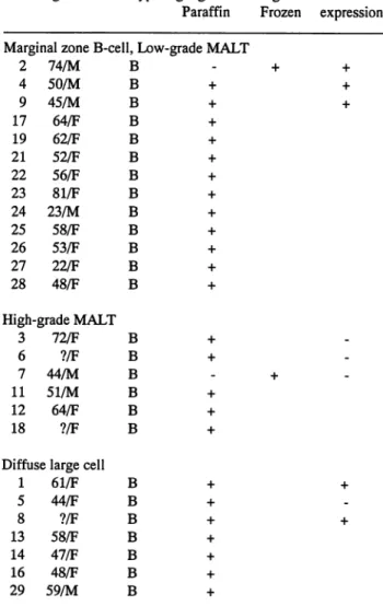
Table 1. Non-Hodgkin's Lymphoma Cases Studied
Cases Age/Sex Phenotype IgH gene rearrangement VacA Paraffin Frozen expression Marginal zone B-cell, Low-grade MALT
2 74/M B -4 50/M B + 9 45/M B + 17 64/F B + 19 62/F B + 21 52/F B + 22 56/F B + 23 81/F B + 24 23/M B + 25 58/F B + 26 53/F B + 27 22/F B + 28 48/F B + High-grade MALT 3 72/F B + 6 ?/F B + 7 44/M B -11 51/M B + 12 64/F B + 18 Diffuj ?/F $e large cell B + 1 61/F B + 5 44/F B + 8 ?/F B + 13 58/F B + 14 47/F B + 16 48/F B + 29 59/M B + + + +
Case numbers 1-9 show gastric lymphoma, case numbers 11-19 thyroid lymphoma, and case numbers 21-29 pulmonary lymphoma.
The length of CDR3 region
The nucleotide sequence of CDR3 is developmentally regulated. The average length of the N-D-N region in the
fetal liver lymphocytes, neonatal and adult peripheral B
cellsis 22 (range 12 to 45),24 (range 9 to 56) and31 (range
13 to 54) nucleotides, respectively, showing a tendency to
increase with age.1 In the low-grade MALT, high-grade
MALT and diffuse large cell lymphomas, the mean length
of the CDR3 region was 47.6 ±10.31 (range 21 to 60),
38.71 ±10.37 (range 27 to 57) and 40.86±3.34 (range 39
Detection of IgH variable region gene in paraffin-embedded tissues from MALT (47)
region was significantly greater in the low-grade MALT lymphoma group than in the other two groups (P<0.05).
The mean length of N-D-N was30.2±8.82 (range 7 to 40),
21.71±7.99 (range 7 to 30) and 24.86±4.10 (range 19 to
32) nucleotides, respectively. These findings indicate that the property of CDR3 in high-grade MALT lymphoma re sembles that in diffuse large cell lymphoma rather than in
grade MALT lymphoma. The maturation stage of
low-grade MALT lymphoma is similar to ontogenetic mature
B-cells, while high-grade MALT lymphoma and diffuse large cell lymphoma is at an ontogenetic early maturation
stage.
Homology search of CDR3 region compared to pub lished rearrangements
By homology search, the lymphoma cell clones of 14 cases (5 of 13 low-grade MALT lymphomas; 4 of 6 high-grade MALT lymphomas; 5 of 7 diffuse large cell lymphomas) showed 60 to 81% homology with autoanti-body-associated lymphocyte clones. The incidence of these autoantibody-associated lymphocyte clones was higher in the high-gradeMALT and diffuse large lymphomas than in low-grade MALT lymphoma. At protein level, 2 cases of low-grade MALT lymphomas showed 71 and 82% homol
ogy in CDR3 to those of autoreactive B cell clones, and 2
high-grade MALT lymphoma's showed 67% homology.
However in diffuse large cell lymphomas homology at pro
tein level was 56 and 57%.
We also investigated the sequences of VH segment in 9
gastric lymphomas. Seven cases (2 from 3 low-grade
MALT lymphomas; 2 from 3 high-grade MALT
lymphomas; 3 from 3 diffuse large lymphomas) showed 85
to 98.3% homology with those of autoantibodies.
Thus, our findings suggest that the cells of MALT lymphoma strongly correlated with autoantigen and that some of diffuse large cell lymphomas may also derived
from the selected autoreactive B-cell clones, even if they did not show histological evidence of MALT lymphoma.
References
1) Sanz I. Multiple mechanisms participate in the generation of diversity of human H chain CDR3 regions. J Immunol
147:1720-1729,1991
2) Wan JH, Trainor KJ, Brisco MJ, Morley AA. Monoclonality in B cell lymphoma detected in paraffin wax embedded sections us ing the polymerase chain reaction. J Clin Pathol 43, 888-890,
1990
3) Yumoto N, Furukawa M, Kurosu K, Mikata A. A particular characteristic of IgH complementarity-determining region 3 sug gests autoreactive B-cell origin of primary gastric B-cell lymphomas. Lab Invest 78: 261-268,1998