Three Cases of Primary Gastrointestinal
Malignant Lymphoma
著者
ONO Nobutaka, TASAKI Kazuhiro, NAKAMURA Naoya,
NOZAWA Yoshihiro, TOMINAGA Kunihiko, HOJO
Hiroshi, ABE Masafumi, WAKASA Haruki
journal or
publication title
鹿児島大学医学雑誌=Medical journal of
Kagoshima University
volume
47
number
Suppl. 2
page range
97-100
URL
http://hdl.handle.net/10232/18321
Med. J. Kagoshima Univ., Vol. 47, Suppl. 2. 97-100, November, 1995
Case Report
Three Cases of Primary Gastrointestinal Malignant Lymphoma
Nobutaka ONO,
Kazuhiro TASAKI, Naoya NAKAMURA, Yoshihiro NOZAWA,
Kunihiko TOMINAGA, Hiorshi HOJO, Masafumi ABE
and Haruki WAKASA
Department of Pathology, Fukushima Medical College, Fukushima, Japan
Introduction
Extranodal lymphoid tissus of the permeable mucosa sites, such as the gastrointestinal tract and bornchi are different in structure and function from peripheral lymph nodes, and it is named as mucosa-associated lymphoid tissue (MALT). Therefore, the clinicopatho-logical features of extranodal lymphomas arising from
these mucosal sites have been considered to be
different from those of nodal lymphomas.
In 1983 and 1984, Isaacson and his colleagues have reported a certain low-grade B-cell lymphoma of the gastrointestinal tract called as MALT lymphomas. The concept of MALT lymphomas extended later to include extranodal lymphomas of the lung, salivary
gland, thyroid, thymus^, breast4' and gallbladder5'.
Recently, a definite nodal B-cell lymphoma called
mantle cell lymphoma, were also reported6'7'. The
histological and clinical features of mantle cell lympho mas are somewhat different from MALT lymphomas.
In this paper, we reported three cases with primary gastrointestinal lymphomas of low grade malignancy and discuss the differential diagnosis of MALT and mantle cell lymphomas.
Case report
Case 1: A 58-years-old female visited Ohara General Hospital because of epigastric discomfort in 1984. An endoscopical examination showed chronic gastritis. She received drug therapy, but the symptom was not improved. A diagnosis of malignant lymphoma was made from biopsied specimen and she subsequently underwent total gastrectomy in 1985. The surgically
Address of Correspondence: Nobutaka ONO,
Department of Panthology, Fukushima Medical College,
Hikarigaoka 1, Fukushima 960-12, Japan
1.2)
resected stomach revealed multiple shallow ulcers in antrum and the histological features were consistent with MALT lymphoma.
y$8Sy
msmmy
m
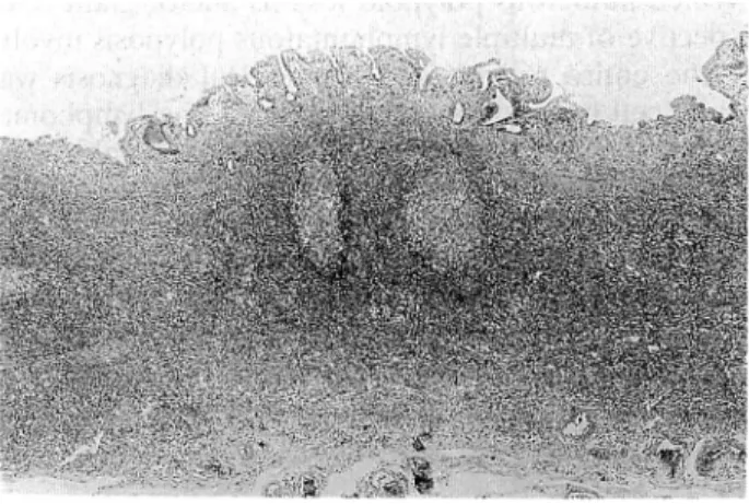
Fig. 1. Case 1. HEX 10. Lymphoma cells infiltrate diffusely in mucosa propria and submucosal layer. Germinal centers surrounded by thin mantle zones were
found.
Fig. 2. Case 1. HEX200. Lymphoma cells have round or
slight indented nuclei and abundant pale cyto plasms, resembled to monocytoid B cells.
(98; Med. J. Kagoshima Univ., Vol. 47, Suppl. 2, November, 1995
m$:>
t ^t*y ffy P
* ... ••'
m
y %.w »•
Fig. 3. Case 1. ALP X 160. Lymphoma cell are positive for
alkaline phosphatase.
Case 2: A 62-years-old female visited Hanawa Hospital because of itching in the anal region and constipation in
July, 1988. Rectal carcinoma was suspected from digital examination and she underwent the resection of the
rectum in August, 1988. The surgically resected rectum revealed numerous polypoid lesions and/or giant folds
suspective of multiple lymphomatous polyposis involv ing the entire rectum and histological diagnosis was
mantle cell lymphoma and/or mantle zone lymphoma. She was treated with one cycle of combination
chemotherapy, but no response was noted and she died
2 years after the onset of her illness.
(HP--Fig. 4. Case 2. HEX5. Lymphoma cells infiltrate massively in mucosa propria of the rectum, resulting in marked
thickening of the wall.
Case 3: A 81-years-old male with a complaint of three years history of gastric ulcer was admitted to Ohara General Hospital in 1989. An endoscopic examination
revealed multiple shallow ulcers in gastric antrum, and malignant lymphoma was detected by a biopsied
specimen and he subsequently underwent gastrectomy in 1990. The surgically resected stomach revealed
multiple shallow ulcers in antrum and the histological
diagnosis was made as MALT lymphoma.
j^V.
Fig. 5. Case 2. HEX200. Lymphoma cells are medium in
size and have scant cytoplasm with round or polygonal nuclei. $?•$&
Sw.3 *A
\.'v V*";- tySte-•'. m i^j-Fig. 6. Case 2. CDS X 160. Lymphoma cells are weakly positive for CD5.
f3M %MW^BWMk' • '•.
*3»«i^&J^!k'~ia~'•••
•'.?: ">?«
P#Miitl
- ^ ..siX "4 .?£??Fig. 7. Case 3. HEX 100. Medium-sized lymphoma cells infiltrate diffusely intermingled with large lympho ma cells. Neumerous lymphoma cells have round or polygonal nuclei and less abundant cytoplasms than
Three Cases of Primary Gastrointestinal Malignant Lymphoma [99] C "-:" '.,, £-. '-•' f~<".'-,_^i'v%y<" \V •' • s:
^<yyf.
*"!,$ £ ~~iy. 'ty^'-ft*^ • JF"""~ l^p"'' IsJi
: 'tV'^r
y y3 • ^J
-..v.. -•.'":".-" v-"N"3,
1
<-^y^ '
~"*[
':';t^-k
''"'"•'"t df-ie'•• ^^**Q.V„,v/-,- - ,,-..1 <• s-r" r-~- . ^ . > i«8fi;' J i <a -•^yC'^i-' y^jy-ras , # ^
Fig. 8. Case 3. CD5X160. Lymphoma cells are weakly positive for CD5.
Table 1, Results of histochemistry
Case 1 Case 2 CD19 + CD20 + CD3 -CDS — CD 10 — CD25 -CD30 — IgM + IgD — ALP + + + + Case 3 + Results 1. Clinical features:
The ages of these patients ranged from 58 to 81 years (mean; 67 years). Two patients were female and one was male. Endoscopically, Case 1 and 3 showed shallow ulcerated or erosive lesions existed in gastric antrum. Case 2 showed numerous polypoid lesions or giant folds through the entire gastrointestinal tract, which is usually called multiple lymphomatous polypo sis. The patient died of malignant lymphoma 2 years after onset, but the prognosis of Case 1 and 3 was
unknown.
2. Histological findings and histopathological diagnosis: Case 1 and 3 showed a diffuse lymphoma cell proliferation in mucosa propria and submucosal layer of the stomach. Lymphoma cells were medium in size and had pale abundant cytoplasm (Case 1, Case 3), containing round to slightly indented nuclei. A few mitotic figures were present and lymphoepithelial lesions were often found. Atrophic germinal centers surrounded by thin mantle zones were observed. The pathologic diagnosis was malignant lymphoma, MALT type, according to the Kiel classification. Case 2 showed a massive proliferation of lymphoma cells
mainly in mucosa propria of the rectum, and a few infiltration to submucosal layer resulting in the thickening of mucosa. Morphologic contours of lymphoma cells were very similar to those of Case 3. Lymphoepilhelial lesions were not found. Naked germinal centers were found in Case 2, and pathologic diagnosis was made as mantle zone lymphoma.
Lymph follicles with atrophic or hypertrophic germinal centers were found in all 3 cases. In Case 1 & 3, germinal centers were surrounded by thin mantle zone, and in Case 2 germinal centers were naked
without mantle zone.
3. Immunohistochemical findings:
Both snap frozen sections and formalin-fixed paraffin
embedded tissuse were used for an immunohistoche
mical and enzymehistochemical analysis.
All 3 cases showed B cell phenotype, showing IgM",
IgD", CD19 ' , CD20+, CD10" and CD3~. CD5 was
expressed in Case 2 and 3, and negative in Case 1. Alkaline phosphatase (ALP) staining was positive for Case 1 and 3, but was negative for Case 2. All 3 cases were negative for CD25 and CD30.4. Genetical analysis:
Southern blotting analysis was performed in Case 2 and 3. Both cases showed the rearrangement of JH,
and germline of bcl-1, bcl-2 and TCR/5.
Discussion
Diagnostic criteria of MALT lymphoma proposed by
Isaacson and his coworkers2"8' are as follows : 1)
neoplastic proliferation of centrocyte-like or small cleaved cell-like cells, 2) neoplastic plasma cell infiltration in the subepithelial layer, 3) remaining of atrophic or hypertrophic lymph follicles, 4) lym phoepithelial lesions, 5) CD5 negative, 6) localized lymphoma lesion for long period, and 7) good prognosis. MALT lymphoma is usually included in low-grade malignancy, but there are some reports of high grade malignancy.
We studied three cases of primary gastrointestinal lymphomas by the use of histologic, immunohistoche mical and genetic methods. Case 1 and 3 showed
localized lesions in the stomach and satisfied above
diagnostic criteria from 1) to 4). Case 1 was thought as a typical or classical MALT lymphoma, because lymphoma cells had abundant pale cytoplasm similar to
monocytoid B cells9' and were positive for CD19,
CD20, IgM, and ALP, but negative for IgD, CD5 and CD10, indicating that the origin seemed to be marginal zone B lymphocytes. Histologic appearance of Case 3 was consistent with MALT lymphoma, but the foci contained large blastoid lymphoma cells. Therefore, Case 3 was diagnosed MALT lymphoma with blastic
transformation namely follicular colonization10'. In
[100] Med. J. Kagoshima Univ., Vol. 47, Suppl. 2, November, 1995
demonstrated some difference from MALT lymphoma, because of the positivity for ALP and CD5. Although MALT lymphomas usually lack of CD5, Case 3 was compatible with MALT lymphoma except CD5-positiv-ity, therefore, Case 3 appeared to be an exceptional case among MALT lymphomas. Morphological fea
tures of Case 2 were similar to those of Case 1 and 3
with exception of scanty cytoplasm. In Case 2, the most striking features different from Case 1 and 3 were the presence of naked germinal centers and the absence of lymphoepithelial lesions. Immunohistochemical data of
Case 2 were different from those of Case 1 and 3:
lymphoma cells showed IgM+, IgD", CD19+, CD20+,
CD5+, CD10" and ALP-. These results suggest that
Case 1 and 3 were MALT lymphoma, and Case 2 was
mantle cell lymphoma11}, corresponding with multiple
lymphomatous polyposis described by Cornes 2\
because the usual phenotype of MALT lymphoma is
SIgM+, IgD", CIgM+/", CD5" and ALP+, whereas
the unusual phenotype of mantle cell lymphoma is
SIgM+, IgD"^, CD5^ and ALP+/". Therefore, IgD,
CD5 and ALP are useful markers to distinguish MALT lymphoma from mantle cell lymphoma.
In conclusion, we have reported two cases with MALT lymphoma and one case with mantle cell lymphoma arising from the gastrointestinal tract. Immunohistochemical and enzymehistochemical methods are able to differentiate MALT lymphoma from mantle cell lymphoma: MALT lymphoma showed the expression of ALP and lack of CD5 and IgD, whereas mantle cell lymphoma showed the expression of CD5 and IgD and lack of ALP.
key words: Malignant lymphoma, gastrintestinal tract, MALT lymphoma, mantle cell lymphoma
References
1) Isaacson PG, Wright DH: Malignant lymphoma of mucosa-associated lymphoid tissue, a distinctive type of B-cell lymphoma. Cancer 1983, 52:1410-6. 2) Isaacson PG, Wright DH: Extranodal malignant
lymphoma arising from mucosa associated lymphoid tissue. Cancer 1984, 53:2515-24.
3) Isaacson PG, Chan JKC, Tang C, Addis BJ:
Low-grade B-cell lymphoma of mucosa-associated lym phoid tissue arising in the thymus. Am J Surg Pathol 1990, 14:342-51.
4) Hugh JC, Jackson FI, Hanson J, Poppema S: Primary breast lymphoma. An immunohistologic study of 20 new cases. Cancer 1990, 66:2602-11. 5) Mosnier JF, Brousse N, Sevestre C, Flejou JF,
Delteil C, Henin D, Potet F: Primary low-grade B-cell lymphoma of the mucosa-associated lymphoid tissue arising in the gallbladder. Histopathol 1992,
20:273-5.
6) Weisenburger DD: Non-Hodgkin's lymphomas of primary follicle/mantle zone origin. Leukemia 1991,
5:26-9.
7) Raffeld M, Sander CA, Yano T, Jaffe ES: Mantle cell lymphoma: an update. Leuk Lymphoma 1992, 8(3): 161-6.
8) Isaacson PG, Spencer J: Malignant lymphoma of mucosa-associated lymphoid tissue. Histopathol
1987, 11:445-62.
9) Nizze H, Cogliatti SB, von Schilling C, Feller AC, Lennert K: Monocytoid B-cell lymphoma: morpho logical variants and relationship to low-grade B-cell lymphoma of the mucosa-associated lymphoid tissue. Histopathol 1991, 18:403-14.
10) Issacson PG, Wotherspoon AC, Diss TC, Pan L: Follicular colonization in B-cell lymphoma of mucosa-associated lymphoid tissue. Am J Surg
Pathol 1991, 15:819-28.
11) Banks PM, Chan J, Cleary ML, Delsol G, de Wolf-Peeters C, Gatter K, Grogan TM, Harris NL, Isaacson PG, Jaffe ES, Mason D, Piled S,
Ralfkiaer E, Stein H, Warnke RA: Mantle cell
lymphoma. A proposal for unification of morpho logic, immunologic, and molecular data. Am J Surg Pathol 1992, 16:637-40.
12) Cornes JS: Multiple lymphomatous polyposis of the gastroinestinal tract. Cancer 1961, 14:249-57.