Malignant Lymphoma Developing from
Long-standing Inflammation and MALT Lymphoma
著者
OHSAWA Masahiko, AOZASA Katsuyuki
journal or
publication title
鹿児島大学医学雑誌=Medical journal of
Kagoshima University
volume
47
number
Suppl. 2
page range
73-76
URL
http://hdl.handle.net/10232/18316
Invited Paper
Malignant Lymphoma Developing
from Long-standing Inflammation and MALT Lymphoma
Masahiko OHSAWA and Katsuyuki AOZASA
Department of Pathology, Osaka University School of Medicine, Suita, Osaka, Japan
Introduction
We summarize here the findings of two types of extranodal lymphomas, which support our proposal for "maligant lymphoma developing in the long-standing inflammation". One type is thyroid lymphoma develop ing chronic lymphocytic thyroiditis (CLTH), an organ-specific autoimmune disease. Another is pleural lymphoma developing in the patients with over 20-years history of pyothorax. This kind of lymphoma mostly overlapped the lymphoma of mucosa-associated lymphoid tissue (MALT).
Thyroid lymphoma
Hashimoto's thyroiditis as a risk factor thyroid lymphoma
Follow-up studies carried out by Holm et al. in
Sweden1^ and by us2) in Japan have comfirmed the
etiologically important role of CLTH in development of thyroid lymphoma. In our study, a total 5592 female patients over 25 years of age with CLTH, diagnosed between 1965 and 1982 at Kuma hospital, Japan, were
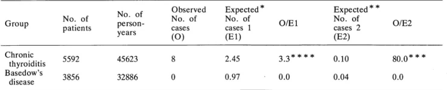
followed up until 1985 (Table 1). Eight new cases of primary thyroid lymphoma were observed (O). Since the expected numbers of patients with malignant lymphoma (El) and thyroid lymphoma (E2) were 2.25 and 0.1, respectively, the O/El and 0/E2 ratios were 3.3(p<0.01) and 80(p< 0.001), respectively. In the reference group including cases of hyperthyroidism, no increased risk of thyroid lymphoma was observed. These findings suggest that the autoimmune reaction present in CLTH may play an important role in the etiology of thyroid lymphomas. We also reported a close relationship between CLTH and thyroid lympho ma on the basis of histology, and all these were
lymphocytic lymphomas'^. Histological findings indica
tive of CLTH included the presence of lymphocytic infiltration usually with the formation of lymphoid follicles containing germinal centers, varing degrees of fibrosis, and oxyphilic changes or squamous metaplasia in the epithelial cells of the thyroid follicles. Histologic al evidence of CLTH in the thyroid tissue apart from lymphomas was confirmed in approximately 80% of patients with or without antibodies against
thyroglobu-lin and/or microsomes.
Table 1. O/El and 0/E2 ratios in patients with chronic thyroiditis and patients with Basedow's disease
Group No. of patients No. of person-years Observed No. of c a s e s (O) Expected * No. of cases 1 (El) O/El Expected* * No. of cases 2 (E2) 0/E2 Chronic thyroiditis Basedow's disease 5592 3856 45623 32886 8 0 2.45 0.97 0.0 0.10 0.04 80.0*** 0.0
*expected number of cases with malignant lymphoma; * *expected number of cases with thyroid lymphoma; * * *p<
0.0001; ****p<0.001
Address for Correspondence: Katsuyuki AOZASA, Department of Pathology, Osaka University School of Medicine, 2-2 Yamadaoka, Suita, Osaka 565, Japan
(74) Med. J. Kagoshima Univ., Vol. 47, Suppl. 2, November, 1995
Histopathology and immunophenotype of thyroid lymphomas
Follicular center cell tumors were the most common
among thyroid lymphomas: 66% in our series3^. In our
10% of cases of thyroid lymphoma, there was diffuse proliferation of atypical small lymphoid cells with a slightly irregular nuclear contour. Immunological marker studies showed that these atypical small lymphoid cells expressed surface properties interme diately between mantle-zone lymphocytes and follicu lar center cells. Immunological and immunohistoche mical studies revealed that thyroid lymphomas were
almost exclusively B-cell derived4).
Pyothorx-associated lymphoma (PAL) In 1987, we reported development of PAL in three patients suffering from long-standing pyothorax, result ing from an artificial pneumothorax for the control of pulmonary tuberculosis, or from tuberculous
pleuritis5). PAL developed in three (2.2%) of 134
patients with chronic pyothorax at one of the hospitals specializing in chest diseases (Kinki Chuo Hospital for Chest Diseases) in Osaka, Japan, during the period 1971-85 (Photo. 1). Meanwhile, our study on malignant lymphomas in the general hospitals of the same district
showed that there were no cases of PAL in over 2000
cases of malignant lymphoma. These findings suggested an etiologically important role for chronic pyothorax (CP) in the development of lymphoma in the pleura cavity.
To obtain more precise information, we carried out a nation-wide study of PAL . The clinical findings are summarized in Table 2. The patients were admitted to hospitals with the histories of CP ranging from 22 to years (mean 33 years), resulting from artificial pneumothorax for the treatment of pulmonary tubercu losis or tuberculous pleuritis. Histologically, all tumors
Photo. 1. Development of lymphoma in the right pleural cavity of a chronic empyma patient.
were lymphocytic lymphoma with the diffuse large cell type being the most common: approximately 80% of all cases. Immunologically, almost all cases showed the B-cell phenotype. Because an autoimmune mechanism was not thought to be likely in the formation and continuation of the phyothorax, these findings sug gested that chronic inflammatory stimulation of a non-autoimmune nature could also be an etiological factor in the development of malignant lymphomas.
To examine risk factors for development of PAL in
patientswith CP, a case-control studywas carried out7).
The factors including onset age of lung tuberculosis and pyothorax, presence of chemotherapy, surgical treat ment, extent of empyema, presence of fistula, history of smoking, and height and weight of patients at first admission were compared in patients with CP alone (70 controls) and CP complicated with lymphoma (42 cases), by which the date of birth and sex were matched. The patients receiving the artifical pneumothorax showed a significant increase in risk for development of PAL (relative risk = 4.92, p<0.05). This finding suggested that chronic non-healing in flammation in the pleural cavity left by artificial
Table 2. Summary of chinical findings in pyothorax-assoicated lymphoma patients
Age at diagnosis
Male: Female ratio
Tuberculosis history lung tuberculosis tuberculous pleuritis 48-81 years(mean 63) 5.2:1 81% 16%
Interval between pyothorax 22-55 years(mean 33) and onset of lymphoma
Presenting symptoms
chest pain 51%
productive cough 54%
and dyspnea
Tumor of chest wall 14%
Diagnosis at admission chronic pyothorax 49% chronic pyothorax 38% complicated with malignancy lung tumor 5% Detection chest X-ray 35% computed tomographic 77% s c a n Definitive diagnosis biopsy 84% autopsy 16%
pneumothorax resulted in the development of PAL. Repeated rentogenographic examinations carried car ried out at the procedure might be causative factor. Role of Epstein-Barr virus (EBV) in pleural lymphomagenesis
Previous study showed the presence of Epstein-Barr virus (EBV) genome together with the expression of latent infection genes in the tumor cells of PAL in five
and four patients, respectively^' '. We examined the
presence of EBV genome in cases with PAL (34 cases) and CP without PAL (16 cases) to evaluate an
association of EBV with PAL10). Combined
polymerase chain reaction (PCR), in situ hybridization method, and immunohistochemistry showed that EBV genome was detected in lymphoma cells in 85% of PAL with almost constant expression of latent membrane protein-1 (Fig. 1, Table 3). On the contrary, EBV genome was detected by PCR in one of 16 CP cases. These findings suggested that neoplastic transformation of infiltrated lymphocytes in CP by the EBV is one of the factors for the development of PAL under the long standing inflammatory circumstances.
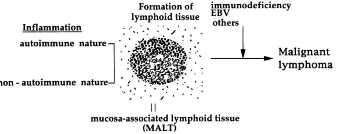
Relationship between lymphocytic lymphoma develop ing from long-standing inflammation and lymphoma of the mucosa-associated lymphoid tissue (MALT)
Lymphomas of the MALT, firstly discribed by
Isaacson et alu), are heterogenous entity in histology,
immunologic character, and primary site of origin. MALT lymphomas are defined as lymphomas arising from the mucosa-associated lymphoid tissue (extranod
al organ), such as Peyer's patch'". Under the normal
conditions, however, the extranodal organs, where the MALT lymphomas develops, such as thyroid, salivary glands, gastrointestinal tract, do not have lymphoid tissue. The lymphoid tissue in these organs is formed through chronic inflammation such as CLTH and Sjogren's syndrome. Recently an association between gastric lymphoma and chronic follicular gastritis caused
by the Hericobacter pylori has been suggestedl2).
M N 1 2 3 4 5 6 7 8 9 JO II 12 13 14 29 hi M 15 16 N 17 18 19 20 21 22 23 24 25 26 27 28 P 119 bp 29 3031 32 33 3435 36 37 383940 41 42 N P 129 bp
Fig. 1. Southern blot analysis of PCR amplified products for Epstein-Barr virus genome. N: negative control, P: positive control(Raji cell line), Cases 1-16: chronic pyothorax(CP), Casesl7-41: Pyothorax-associated lymphoma(PAL), The positive results are shown by the band at 129 base paire in all PAL and one of CP(Lane 13)
Table 3. Summary of Epstein-Barr virus study
Pyothorax-associated lymphoma
Chronic pyothorax ': a few lymphocytes
PCR
EBV genome EBNA2A EBNA2B ISH LMP-1
26/26 1/16 14 0 10 0 28/33 0/1 30/34 1 / 1
(76] Med. J. Kagoshima Univ., Vol. 47, Suppl. 2, November, 1995
Formation of immunodeficiency
lymphoid tissue
"
Inflammation
autoimmune nature-.
non - autoimmune nature-1
Malignant
lymphoma
II
mucosa-associated lymphoid tissue
(MALT)
Fig. 2. Development of extranodal lymphoma from chronic inflammation of
an autoimmune and non-autoimmune nature
These findings indicate that the MALT lymphomas, except for developing Peyer's patch, can be categorised as lymphoma developing in the long-standing in flammation (Fig. 2). Doctor Isaacson agreed with this
13)
v i e w
Key words: Thyroid lymphoma, pyothorax-associated lymphoma, MAJLT lymphoma, chronic inflammation, Epstein-Barr virus
References
1) Holm FM, Blomgren H, Lowhagen H. Cancer risks in patients with chronic lymphocytic thyroiditis. N Engl J Med 1985; 312: 601-4.
2) Kato I, Tajima K, Suchi T, Aozasa K, Matsuzuka F, Kuma K et al. Chronic thyroiditis as a risk factor of B-cell lymphoma in the thyroid gland. Jpn J Cancer
Res 1985; 76: 1085-90.
3) Aozasa K, Inoue A, Tajima K, Miyauchi A, Matsuzuka F, Kuma K. Malignant lymphomas of the thyroid gland; analysis of 79 patients with emphasis on histologic prognostic factors. Cancer
1986; 58: 100-4.
4) Aozasa K, Ueda T, Katagiri S, Matsuzuka F, Kuma K, Yonezawa T. Immunologic and immunohistolo gic analysis of 27 cases with thyroid lymphomas.
Cancer 1987; 60: 969-73.
5) Iuchi K, Ichimiya A, Akashi A, Mizuta T, Lee Y-E, Tada N et al. Non-Hodgkin's lymphoma of the pleural cavity developing from long-standing pyoth
orax. Cancer 1987; 60: 1771-75.
6) Iuchi K, Aozasa K, Yamamoto S, Mori T, Tajima K, Mukai K et al. Non-Hodgkin's lymphoma of the pleural cavity developing from long-standing pyoth orax: summary of clinical and pathological findings in thirty-seven cases. Jpn J Clin Oncol 1989;
19:249-57.
7) Aozasa K, Ohsawa M, lochi K, Tajima K, Komatsu H, Shimoyama M. Artificial pneumothorax as a risk factor for development of pleural lymphoma. Jpn J
Cancer Res 1993; 84:55-7.
8) Fukayama M, Ibuka T, Hayashi Y, Ooba T, Koike M, Mizutani S. Epstein-Barr virus in pyothorax-associated pleural lymphoma. Am J Pathol 1993;
143:1044-9.
9) Sasajima Y, Yamabe H, Kobashi Y, Hirai K, Mori S. High expression of Epstein-Barr virus latent protein EB nuclear antigen on pyothorax-associated lymphomas. Am J Pathol 1993; 143: 1280-5. 10) Ohsawa M, Tomita Y, Kanno H, Iuchi K,
Kawabata Y, Nakajima Y et al. Role of Epstein-Barr virus in pleural lymphomagenesis. Mod Pathol 1995 (in Press).
11) Isaacson PG, Spencer J. Maligant lymphoma of mucosa-associated lymphoid tissue. Histopatholo gy 1987; 11: 445-62.
12) Wotherspoon AC, Ortiz-Hidalgo C, Falzon MR, Isaacson PG. Helicobacter pylori-associated gastri tis and primary B-cell gastric lymphoma. Lancet 1991; 338(8776):1175-6.
13) Isaacson PG, Falzon M. Malignant lymphoma of the mucosa-associated lymphoid tissue. Am J Surg Pathol 1992; 16:91-2(Authors' response).