Radiographic and endoscopic diagnosis of
gastric malignant lymphoma, especially
mucosa-associated lymphoid tissue (MALT) type
lymphoma
著者
AOZAKI Shinichiro, OTSUJI Masato, NISHIMATA
Hiroto, NISHIMATA Yoshito, MISONO Toshiaki
journal or
publication title
鹿児島大学医学雑誌=Medical journal of
Kagoshima University
volume
51
number
Suppl.
page range
22-23
URL
http://hdl.handle.net/10232/18350
Med. J. Kagoshima Univ., Vol. 51. Suppl. 22-23, July, 1999
Radiographic and endoscopic diagnosis of gastric malignant lymphoma,
especially mucosa-associated lymphoid tissue (MALT) type lymphoma
Aozaki Shinichiro1, Otsuji Masato2, Nishimata Hiroto3, Nishimata Yoshito3and Misono Toshiaki4
'Department of Internal Medicine, Sendai Saiseikai Hospital, Sendai, Kagoshima Prefecture
2Department of Internal Medicine, Imamura Hospital, Kagoshima
3Nanppu Hospital, Kagoshima
4Second Department of Internal Medicine, Kagoshima University Faculty of Medicine
Introduction
According to the progress in diagnosis of gastric le sions by means of X-ray and endoscopic images, we have met several chances to diagnose gastric malignant lymphomas (gML). But, an enough information for the diagnosis of the early phase gMLs has not yet been re ported. This paper reviewed our cases of gMLs and tried to discuss the diagnosis of the early phase gMLs.
Material and method
Thirty-two cases, of which the stomachs were
resected under the diagnosis of primary gML, were re
viewed from a viewpoint of clnicopathology.
The patients are 19 males and 13 females. Mean
age of these patients was 56.4 years. The intragastric loca tion of the primary gMLs was expressed as cardia and fun dus (C), body (M) or antrum (A). The macroscopic figures
of these primary gMLs were categorized in superficial, ul cerative, protruded, excavated, and giant folds types ac
cording to Sano's macroscopic classification1. The resected gastric lesions could be reviewed
histopathologically in 24 cases and were categorized ac
cording to the concept of mucosa-associated lymphoid tis sue (MALT) type lymphoma of Issacson2"4. The 24 cases comprise 12 low grade MALT type, 5 high grade MALT type with low grade component, 3 high grade MALT type without low grade component, and diffuse large (B-) cell type. The other cases, informed to involve deeper than the
proper muscular layer, were treated as high grade, consid
ering the clinical information. In order to present differ ences in X-ray and endoscopic images between early gMLs (Group A) (12 low grade MALT type) and progressive cases (Group B) (the others), comparison in each feature of the image diagnosis was performed, including 5 cases of low grade MALT type under follow-up.
Result and discussion
Macroscopic figure and depth of invasion: Cases of Group A comprise 11 superficial type (1 m, 10 sm) and one
ulcerative type (sm). Cases of Group B comprise 2 super ficial type (2 sm), 1 ulcerative type (sm), 16 excavated type (deeper than mp), and 1 giant folds type (ss). When a pri
mary gML involving up to sm is treated as an early case, the all cases of Group A were of the early case. Most cases of Group B were progressive cases.
Location of the lesion: Cases of Group A were 2 M, 5 A,
one case involving two regions (A>M) and one case in
volving the all regions. Cases of Group B were 3 C, 4 M, 9
A, 3 cases involving 2 regions (2 M>A, 1 M>C), one case involving the all regions. Many cases of the both groups involved the A region. There were 3 cases of Group A, manifesting multiple lesions (5, 3 and 2 lesions).
Image findings: Group B cases categorized as excavated
type presented figures resembling Borrman 2 or 3 type gas tric cancer. In the differential diagnosis from gastric can cer, smooth margin of depression, findings of a submucosal tumor in parts, and preservation of extensibility of the stomach in spite of the largeness of the lesion were charac
teristic for the excavated type. The giant folds type showed
thick and linear folds and maintained well extensibility,
which can differentiated the giant folds type from scirrhous type of gastric cancer. Group A cases of 12 low grade MALT type and the 5 cases under follow-up were 16 super ficial and one ulcerative type. The superficial type cases could be divided in three subcategories by image diagnos
tic features.
1) Flat type of granular mucosal surface (4 cases): The lesion involved wide areas and showed obscure demarca
tion of the lesion. In X-ray images, patterns of various-sized granular protrusion with multiple small ulcers,
Radiographic and endoscopic diagnosis of gastric malignant lymphoma
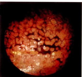
[23]Figure 2 Figure 3 Figure 4
Figure 2. The mucosa easy to bleed
Figure 3. Uneven mucosa with spontaneous bleeding before dye spray
Figure 4. By dye spraying method, various-sized granules with sporadically distributed sulcus-like erosions can be recognized easily.
sion, and irregular pattern of areae gastricae were recog nized. Endoscopy showed diffuse granular mucosa with multiple small ulcers or erosion, and the mucosa easy to bleed. The granular mucosa could be observed clearly in cases of a small amount of air in the stomach, in the sight along longitudinal axis, and by employing dye spraying
method.
2) lie-like depression (12 cases): Localized lesion was seen in each stomach. In 3 cases multiple lesion could be recognized. In X-ray images, the lesion was enhanced as
irregularity in or loss of pattern of areae gastricae. In some
cases complication of multiple ulcers or erosion was noted
in the depression. Endoscopy revealed discolored shallow depression with or without sporadic petechiae or granular
protrusion of the mucosa.
3) Flat elevation (1 case): The flat elevation comprises
fused thick folds of the mucosa.
One case of ulcerative type associated lie-like depression around the ulcer.
Conclusion
Most of early primary gML are thought to be of MALT type. In order to diagnose these early primary gMLs, care ful observations of the images are necessary, based on the understanding of characteristics of the superficial type of
the gMLs.
Reference
1. Sano R. Clinicopathology of gastric diseases. Igaku-Shoin 1982 pp. 258 (in Japanese)
2. Issacson P, Weight DH. Malignant lymphoma of mucosa-asso
ciated lymphoid tissue: A distinct type of B-cell lymphoma. Can
cer 1983: 52:1410-16
3. Issacson P, Weight DH. Extranodal malignant lymphoma aris
ing from mucosa-associated lymphoid tissue. Cancer 1984: 53: 2515-24
4. Issacson P. Gastrointestinal lymphoma. Hum Pathol 1994: 25: 1020-29