A Case of Adult T-Cell Leukemia/Lymphoma in
Stomach
著者
HASUI Kazuhisa, TASHIRO Yukie, KITAMURA
Hiroshi, SHIMIZU Takeshi, TANAKA Sadao, SATO
Eiichi
journal or
publication title
鹿児島大学医学雑誌=Medical journal of
Kagoshima University
volume
47
number
Suppl. 2
page range
107-110
URL
http://hdl.handle.net/10232/18324
Med. J. Kagoshima Univ., Vol. 47, Suppl. 2. 107—110, November, 1995
Case Report
A Case of Adult T-Cell Leukemia/Lymphoma in Stomach
Kazuhisa HASUI1, Yukie TASHIRO1'2,
Hiroshi KITAMURA2, Takeshi SHIMIZU2, Sadao TANAKA2
and Eiichi SATO1'Second Department of Pathology, Faculty of Medicine Kagoshima University, Kagoshima, Japan
2Department of Pathology, Kagoshima-shi Medical Association's Hospital, Kagoshima, Japan
Summary
A 56 year old Japanese man was HTLV-1 carrier suffering from peripheral T-cell lymphoma pleomor phic small cell type manifesting multiple gastric lymphomatoid polyposis. The first symptom was epigastralgia and within one year clinical course, leukemia, hypercalcemia and lymph nodes swelling were not noted. A diagnosis of adult T-cell leukemia/lymphoma (ATLL) was based on immunohis tochemistry and DNA-RNA in situ hybridization of HTLV-1 pX Tax and Env. A small number of
lymphoma cells reacted with monoclonal antibody 6C2 against HTLV-1 gp46 Env and its precursor and the
most lymphoma cells were labeled by monoclonal antibody HML-1 against a homing receptor. Unusual
gastric involvement of this ATLL might be explained by the nature of the lymphoma cells expressing homing receptor.
Key words: ATL, Stomach, Immunohistochemistry, In-situ-hybridization, HTLV-1, HML-1
Introduction
Gastrointestinal involvement of Adult T-cell
leukemia/lymphoma (ATLL) has been reported1'2'3).
We also experienced unusual gastric lymphomatoid polyposis in a HTLV-1 carrier. Histopathological diagnosis of ATLL was based on the results of DNA-RNA in-situ-hybridization (ISH) of HTLV-1 proviral DNA env and pX Tax regions and of
paraffin-immunohistochemistry for HTLV-1 gp46 Env4).
Here, we report a case with the trial of histopatholo gical diagnosis of ATLL.
Address for Correspondence: Kazuhisa HASUI,
Second Department of Pathology, Faculty of Medicine Kagoshima University, Sakuragaoka 8-35-1, 890 Kagoshi
ma, Japan
Case
Patient was a 56 year old Japanese man. The first symptome was epigastralgia. The first gastroendoscopic findings were those of hemorrhagic gastritis. After one month, he had epigastralgia again. The second gastroendoscopic and radiographic examinations showed a slightly elevated (Ila-like in Japanese classification of endoscopy) lesion on the anterior wall of the pyloric antrum. The endoscopic biopsy specimen was diagnosed as malignant lymphoma (ML). Because of gradual growth of the ML, gastorectomy was performed. In the pre-operative examinations a small
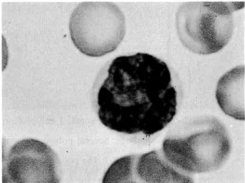
number of atypical lymphocytes (Fig. 1) and
anti-HTLV-1 antibodies were noted in his peripheral blood. The other laboratory data were within the normal range, including serum level of calcium and LDH. In
Fig. 1. Atypical lymphocyte in the peripheral blood. This atypical lymphocyte has lobulated nuclei but is small in comparison with the typical flower cells
[108) Med. J. Kagoshima Univ.. Vol. 47. Suppl. 2. November, 1995
Fig. 2. Adult T-cell leukemia/lymphoma, T-cell plemorphic lymphoma, small cell type, manifesting polypoid lesions in
stomach
a) Macroscopic findings: Several polypoid lesions (7mm in the longest axis, in max.) in the anterior wall of the
pylorus.
b) Low power view (H.E.): The polypoid lesion shows lymphocytic stroma. The glands present intact appearances.
c) Lymphocytic stroma (H.E.): Small lymphocytes locate in the interstitium and in lymph vessels. No obvious lymphoepithelioid lesions are seen.
d) High power view (H.E.): The small lymphocytes have convoluted and hyperchromatic nuclei. Among them there
are a few large or giant lymphoid cells as a hallmark for T-cell pleomorphic lymphoma.
e) HML-1 (frozen-immunohistochemistry): Most of the small lymphocytes show strong positive stains along cell surface.
f) 6C2 (HTLV-1 gp46 Env) (paraffin-immunohistochemistry): There are some lymphocytes revealing cytoplasmic
A Case of ATLL in Stomach [109]
the clinical course intestinal multiple lymphomatoid polyposis was diagnosed by means of endoscopy. Two
months before his death, serum LDH had been increasing up to 1848 W-U. No obvious leukemia was
noted. In one year form the first symptome he died suddenly of hemorrhage in a rapidly grown recurrent gastric lymphoma. Autopsy was not performed.
Pathology
The resected stomach showed multiple Ila-like mucosal polypoid lesions in the anterior wall (Fig. 2a). The polypoid lesions in the pyloric glandular mucosa (Fig. 2b) showed dense lymphocytic stroma comprising
small to medium-sized lymphocytes (Fig. 2c). A few large lymphoid cells having convoluted nuclei were seen among the lymphocytes (Fig. 2d). So-called lymphoepithelial lesions were not found. A few
atypical large or giant lymphoid cells were seen in parts
and the small to medium-sized lymphocytes had convoluted nuclei (Fig. 2d).
Frozen-immunohistochemistry showed that the lym phocytes were positive for CD2, CD3, CD4, CD25,
HLA-DR and HML-1 (Fig. 2e) and negative for CD1,
CD7, CD8, CD15 (Leu Ml, Leu M3, Leu7), CD20, CD22, CD30 and Ber H2. Seventy to 80% of the lymphocytes were positive for Ki-67. Paraffin-immuno histochemistry showed that the lymphocytes were
positive for CD3, UCHL-1, MT-1 and LN-2 and
negative for MB-1, L26, LN-1, LN-3, Leu Ml, Ber H2 and antibodies for immunoglobulin heavy and light chains. In paraffin-immunohistochemistry with micro
wave pretreatment5) the lymphocytes were weakly
positive for CD25.
Anti-HTLV-1 gp46 Env antibody (6C2, Cellular Product Inc.) reacted positively with some lymphocytes
(Fig. 2f) in its paraffin-immunohistochemistry4).
DNA-RNA in situ hybridization employing HTLV-1
pX Tax and Env biotinylated probes produced by
means of polymerase chain reaction 6,7) showed weak
signals only in a few lymphocytes.
The gastric lesion was diagnosed histopathologically as peripheral T-cell malignant lymphoma, pleomorphic
small cell type and etiologically as adult T-cell
leukemia/lymphoma (ATLL) manifesting multiple lymphomatoid polyposis in the stomach. The all regional lymph nodes of the stomach were free from
the malignant lymphoma.
Discussion
The definite diagnosis of ATLL is made by proving monoclonal integration of HTLV-1 proviral genome in
neoplastic peripheral T-cells. In frozen-immunohis tochemistry the most ATLL cells have interleukin 2 receptor (IL2R, CD25) on their surface. In histoche mistry using routine paraffin sections, methods for diagnosis of ATLL have been developing. In
paraffin-immunohistochemistry with microwave antigen-retriev
al pretreatment5) IL2R (CD25) could be visualized in
ATLL cells. Monoclonal antibody 6C2 was introduced
to detect HTLV-1 gp46 Env and its precursor in
paraffin-immunohistochemistry4) and the antibody 6C2
labeled some lymphoma cells in this case (Fig. 2f). We
succeeded to detect signals of HTLV-1 proviral
DNA6'7) but in some cases such as this case only a few
ATLL cells showed the signals. The histochemicaldetection of proteins and signals of HTLV-1 proviral
DNA means HTLV-1 infection in ATLL cells but can not prove monoclonal integration of HTLV-1 proviral
DNA in ATLL cells. Recently inverse polymerase
chain reaction (IPCR) method to detect monoclonal integration of proviral DNA of HTLV-1 has been
developed by M. Matsuoka and K. Takatsuki (Kuma-moto Univ.). We expect application of the IPCR method to a small amount of DNA extracted from
paraffin sections.
Unusual gastric involvement of this ATLL might be explained by a horming receptor recognized by HML-1 on the lymphoma cells, although the antigen recog nized by HML-1 may be one of the activating antigens
in ATLL8). Through the stay of ATLL cells having the
homing receptor in the mucosa, the ATLL cells proliferate to form a tumor in the mucosa and do not recirculate into the other organs.
Acknowledgement
Authors thank Dr. M. Tokunaga (Director Dr., Department of Pathology, Kagoshima City Hospital) for his examination by means of frozen-immunohis tochemistry of this case.
References
1) Utsunomiya A, Hanada S, Terada A^ Kodama, M, Uematsu T et al. Adult T-cell leukemia cell infiltration into the gastrointestinal tract. Cancer 1988, 61:824-8.
2) Obata S, Matsuzaki H, Nishimura H, Kawakita M and Takatsuki K. Gastroduodenal complications in patients with adult T-cell leukemia. Jpn J Clin Oncol. 1988, 18:335-42.
3) Aozaki S, Yamauchi K, Iriki S, Yasuda M, Nakama M et al. A case of adult T-cell leukemia presenting multiple lymphomatoid polyposis-like infiltration in intestine. Stomach and Intestine (Tokyo) 1991,
26:1297-303 (in Japanese).
4) Hasui K, Sato E, Katahira K, Setoyama M and
Kanzaki T. A case of cutaneous type adult T-cell leukemia/lymphoma with difficult differential di agnosis from mycosis fungoides. Lymphomas of the skin 1995, vol. XIV: 82-91 (in Japanese). 5) Merz H, Rickers O, Schrimel S, Orscheschek K and
Feller AC. Constant detection of surface and cytoplasmic immunoglobulin heavy and light chain
[no] Med. J. Kagoshima Univ., Vol. 47, Suppl. 2, November, 1995
expression in formalin-fixed and paraffin-embedded
material. J Pathology, 1993, 170:257-64.
6) Hasui K and Sato E. Human T-cell leukemia virus type (HTLV-1) infection in malignant lymphomas in
a HTLV-1-endemic area, Kagoshima, Japan: HTLV-1 in-situ-hybridization (ISH) analysis em
ploying a highly biotinylated concatamer probe synthesized by polymerase chain reaction (PCR) for
HTLV-1 proviral DNA pX Tax region. DENDRI
TIC CELLS 1994, 4:125-37.
7) Hasui K, Sato E, Sueyoshi K et al. Cold in-situ-hybridization of HTLV-1. DENDRITIC CELL 1995, 5:27-35.
8) Kitajima S, Tokunaga M. Goto M and Sato E,
Utsunomiya A et al. Monoclonal antibody HML-1 reactivity with adult T-cell leukemia/lymphoma and other lymphomas. Histopathology 1944, 25:229-36.