Cutaneous T- Cell Lymphoma with Monoclonal
Plasmacytosis
著者
MIMURA Rokuro, MORI Yoshitaka
journal or
publication title
鹿児島大学医学雑誌=Medical journal of
Kagoshima University
volume
47
number
Suppl. 2
page range
171-173
URL
http://hdl.handle.net/10232/18339
Med. J. Kagoshima Univ., Vol. 47, Suppl. 2. 171-173, November, 1995
Case Report
Cutaneous T- Cell Lymphoma with Monoclonal Plasmacytosis
Rokuro MIMURA and Yoshitaka MORI
Department of Pathology, Hyogo College of Medicine, Nishinomiya, Japan
Summary
A 75-year-old man with a well-established cutaneous T-cell lymphoma(mycosis fungoides) had a monoclonal gammopathy IgA- A type. Histologically infiltrate of plasma cells with an IgA- A type was coincidently present around atypical lymphocytes in the dermis. Some cases were repoted as Sezary's syndrome or mycosis fungoides or cutaneous T-cell lymphoma in association with a monoclonal gammopathy. But proliferation of plasma cells related mycosis fungoides has not been described. This association suggests the production of cytokine by lymphoma cells.
Key words: Cutaneous T-cell lymphoma, mycosis fun goides, monoclonal plasmacytosis, monoclonal gam mopathy
Introduction
Cutaneous T-cell lymphomas(mycosis fungoides and Sezary's syndrome) primarily involve the skin. Most of the neoplastic T-cells are typical of CD4-positive helper/inducer T-cells. Some cases were reported as
Sezary's syndrome1"4^ or mycosis fungoides5"8^ or
cutaneous T-cell lymphoma9) in association with a
monoclonal gammopathy. Some of these cases compli
cated myeloma,1'2,7* which showed diffuse proliferation
of atypical plasma cells in the bone marrows. But most of them had benign monoclonal gammopathy unrelated to the lymphoma. We presented a patient with mycosis fungoides in association with a monoclonal gammo pathy and plasmacytosis. This association lends support to the relation between T-heper-cell malignant prolifer ation and plasma cell proliferartion.
Report of a case
A 66-year-old man presented in 1982 with a itching erythematous rash on the face, and was diagnosed as eczematous dermatitis. In the ensuing 8 years the
Address for Correspondence: Rokuro MIMURA, Department of Pathology, Hyogo College of Medicine, Mukogawa-cho 1-1, Nishinomiya 663, Japan
patient had a recurrent skin rash. In 1990, erythema
tous nodules developed on the face and the trunk, and were biopsied. The diagnosis was cutaneous T-cell lymphoma with monoclonal plasmacytosis. There were also palpable lymphadenopathies in the bilateral
cervical and axillary regions. Biopsies of the enlarged
lymph nodes performed also showed infiltration of neoplastic T-cells and plasma cells.
Bone radiograph revealed no punched out lesion. A
computed tomographic scan of his chest and abdomen was performed, which failed to reveal any evidence of
mediastinal and retroperitoneal lymphadenopathies. His laboratory analyses disclosed the following
values: red blood cell count, 433xl04/^l; white blood
cell count 47xl02//H, with a differential cell count of
22% band cell forms, 47% segmented forms, 2% eosinophils, 8% monocytes, and 21% lymphocytes;and platelet count 20xl04/^l.
Serum immunoglobulin assay showed IgG of 1510
mg / dl(normal, 750 to 1700); IgA, 1890mg / dl(normal,
100 to 440); and IgM, 143mg/dl(normal, 160 to 295). Serum protein electrophoresis levels showed M-protein(IgA- A), and there was no evidence of Bence-Jones protein in the urine. The titer of anti-HTLV-1 antibody was less than x5. The results of the remainder of the patient's biochemical profile were within normal limits. The patient was treated with combination chemotherapy. However, he died of pneumonia on January in 1991.
Histology.
Several specimens of facial erythematous rash showed features of typical mycosis fungoides: The epidermis showed slight acanthosis and there was slight
hyperkeratosis(Fig. 1). In the upper dermis infiltration
of small-sized lymphocytes with irregular shaped nuclei was present, and within the epidermis small numbers of
atypical, irregular lymphcytes were found (Pautrier's
microabscess). In addition to the epidermis, follicular epithelium was involved. In the periphery of the dermal infiltate plasma cells also were present(Fig. 2).
A biopsy specimen of skin erythematous nodule revealed a massive dermal cellular infiltrate consisting of lymphocytes that were medium-sized posessing irregular nuclei and were surrounded by plasma
[172] Med. J. Kagoshima Univ., Vol. 47, Suppl. 2, November, 1995
cells(Fig. 3). And a heavily infiltrate of atypical
lymphocytes was seen in the epidermis(Fig. 4).
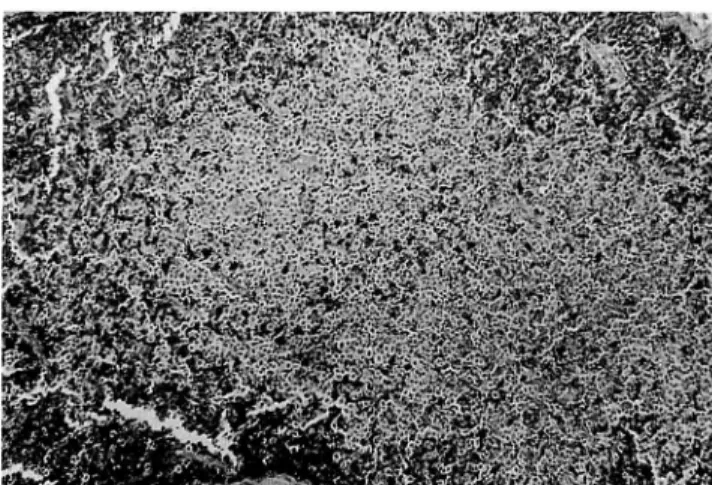
The lymph node biopsy specimen disclosed that the cortical architecture of the lymph node was destroyed and a marked infiltration of large numbers of
Fig. 1. Skin: The epidermis shows slight hyperkeratosis and mild acanthosis. A slightly perivascular infiltrate is seen in the superficial dermis.
Fig. 3. Skin nodule: In the dermis was occupied with a intense cellular infiltrate, that is composed of atypical lymphocytes and plasma cells.
Fig. 5. Lymph node: A infiltration of the paracortical region by pleomorphic lymphocytes.
pleomorphic medium-sized lymphocytes was seen in the paracortical region (Fig. 5). A proliferation of plasma cells was also present(Fig. 6). The post capillary venules appeared to be accentuated or increased in number, but no features of dermatopathic
***** ^-s •*.
C*Fig. 2. Skin: Within the superficial dermis is a lymphocytic infiltrate and plasma cells also are found.
Fig. 4. Skin nodule: Within the epidermis (a hair follicle) are collection of atypical lymphocytes. And they shows nuclear hyperchromatism and irregularity.
Cutaneous T-Cell Lymphoma with Moonoclonal Plasmacytosis (173)
m ' - • . . - :
m y y y m y y m y y
- • , , ,_ .?.
*./** &t.
> W-,: •<•••:% x^xa~< c &y •4,
Fig. 7. Immunostain of skin nodule: The proliferative plasma cells are positive with anti-lambda anti body.
lymphadenopathy were seen.
Immunostain performed on skin and lymph node specimens showed that the pleomorphic lymphocytes had T cell marker(UCHL-l) and plasma cells had monoclonal intracytoplasmic immunoglobulin (IgA- A)
(Fig. 7).
Comment
We presented a patient wth cutaneous T-cell lymphoma(mycosis fungoides) in association with a monoclonal gammopathy and plasmacytosis. Histologi cally skin biopsy specimens showed typical features of mycosis fungoides: The epidermis showed slight acanthosis and there was slight hyperkeratosis. In the upper dermis infiltration of small-sized lymphocytes with irregular shaped nuclei was present, and within the
epidermis Pautrier's microabscess were found. In
addition to the epidermis, follicular epithelium was
involved.
The lymph node biopsy specimen disclosed that the cortical architecture of the lymph node was destroyed and a marked infiltrartion of large numbers of pleomorphic medium-sized lymphocytes was seen in the paracortical region. And the post capillary venules appeared to be accentuated or increased in number, but no features of dermatopathic lymphadenopathy were seen. The histological appearance of the lymph node was in accordance with previous reports in Sezary's syndrome rather than mycosis fungoides.
Characteristic feature in this patient was that infiltrate of plasma cells with the same type of serum immunoglobulin(IgA-lambda)was coincidently present around atypical T-lymphocytes both in the skin and in the lymph node.
The frequency of monoclonal gammopathy in the
general population is related to age and the incidence
of monoclonal gammopathy unrelated to the lympho
ma has been noted as high as 4.8% over the age of 80
years. The occurrence of monoclonal gammopathy has beem previously reported in 17 cases of cutaneous T-cell lymphoma. In three of 17 cases the monoclonal gammopathy was related to myeloma, and one showed atypical plasmacytosis in the bone marrow. But in 13 of 17 cases no proliferation of plasma cell were reported and most of them had benign monoclonal gammopathy unrelated to the lymphoma.
The importance of this interesting association between cutaneous T-cell lymphoma and monoclonal gammophthy is controversial. We presented a patient with mycosis fungoides in association with a monoclon al gammopathy and plasmacytosis. This association lends support to the relation between T-helper-cell malignant proliferation and plasma cell proliferation.
Reference
1) Kovary PM, Suter L, Macher E, Niedorf H,
Grundmann E, Lukitsch O, et al. Monoclonal
gammopathies in Sezary syndrome: A report of four new cases and a review of the literature. Cancer
1981; 48: 788-92.
2) Venencie PY, Winkelmann PK, Puissant A, Kyle RA. Monoclonal gammopathy in Sezary syndrome: Report of three cases and review of the literature.
Arch Dermatol 1984; 120: 605-8.
3) Shimamura R, Kaneko T, Morioka E, Shibuya T, Ishibashi H, Niho Y. Sezary syndrome associated with hepatocellular carcinoma and monoclonal gammmopathy-A case report. Rinsho Ketsueki
1989; 30: 1067-73.
4) Borish L, Dishuck J, Cox L, Mascali JJ, Williams J, Rosenwasser LJ. Sezary syndrome with elevated serum IgE and hypereosinophilia: Role of dysregu-lated cytokine production. J Allergy Clin Immunol
1993; 92: 123-31.
5) Palermo A, Moretti S, Difonzo EM, Fattorossi. Mycosis fungoides with monoclonal gammopathy, hypereosinophilia, and hyper-IgE. Dermatologica
1984; 169: 194-6.
6) Venencie PY, Winkelmann PK, Friedman SJ, Kyle RA, Puissant A. Monoclonal gammopathy and mycosis fungoides: Report of four cases and review
of the literature. J Am Acad Dermatol 1984; 11:
576-9.
7) Ruffieux C, Stalder M, Tran-Thang C. Mycosis fungoides and monoclonal gammopathy. Fortuitous or causal association? Dermatologica 1984; 168:
170-4.
8) Lawrence CM, Marks JM, Burridge A, Moss C, Ive
FA, McGibbon D, et al. The nature of mycosis
fungoides. Q J Med 1986; 58: 281-93.
9) Joyner MV, Cassuto JP, Dujardin P, Barety M, Duplay H, Audoly P. Cutaneous T-cell lymphoma
in association with a monoclonal gammopathy. Arch Dermatol 1979; 115: 326-8.