A Case of Malignant Lymphoma of Breast
著者
SUEYOSHI Kazunobu, YONEZAWA Suguru, SATO
Eiichi
journal or
publication title
鹿児島大学医学雑誌=Medical journal of
Kagoshima University
volume
47
number
Suppl. 2
page range
129-131
URL
http://hdl.handle.net/10232/18330
Med. J. Kagoshima Univ., Vol. 47, Suppl. 2. 129-131, November, 1995
Case Report
A Case of Malignant Lymphoma of Breast
Kazunobu SUEYOSHI,
Suguru YONEZAWA and Eiichi SATO
Second Department of Pathology, Faculty of Medicine Kagoshima University, Kagoshima, Japan
Summary
A case of malignant lymphoma of the breast in a 66 year old woman is presented. She noticed a mass of her right breast. A simple mastectomy was performed. The specimen showed multifocal vague nodular prolifera tion of atypical lymphoid cells having medium to large-sized nuclei. Some giant bizarre atypical lymphoid cells and lymphoepitheHal lesion (LEL) were noted. Im munohistochemically, the atypical lymphoid cells were positive for L26 and MxPanB as markers of B-cell. The lesion was diagnosed as B-cell lymphoma, diffuse, pleomorphic type (LSG classification) and centroblas tic lymphoma polymorphous subtype (updated Kiel classification).
Key words: breast, lymphoma, gene rearrangement
immunohistochemistry
Introduction
Malignant lymphoma of the breast, primary as well as secondary, is rare. Hugh et al. described that two clinicopathoiogic types of breast lymphomas. The first affects pregnant or lactating women who have bilateral masses These tumors are rapidly fatal, and correspond to Burkitt's type lymphoma. The second affects primarily older women who have unilateral mass.
Histologically, most of these lymphoma can be grouped
into large cell B-cell lymphoma and monocytoid B-cell lymphoma, but T-cell lymphoma of breast is rare. Cases of monocytoid B-cell lymphoma of the breast were categorized as mucosa-associated lymphoid tissue (MALT) associated lymphoma. It is considered that pathogenesis of the breast lymphoma is associated with
inflammation especially lymphocytic mastopathy1'.
Moreover, some cases of breast lymphoma were
Address for Correspondence: Kazunobu SUEYOSHI, Second Department of Pathology, Faculty of Medicine Kagoshima University, Sakuragaoka 8-35-1, Kagoshima 890 Japan
positive for estrogen receptor (ER), immunohistoche mically.
In this report, the histologic findings and gene rearrangement study of immunoglobulin heavy chain (IgH) of this rare lesion are described.
Case Presentation
The case examined was 66 year old woman. One
month before the admission, she noticed a mass of her right breast. On physical examination, no lymphadeno
pathy was found. A simple mastectomy was performed.
Pathological findings
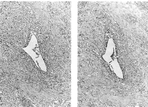
On H&E staining, the specimen showed mulitifocal vague nodular proliferation of atypical lymphoid cells at a low-power magnification (Figure 1). The atypical
lymphoid cells infiltrated into the peripheral adipose
tissues and the pectoral muscle. In the center of the vague nodular lesion, there was the mammary duct
with abundant lymphoid cell infiltration indicating
lymphoepitheHal lesion (LEL) (Figure 2-a). On silver
""Av- "•'• ,' ,,' 'i ,
-w : "»;
mmyyti' ,
-
8 % •-"
Fig. 1. A low-power view of malignant lymphoma of breast showing multifocal vague nodular growth of lym phoma cells infiltrating into the peripheral adipose tissue (HE).
[130] Med. J. Kagoshima Univ., Vol. 47. Suppl. 2, November, 1995
^£iy^i^y$!
-.vW .•••'•••
.••
^
>^"i-'-vv
&^f^^ss# l&L; • :•-.- -.. ... .: t-VV V- y^^iMyy^ -.".••• „v.Fig. 2. lymphoepitheHal lesion (LEL). (a) HE. (b) Silver impregnation.
j • i . « i - •' 2 •A* ♦* ?«*« • '*--. /?• *J * .-^ *«* :•- * .v * ^ „.« V
it-Fig. 3. A high-power view of malignant lymphoma of breast showing marked pleomorphism 'and a bizarre giant tumor cell (Giemsa stain).
impregnation, LEL had well-circumscribed reticulin fibers around the mammary duct with intraepithelial lymphoid cells (Figure 2-b). On Giemsa staining, the atypical lymphoid cells had pleomorphism at a high-power magnification. The atypical giant cells with bizarre, multilobated, hyperchromatic nuclei were also noted (Figure 3). The nuclei of the most of atypical
cells were twisted or indented shaped and had some
marginally situated nucleoli. Some nuclei were round and had fine chromatin with or three marginally situated nucleoli. The cytoplasm was basophilic and abundant. Mitotic activity was frequent. Small amount of immunoblasts and plasma cells were also observed. Judging from these findings, this case was classified into centroblastic lymphoma, polymorphous subtype according to the updated Kiel classification, and
malignant lymphoma, diffuse, pleomorphic type according to the LSG classification.
Immunohistochemical findings
These atypical lymphoid cells were positive for L26, MxPanB, and immunoglobulin kappa light chain as markers of B-cell. Staining for CD3 as marker of T-cell was positive for the most of background small lymphocytes. LN-3 as marker of HLA-DR was strongly reacted with neoplastic cells as well as mammary ductal epithelium.
Detection of rearrangement of immunoglobulin heavy chain gene
To identify the monoclonarity of the atypical lymphoid cells, we examined the presence of rearrange ment of IgH, using the paraffin-embedded section by
polymearse chain reaction (PCR). The PCR process2',
using the primers (JH1/JH2) (Table 1) specific to IgH,
were performed on automated thermal cycler
(Perkin-Elmer-Cetus, Norwalk, CT, USA) with denaturation at 94°C for 30s, annealing at 55°C for 30s, and polymeriza tion at 72°C for 30s, during 50cycles. The reaction volume was 50 M 1, where 2.5units of Taq DNA polymerase were present. The amplified products were electrophoresed on 4% agarose gel.
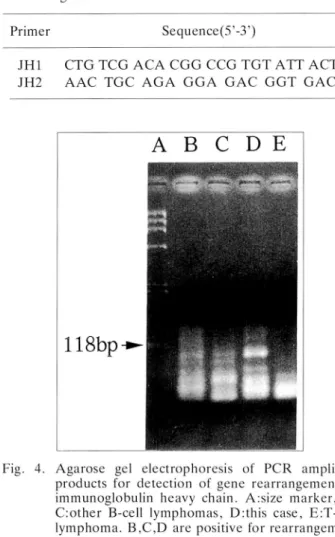
The results were shown in Figure 4.. One rearrange ment band, approximately lOObp, of IgH in this case could be detected, this finding suggest that this lesion was consistent with monoclonal proliferation of neoplastic B-cells.
A Case of Malignant Lymphoma of Breast [131]
Table 1. Primers specific to immunoglobulin heavy chain
gene
Primer Sequence(5'-3')
JH1 CTG TCG ACA C G G CCG TGT ATT ACT G
JH2 A A C TGC A G A G G A G A C G G T G A C C Fig. 4.
A
B
C
D
E
, — .118bp-»-4K.
Agarose gel electrophoresis of PCR amplified products for detection of gene rearrangement of immunoglobulin heavy chain. A:size marker, B, C:other B-cell lymphomas, D:this case, E:T-cell lymphoma. B,C,D are positive for rearrangement bands, and E is negative.
Discussion
We demonstrated a case of B-cell lymphoma of the breast, that was classified into centroblastic lymphoma, polymorphous subtype, according to the updated Kiel classification. Malignant lymphoma of the breast is uncommon, representing 0.004 to 0.5% of all breast
malignant tumors1'. This rare lesion among breast
malignant tumors may be made erroneous diagnosises as other malignant tumors such as poorly differentiated carcinoma or inflammatory disorders, therefore, it is important that the characteristic features of breast
lymphoma are picked up, and we should more carefully carefully exmine the lesion such as this case pathohisto-logically, immunohistopathohisto-logically, and genetically.
The characteristic findings in 14 cases of breast lymphoma that we recently examined are multifocal vague nodular proliferation of lymphoma cells and periductal infiltration of lymphoma cells. These findings were observed in this case, and lymphoepithe Hal lesion that is peculiar to mucosa-associated lymphoid tissue (MALT) associated lymphoma is also
noted.
Immunohistochemically, many T-cells and his tiocytes infiltrated into the background of this lesion, and the neoplastic B-cells as well as the mammary ductal epithelium infiltrated by the tumor cells were strongly positive for LN-3 as marker of HLA-DR. These findings suggest that breast lymphoma is
associated with inflammation or the other
immunoreac-tive disorders of breast. Especially, the presious studies demonstrate that lymphocytic mastopathy is associated
with pathogenesis of breast lymphoma1'.
The gene rearrangement of IgH was demonstrated in this case which could detect monoclonal rearrangement band. The study using PCR is useful for the paraffin-embedded tissue of the rare case saving no fresh frozen
material such as this case. However, monoclonal
rearrangement band of IgH gene is observed in the case of B-cell lymphoma as well as the case of reactive proliferation of B-cells, thus this method using PCR should be used to assist the pathohistological examina tion, especially when a lesion is composed of mixed
proliferation of varied cells.
References
1) Aozasa, T., Ohsawa, M., Saeki, K., Horiuchi, K,
Kawano, K, and Taguchi, T. (1992): Malignant lymphoma of the breast. Immunologic type and association with lymphocytic mastopathy. Am. J.
Clin. Pathol., 97, 699-704.
2) McCarthy, K. P., Sloane, J. P., and Wiedemann, L. M. (1990): Rapid method for distinguishing clonal from polyclonal B cell populations in surgical biopsy specimens. J. Clin. Pathol., 43, 429-32.
3) Hugh, J. C, Jackson, F. I., Hanson, J., and Poppema, S. (1990): Primary breast lymphoma. An immunohistologic study of 20 new cases. Cancer,