【症例報告】 Case Report
患者由来抗 E 出現後に Evans 症候群を発症した同種造血幹細胞移植の 1 例
山崎 理絵1) 上村 知恵1) 五十嵐靖浩1) 藤村 亮介1) 森 文香1)
中山 瞳2) 甲田 祐也2) 加藤 淳2) 森 毅彦2) 田野崎隆二1)
同種造血幹細胞移植後
Evans
症候群発症の前に,患者由来不規則抗体の陽性化を認めた症例を経験したため報告 する.【症例】28歳女性,O型Rh(+).慢性骨髄性白血病(急性転化)に対し,HLA
一致非血縁ドナー(男性,A
型Rh
(+))より骨髄移植を施行した.移植後急性移植片対宿主病および器質化肺炎を合併し,ステロイドにより 加療し改善した.1
カ月に1
度不規則抗体スクリーニングを実施していたが,ステロイド減量中の移植後1
年の定期 外来受診時,はじめて抗E
が陽性となった.その1
カ月後に高度の貧血および血小板減少を認め,Evans症候群と 診断された.ドナーおよび患者のRh
血液型はDCcEe
およびDCCee
であり,発症時に患者由来B
細胞の残存を認 めた.患者残存B
リンパ球による同種抗体産生をきっかけに,赤血球・血小板に対する自己抗体が産生されたもの と考えられた.【結語】同種造血幹細胞移植後に不規則抗体が出現した場合は,同定を速やかに行うとともに,病態 変化の可能性を念頭に入れて,注意深く経過を追う必要がある.キーワード:免疫性溶血性貧血,Evans症候群,同種造血幹細胞移植,不規則抗体
はじめに
同種造血幹細胞移植(allogeneic hematopoietic stem
cell transplantation:allo-HSCT)後には,原病,薬剤,
感染,ドナー・患者間の血液型不一致,微小血管血栓 など,様々な要因により溶血をきたす1).自己抗体ある い は 同 種 抗 体 に よ る 免 疫 性 溶 血 性 貧 血(immune-
mediated hemolytic anemia)もその一つである.自己
免疫性溶血性貧血(autoimmune hemolytic anemia;AIHA)は allo-HSCT
後1.5〜4.4% に発症すると報告さ
れている2)〜4).しかし,
AIHA
に特発性血小板減少性紫斑病(idiopathic thrombocytopenic purpura;ITP)を 合併する
Evans
症候群のallo-HSCT
後の発症について は数例報告があるのみである5)6).今回我々は,非血縁 者間骨髄移植1
年後にEvans
症候群を合併し,リツキ シマブ投与後のステロイド治療により軽快した症例を 経験した.本症例では,症状出現前に定期的な不規則 抗体スクリーニング検査で抗E
が陽性化した.AIHA 発症との関連性について考察し報告する.症 例
患者:28歳女性
O
型Rh(+)不規則抗体無し.
既往歴・妊娠歴:なし.
経過:某年
8
月より動悸や頭痛を自覚するようになり近医受診,血液検査で白血球
105,000/μ l
,芽球44%,
ヘモグロビン(Hgb)
9.2g/d l
,血小板6.5
万/μl
を指摘 され,当院紹介受診となった.骨髄検査で芽球95.7%,
Major BCR-ABL
陽性,染色体検査では,解析した20
細胞全てに(9;22)t
(q34;q11.2)を認め,慢性骨髄性 白血病急性転化期(リンパ芽球性)と診断された.プ レドニゾロン(PSL)先行投与後にダサチニブ140mg/
日にて加療し,翌年
2
月にHLA
一致非血縁ドナー(男 性,A型Rh
(+))より骨髄移植を施行した.前処置は 全身放射線照射12Gy+シタラビン 8g/m
2+シクロホス ファミド120mg/kg,移植片対宿主病(graft-versus- host disease:GVHD)予防は,タクロリムス+メトト
レキサートとして,Day 17
に生着を確認した.生着期 に急性GVHD(Grade II:皮膚)を認めたが,PSL 1
mg/kg/日で改善した. Day100
前後に器質化肺炎を発症し,メチルプレドニゾロン
2mg/kg/日で加療開始し,
改善を認めた.
Day130
よりステロイドの減量を開始した.
Day 138
を最後に赤血球輸血(O型)が不要となり,血小板輸血も
Day 152
で不要となったが,Hgb値は8 g/d l
台後半で推移していたため,1カ月ごとの外来受 診時に輸血に備えて不規則抗体スクリーニングを行っ ていた.PSL 4mg/日まで減量していた外来受診日
(Day335)
の不規則抗体スクリーニングで抗E
が検出された.1)慶應義塾大学医学部輸血・細胞療法センター 2)慶應義塾大学医学部血液内科
〔受付日:2020年3月17日,受理日:2020年5月14日〕
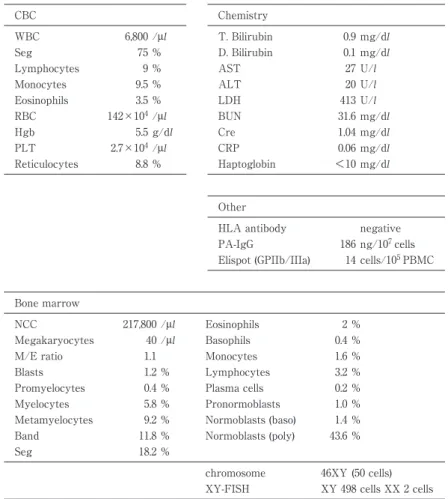
Table 1 Laboratory data on admission
CBC Chemistry
WBC 6,800 /μl T. Bilirubin 0.9 mg/dl
Seg 75 % D. Bilirubin 0.1 mg/dl
Lymphocytes 9 % AST 27 U/l
Monocytes 9.5 % ALT 20 U/l
Eosinophils 3.5 % LDH 413 U/l
RBC 142×104/μl BUN 31.6 mg/dl
Hgb 5.5 g/dl Cre 1.04 mg/dl
PLT 2.7×10⁴/μl CRP 0.06 mg/dl
Reticulocytes 8.8 % Haptoglobin <10 mg/dl
Bone marrow
NCC 217,800 /μl Eosinophils 2 %
Megakaryocytes 40 /μl Basophils 0.4 %
M/E ratio 1.1 Monocytes 1.6 %
Blasts 1.2 % Lymphocytes 3.2 %
Promyelocytes 0.4 % Plasma cells 0.2 %
Myelocytes 5.8 % Pronormoblasts 1.0 %
Metamyelocytes 9.2 % Normoblasts (baso) 1.4 %
Band 11.8 % Normoblasts (poly) 43.6 %
Seg 18.2 %
chromosome 46XY (50 cells) XY-FISH XY 498 cells XX 2 cells FISH: Fluorescence in situ hybridization; PA-IgG: Platelet-associated IgG; M/E ratio:
myeloid/erythroid ratio
Other
HLA antibody negative
PA-IgG 186 ng/107 cells Elispot (GPIIb/IIIa) 14 cells/105 PBMC
後方視的に確認したところ移植前後に計
8
回E
抗原陽 性の赤血球製剤が輸血されており,最後のE
抗原陽性輸血は
Day119
であったが,その後のスクリーニングで不規則抗体は一貫して陰性であった.臨床症状はなく,
血液検査も変化なかったため,PSL 4mg/日を
2mg/
日へ減量し経過観察となった.
Day 356
労作時呼吸苦に て 来 院,白 血 球5,500/μ l
,Hgb 5.5g/dl
,血 小 板2.7
万/μl
と2
系統の血球減少を認め入院となった.入院時身体所見:体重
50.7kg,血圧 86/51mmHg,脈
拍93/min,体温 37.2℃,酸素飽和度 98%.眼瞼結膜貧
血様,眼球結膜黄染認めず.表在リンパ節触知せず,心音整,肺野清.肝脾触知せず.下腿浮腫なし.
入院時検査所見(Table 1):末梢血では高度な貧血,
血小板減少を認め,網状赤血球の増加,間接ビリルビ ン,
LDH
の上昇から溶血が疑われた.ハプトグロビン は感度以下,エリスポット検査でGlycoprotein
(GP)IIb/
IIIa
抗体産生細胞を認めた.骨髄は軽度過形成で赤芽球 系細胞および巨核球の増加を認め,原病の再発所見は 認めなかった.骨髄のXY-FISH
にて500
細胞中2
細胞 の患者由来細胞を認めた.輸血検査所見:血液型オモテ検査では,A抗原陽性
であったが一部にフリーセルを認め,O型血球の残存 が疑われた.フローサイトメトリー解析では,A型血 球は
90.2% であった(Fig. 1).不規則抗体スクリーニ
ングで抗E
陽性,広スペクトラム抗血清による直接クー ムス試験にてIgG
および補体が赤血球表面に存在する ことが確認された.赤血球自己抗体解離試験後の血清 は,各種血球に広範囲に反応したが,特にE
抗原陽性血球に
3+から 4+と強く反応した.移植後の Rh
血液型は,カラム凝集法にて
DCcEe
であった.患者のRh
血液型は,保存していた患者の移植前末梢血単核球を 用いてPCR-SSP
法にてRHCE
遺伝子を解析し7),DCCee
と判定した.末梢血
B
リンパ球由来解析:追加検討として後日デ ジタルPCR
で入院時の患者末梢血中のB
リンパ球の由 来解析を行った.単核球よりCD3
陽性細胞およびCD
19
陽性細胞を単離し,DNA
を抽出した.QuantStudio
3D Digital PCR System
(Thermo Fisher Scientific)に よりSRY
(sex determining region on Y)陽性細胞を男 性由来細胞としてドナー由来細胞の割合を解析するこ ととし,VIC
標識TaqMan Copy Number Reference
Assay human RNase P
を全細胞数カウントに,FAMFig. 1 Flow cytometry analysis of red blood cells on admission. In the histograms, FITC-derived fluorescence is displayed on the X axis and the number of cells on the Y axis. Fig. 1-A and 1-B show controls. Two populations of cells, group A and group O, were detected (Fig. 1-C). Almost 10% of the patientʼs red blood cells did not ex- press A antigens. These cells were considered to be remaining recipient-derived red cells.
Table 2 Digital PCR analysis of patient lymphocytes on admission
Sample Cell count*/μl SRY copies/μl Ratio of SRY-positive cells
male control 169.10 170.72 1.01
patient CD3-positive cells 184.57 176.57 0.96
patient CD19-positive cells 199.81 177.50 0.89
patient PBMC 118.01 112.02 0.95
PBMC: peripheral blood mononuclear cells; SRY: sex-determining region on Y
*Cell count was calculated by one-half copies of RNase P.
標識
Taqman
Ⓡgene expression assays SRY
をドナー由 来細胞のカウントに用いた.CD19
陽性細胞の89% が
ドナー由来のSRY
陽性細胞であり,約10% の患者由
来B
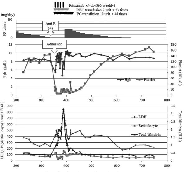
細胞が残存していることが確認できた(Table 2). なお,本検討は慶應義塾大学医学部倫理委員会の承認 を受け,患者より同意を取得の上実施している.入院後経過(Fig. 2):直接
Coombs
試験が陽性であ ること,およびGP IIb/IIIa
抗体産生細胞の存在からEvans
症候群と診断した.抗E
が患者由来の同種抗体である可能性を考え,輸血は
E
抗原陰性血で対応した.ステロイド加療中の発症であったため
Day366
よりリ ツキシマブ(375mg/m2)を週1
回,計4
回投与した.効果不十分であったため,
Day396
よりPSL 0.5mg/kg/
day
を追加したところ速やかにデータの改善を認めた.Day416
より輸血が不要となり,Day426に退院,ステロイドは漸減し
Day650
に中止した.Day713
には不規 則抗体も陰性化した.考 察
今回我々は,同種骨髄移植後に
Evans
症候群を合併 した症例を経験した.本症例で特記すべきは,発症時患者
B
リンパ球が残存しており,ドナー特異血液型抗 原であるE
に対する不規則抗体が同定されている点で ある.今回の溶血および血小板減少の機序を推測する.溶 血および血小板減少の原因抗体は,ドナー由来自己抗 体と思われ,ステロイド減量後の免疫能亢進に伴い,
自己抗体が産生されたと考えられる.同種移植後には 自己免疫疾患が起こりやすいことが知られるが,その 中でも
AIHA
の発症率は高い.ChenらはB
リンパ球 とT
リンパ球の免疫再構築のアンバランスにより,oli- goclonal
な抗体産生が促進されることがAIHA
発症の 原因と考察している8).最初に検出された抗
E
もドナー由来自己抗体で,発 症の初期段階を見ていた可能性がある.Issitt
らは,直 接クームス陽性患者に検出される同種抗体の約半数は,特定の抗原に対する特異性を示してはいるが,実は別 の抗原を認識している
mimicking
自己抗体であると報 告している9).しかし本症例に関しては,E
抗原を認識 していることが明らかであること,E
抗原がドナーの特 異抗原であったこと,患者B
細胞の残存が確認されて いることから,最初に検出された抗E
は患者由来同種Fig. 2 Clinical course
Treatments including transfusions are shown in the top chart. Changes in hemoglobin titers and platelet counts are shown in the middle chart and those in reticulocyte counts, LDH, and total bilirubin values are shown in the bottom chart.
Abbreviations; PSL: prednisolone; RBC: red blood cell; PC: platelet concentrate; Hgb: hemoglobin
抗体であったと推測した.
また本症例では臨床的に明らかな溶血を認めなかっ たものの,器質化肺炎の加療中より
LDH
と網状赤血球 の軽度上昇を伴う貧血が持続していた.移植後早期か ら抗E
産生患者由来B
細胞は存在しており,ステロイ ド減量に伴い抗体産生が顕著となり,抗E
が検出でき るようになった可能性もある.同種移植例については,AIHAだけでなく同種抗原 を起因とする移植後溶血反応についてもいくつか報告
がある.
Sokal
らは,患者由来抗D
によるドナー赤血球の溶血を認めた後に,AIHAをきたした症例を報告し た10).
Wang
は移植後AIHA
の患者の58% に Rh
抗原 やKell
抗原などに対する同種抗体が認められたと報告 し,残存する患者免疫機構がどの程度関与しているの かについては,まだわかっていないことが多いとして いる4).Franchini
11)らは同種移植後の不規則抗体の出現率について,
1〜8.7% 程度と報告しており,治療抵抗性
の溶血反応につながる可能性を示唆している.また,同種移植後に限らず,赤血球自己抗体保有患 者が同種抗体を同時に保有する割合は
8〜40% で通常
の同種抗体の検出頻度より高いと報告されており12)13), 全般的な免疫能の亢進により自己抗体と同種抗体が同 時に存在することは珍しくない.自己抗体陽性患者への輸血に際しては,基本的に患 者と一致する赤血球製剤を選択することが推奨される が,自己抗体と同種抗体の両方を持つ患者に対しては,
同種抗体に対する抗原陰性血液の輸血を優先すること がガイドライン14)上推奨されている.本症例においては 同種抗体である可能性が否定できないことから,この ガイドラインに則り,最初から
E
抗原陰性血の輸血を 行った.同種移植後のAIHA
症例においては,このよ うに複雑な病態を呈する可能性があり,あらゆる側面から検討してより安全な輸血を選択することが必要と 考える.
直近の輸血など明らかな誘因なく,同種移植後に突 然不規則抗体が陽性化した場合には,抗体の特異性,
ドナーと患者の赤血球抗原との反応性,直接クームス 試験などを確認するとともに,その後の溶血イベント の発症に注意する必要がある.
著者のCOI開示:本論文発表内容に関連して特に申告なし
謝辞:RHCE遺伝子解析を実施してくださった日本赤十字社中
央血液研究所血液型チーム小笠原健一先生,伊佐和美先生のご協 力に深謝いたします.
文 献
1)Holbro A, Passweg JR: Management of hemolytic ane- mia following allogeneic stem cell transplantation. He- matology Am Soc Hematol Educ Program, 2015: 378―
384, 2015.
2)Sanz J, Arriaga F, Montesinos P, et al: Autoimmune hemolytic anemia following allogeneic hematopoietic stem cell transplantation in adult patients. Bone Mar- row Transplant, 39: 555―561, 2007.
3)González-Vicent M, Sanz J, Fuster JL, et al: Autoim- mune hemolytic anemia (AIHA) following allogeneic he- matopoietic stem cell transplantation (HSCT): A retro- spective analysis and a proposal of treatment on behalf of the Grupo Español De Trasplante de Medula Osea en Niños (GETMON) and the Grupo Español de Trasplante Hematopoyetico (GETH). Transfus Med Rev, 32: 179―
185, 2018.
4)Wang M, Wang W, Abeywardane A, et al: Autoimmune hemolytic anemia after allogeneic hematopoietic stem cell transplantation: analysis of 533 adult patients who underwent transplantation at Kingʼs College Hospital.
Biol Blood Marrow Transplant, 21: 60―66, 2015.
5)Ryeon Lee S, Lee SY, Nam MH, et al: Successful control of steroid-intolerant Evansʼ syndrome associated with allogeneic peripheral blood hematopoietic stem cell transplant by rituximab. Leuk Lymphoma, 52: 528―530, 2011.
6)Faraci M, Zecca M, Pillon M, et al: Autoimmune hemato- logical diseases after allogeneic hematopoietic stem cell transplantation in children: an Italian multicenter expe- rience. Biol Blood Marrow Transplant, 20 : 272―278, 2014.
7)Kikuchi G, Kurita R, Ogasawara K, et al: Application of immortalized human erythroid progenitor cell line in se- rologic tests to detect red blood cell alloantibodies.
Transfusion, 58: 2675―2682, 2018.
8)Chen FE, Owen I, Savage D, et al: Late onset haemolysis and red cell autoimmunisation after allogeneic bone marrow transplant. Bone Marrow Transplant, 19: 491―
495, 1997.
9)Issitt, PD, Combs MR, Bumgarner DJ, et al: Studies of an- tibodies in the sera of patients who have made red cell autoantibodies. Transfusion, 36: 481―486, 1996.
10)Sokol RJ, Stamps R, Booker DJ, et al: Posttransplant immune-mediated hemolysis. Transfusion, 42: 198―204, 2002.
11)Franchini M, Gandini G, Aprili G: Non-ABO red blood cell alloantibodies following allogeneic hematopoietic stem cell transplantation. Bone Marrow Transplant, 33:
1169―1172, 2004.
12)Branch, D. R., Petz, L. D.: Detecting alloantibodies in pa- tients with autoantibodies. Transfusion, 39: 6―10, 1999.
13)山口 瞳,杉本達哉,前沢由美子,他:赤血球自己抗体 陽性患者への赤血球輸血の解析.日本輸血細胞治療学会 誌,63(2):95―104, 2017.
14)日本輸血・細胞治療学会ホームページ:赤血球型検査
(赤血球系検査)ガイドライン(改訂2版).
http://yuketsu.jstmct.or.jp/wp-content/uploads/2016/
10/5bc721e299263f6d44e2215cbdffbfaf.pdf(2020 年 4 月20日現在).
EVANS SYNDROME AFTER ALLOGENEIC STEM CELL TRANSPLANTATION FOLLOWING DEVELOPMENT OF RECIPIENT-DERIVED ANTI-E
Rie Yamazaki
1), Tomoe Uemura
1), Yasuhiro Igarashi
1), Ryosuke Fujimura
1), Ayaka Mori
1), Hitomi Nakayama
2), Yuya Koda
2), Jun Kato
2), Takehiko Mori
2)and Ryuji Tanosaki
1)1)
Center for Transfusion Medicine and Cell Therapy, Keio University School of Medicine
2)
Division of Hematology, Department of Medicine, Keio University School of Medicine
Abstract:
Immune-mediated hemolytic anemia including Evans syndrome, is a known to be a complication of allogeneic he- matopoietic stem cell transplantation (allo-HSCT). We recently experienced a 28-year-old female patient with chronic myelogenous leukemia (CML) in blastic crisis who developed Evans syndrome after allogeneic bone marrow trans- plantation from an unrelated male donor (blood type A, Rh+). She developed acute graft-versus-host disease (GVHD) and organizing pneumonia, which was treated with steroid therapy. One year after allo-HSCT, anti-E appeared during a regular checkup for irregular antibody following withdrawal of the steroid. One month later, she developed severe hemolytic anemia and thrombocytopenia. The Rh blood types of the donor and recipient were DCcEe and DCCee, re- spectively. At the onset of hemolysis, recipient-derived B cells remained in her peripheral blood. These observations led us to speculate that allo-antibody production by the remaining recipient-derived B cells triggered the generation of autologous antibodies against red blood cells and platelets, resulting in Evans syndrome. We suggest that an irregu- lar antibody screening test might be helpful in predicting hemolytic events after allo-HSCT.
Keywords:
Immune-mediated hemolytic anemia, Evans syndrome, allogeneic stem cell transplantation, irregular antibodies
!2020 The Japan Society of Transfusion Medicine and Cell Therapy Journal Web Site: http:!!yuketsu.jstmct.or.jp!