東北医科薬科大学 審査学位論文(博士)
氏名(本籍) チャン ゴウェイ
張 国偉(中国)
学位の種類 博士(薬科学)
学位記番号 博薬科第18号
学位授与の日付 平成31年3月8日
学位授与の要件 学位規則第4条1項該当
学位論文題名 Functional analysis of N-acetylglucosaminyltransferase- I: a novel regulator of epithelial-mesenchymal transition
論文審査委員
主査 教 授 細 野 雅 祐
副査 教 授 関 政 幸
副査 教 授 顧 建 国
Functional analysis of
N-acetylglucosaminyltransferase-I: a novel regulator of epithelial-mesenchymal transition
東北医科薬科大学大学院薬学研究科
張 国偉
平成 31 年 3 月
Functional analysis of
N-acetylglucosaminyltransferase-I: a novel regulator of epithelial-mesenchymal transition
Contents
Introduction ……… 1
Materials and Methods ……… …… ……… 4
Results ……… 10
Discussion……… 25
References……… 30
Abbreviations……… ……… ……… 41
Acknowledgements……… 42
1. INTRODUCTION
The epithelial-to-mesenchymal transition (EMT) is a cellular trans-differentiation process that allows polarized epithelial cells to undergo multiple biochemical changes, and enables them to acquire a mesenchymal identity that results in a loss of cell-cell adhesion. EMT is integral in cell adhesion, migration and wound healing, and pathologically contributes to cancer metastasis and resistance to therapy (1). This switch in cell behavior is executed mainly by specific transcription factors (e.g., Snail, TWIST, and ZEB families) at transcriptional levels (2). Mesenchymal-to-epithelial transition (MET) is a reversible biological process that involves the transition from motile, spindle-shaped mesenchymal cells to planar arrays of polarized cells that acquire cell-cell adhesion. Recent studies have shown that MET participates in the establishment and stabilization of distant metastases by allowing cancerous cells to regain epithelial properties and integrate into distant organs (3). Several studies have focused on the sequential EMT-MET process that underlies the fate of cells (4, 5) and the flexible transitions are related to different stages of metastasis (6). Details of the mechanism involved in this process, however, awaits elucidation.
An important hallmark of EMT is the attenuation of the epithelial marker E-cadherin, and the enhancement of mesenchymal markers such as N-cadherin (7). E-cadherin is a central component of cell-cell adhesion junctions, the loss of which facilitates cell migration and alters integrin-mediated cell adhesion (8). Integrins control cell interactions with the extracellular matrix (ECM), which is critical to EMT progression (9, 10). In fact, TGFβ-induced EMT enhances the expressions of αVβ6 and αVβ3 integrin, which in turn alters cell-matrix adhesion to promote the EMT process (11, 12).
Ectopic expression of β4 integrin upregulates cell proliferation and induces EMT (13).
Both E-cadherin and integrin are glycoproteins. Aberrant expression of N-glycans have been observed in several EMT models (14). Previous studies by our group, as well as those of others, have found that alteration of N-glycans by manipulation of the
that include cell adhesion molecules and surface receptors such as E-cadherin, integrins and epithelial growth factor receptors (EGFR). Expression of tumor-associated glycans is involved in the dysregulation of glycogenes, which comprise glycosyltransferases and glycosidases. For example, overexpression of N-acetylglucosaminyltransferases V (GnT-V) promoted E-cadherin destabilization, while expression of GnT-III stabilized E-cadherin (15). TGF-β-induced EMT increased the α1,6-fucosylation expression catalyzed by α1,6 fucosyltransferase (FUT8), and overexpression of FUT8 promoted EMT progression (16, 17). In addition, overexpression of GnT-III inhibited integrin α3β1-mediated cell adhesion and migration (18, 19) as well as the binding of α5β1 integrin to fibronectin (FN) (20). Thus, many studies have envisaged the possibility of using glycan-based therapies for cancer treatment (21-23).
There are three main types of glycans: high mannose, hybrid and complex glycans.
High-mannose is just two N-acetylglucosamines with many mannose residues. Hybrid oligosaccharides contain a mannose residue on one side of the branch, while on the other side an N-acetylglucosamine initiates a complex branch. Complex oligosaccharides contain almost any number of the other types of saccharides, including more than the original two N-acetylglucosamines. Thus far, most studies have focused on complex N-glycans and little is known about the functions of high-mannose types of N-glycans. In fact, high-mannose types of N-glycans have been detected, at relatively lower levels in glycoproteins expressed either on the cell surface or in secretions (24).
Interestingly, the expression levels of high mannose N-glycans are known to increase in breast cancer processes (25), but these are decreased in gastric cancers (26).
Swainsonine (SW), an inhibitor of Golgi α-mannosidase II, is known to block the processing of high-mannose N-glycans to complex N-glycans, but not hybrid types of N-glycans (27, 28). Treatment with SW inhibited tumor growth and metastasis (29, 30), although the underlying mechanism for the function of SW remains unclear. The
carcinoma have been discouraging (31). Thus, the discovery of new therapeutics based on high-mannose N-glycans is urgent.
N-Acetylglucosaminyltransferase I (GnT-I) is a key enzyme in the cis-Golgi cisternae that transfers N-acetylglucosamine (GlcNAc) onto the N-glycan core (containing five mannose) after the mannose branch is trimmed by α-mannosidase I (Fig.1). The addition of GlcNAc by GnT-I is required for the action of α-mannosidase II and N-acetylglucosaminyltransferase-II (GnT-II), and then GnT-III, GnT-IV or GnT-V in a catalytically ordered pathway (32, 33). Deficiency of GnT-I leads to loss of the downstream N-glycosylation pathway and results in the presence of high-mannose types of N-glycans only (34-36). GnT-I is known to be crucial for embryo development (37), and important for cytokine signaling (34). The knockdown of GnT-I using shRNA suppresses cell migration, invasion and tumor growth (38).
In the present study, we generated GnT-I knockout cells (KO) using a CRISPR/Cas9 system, and found that GnT-I worked as a switch to activate EMT, and up-regulated integrin-mediated functions. Clearly, the mesenchymal markers were decreased in the KO cells, but they were increased in the restoration of GnT-I genes in KO cells (Rescued cells, RES). Furthermore, we found that the expression of GnT-I was closely related to drug resistance and colony formation. Thus, our study clearly demonstrated the dynamic regulation by GnT-I was critical for cell behaviors, and targeting GnT-I could be a potential therapeutic strategy for cancer treatment.
Figure 1. The role of GnT-I in biosynthesis of N-linked glycosylation.
2. MATERIALS AND METHODS 2.1 Antibodies and reagents
In the present study, experiments were performed using the following antibodies:
antibody against human β1 integrin subunit (P5D2) was obtained from Developmental Studies Hybridoma Bank, University of Iowa; rabbit mAb against GnT-I (ab180578) was from Abcam; Mouse mAb against Smad2 (3103), rabbit mAbs against Vimentin (5741), ZEB1 (3396), Claudin-1 (13255), Snail (3879), p-EGFR (4267), EGFR (3777) and p-Smad2 (3108) were from Cell Signaling Technology; mouse mAbs against N-cadherin (610920), β1 integrin (610468), focal adhesion kinase (FAK, 610088) and p-FAK (611807) were from BD Biosciences; mouse mAbs against α5 integrin (328002), αV integrin (327902), β3 integrin (336402), and β5 integrin (345202) were from Biolegend; mouse pAb against α3 integrin (Sc-6592) was from Santa Cruz Biotechnology; mouse mAb against β4 integrin (MAB2059) was from Millipore; mouse mAb against α-tubulin (T6199) and α-SMA (A2547) were from MilliporeSigma. Alexa Fluor® 647 goat anti-mouse IgG and streptavidin were obtained from Thermo Fisher Scientific (Waltham, MA USA). The peroxidase-conjugate goat antibodies against mouse and rabbit IgG were obtained from Cell Signaling Technology. The Sulfo-NHS-SS-biotin was from Thermo Scientific. Biotinylated Datura stramonium agglutinin (DSA, B-1185), Phaseolus vulgaris leucoagglutinin (L-PHA, B-1115), Phaseolus vulgaris erythroagglutinin (E-PHA, B-1385), Concanavalin A (ConA, B-1005), Maackia amurensis agglutinin (MMA, B-1265) and Aleuria aurantia lectin (AAL, B-1395) were purchased from Seikagaku Corporation (Tokyo, Japan).
2.2 Cell culture
The HeLa (Riken, Japan), 293T (Riken, Japan) and MDA-MB-231 (ATCC) cell lines were cultured in Dulbecco’s modified Eagle’s medium (DMEM) with 10% fetal bovine
2.3 Genomic deletion of GnT-I-KO in HeLa cells
The pSpCas9(BB)-2A-GFP (PX458) plasmid was a gift from Dr. Feng Zhang (Addgene plasmid # 48138) (39). The Target site on human GnT-I exon 1 (CGAAAGTACTCGAAGAAGTC), which was chosen from the genome-scale CRISPR-Cas9 knockout library (40), was cloned into PX458. GnT-I-KO cell lines were created by electroincorporating cells with vector containing the target sequence according to the manufacturer’s recommendation (Amaxa®cell line Nucleofector®kit R).
After 3 days of transfection, GFP-positive cells were sorted using FACSAria II (BD Bioscience). Approximately 10 days thereafter, FACS sorting was performed using L-PHA, which preferentially binds to the Galβ4GlcNAcβ6 (GlcNAcβ2Manα3) Manα3) structure to detect GnT-I deletion (KO). The process was repeated three times to obtain the GnT-I-KO cell line.
2.4 Establishment of GnT-I rescued HeLa cells
The full open reading frame of human GnT-I was cloned into the pENTR/D-TOPO vector, and then transferred into the CSII-EF-Rfa destination vector by LR recombination according to the manufacturer’s instructions (Invitrogen, CA, USA). To prepare lentivirus, 293T cells were co-transfected with CSII-EF-Rfa-GnT-I and lentiviral packaging vectors (pCAG-HIVgp and pCMV-VSV-G-RSV-Rev). The culture supernatants were collected 48 hrs after infection, and the virus was introduced into GnT-I-KO HeLa cells via addition of the supernatant to the culture medium. The cells highly reacted with L-PHA were selected utilizing FACSAria II for GnT-I-RES cells, and the stable GnT-I-RES cell line was used for subsequent studies.
2.5 Western blot
Total cell lysates were prepared with lysis buffer containing 20 mM Tris-HCl (pH 7.4), 100 mM NaCl, 1% Triton X-100, protease inhibitors and phosphatase inhibitors
protein assay kit (Pierce). An equal amount of protein from each sample was subjected to SDS-PAGE and transferred to PVDF membranes (Millipore). The membranes were detected with primary and secondary antibodies, and visualized by chemiluminescent detection (Millipore).
For peptide-N-glycosidase F (PNGase F) treatment, the indicated cell lysates were treated with PNGase F at 37 °C for 1 h according to the manufacturer’s instruction, and then subjected to western blot.
2.6 Flow cytometric analysis
After detachment, cells were washed with ice-cold PBS and stained with primary antibodies or lectins in PBS containing 1mM EDTA and 0.1% BSA (m/v) at 4 °C for 20 minutes, followed by incubation with Alexa Fluor 647 conjugated secondary antibodies according to the primary antibodies or lectins (Invitrogen) for 20 min at 4 °C.
Unstained cells were included as controls, and dead cells and debris were excluded from the analysis, based on scatter characteristics. Data for at least 10,000 live events per sample were acquired via FACSCalibur flow cytometry (BD Biosciences). After incubation with doxorubicin for 1 day, the indicated cells were detached and washed with ice-cold PBS, and then subjected to flow cytometry. The fluorescence was measured with a FL2 (Phycoerythrin) bandpass filter to monitor doxorubicin cellular uptake. Untreated cells were used as controls, and dead cells and debris were excluded from the analysis, based on scatter characteristics.
2.7 Cell migration assay
For in vitro wound-healing assay, cells were seeded in 6-well plates precoated with 10 µg/ml FN (Sigma-Aldrich) and grown into a confluent monolayer. Then the cells were scratched via a pipette tip. Images of the wound area were photographed after 8 hours,
Transwell chamber system (BD BioCoatTM control inserts, 8 μm pore size inserts). The bottom side of the chamber was coated with 10 µg/ml FN, and blocked with 1% BSA.
Cells were seeded in the upper FN-pretreated chamber at 4×10⁴ cells in 0.2 ml serum-free DMEM media. Then, 0.6 ml of complete medium containing 10% FBS was placed into the bottom well. After incubation for 8 h, migrated cells on the lower surface were fixed, and then stained with crystal violet stain and counted under a light microscope. Three independent experiments were performed for each assay.
2.8 Soft agar assay
Cells were suspended in DMEM containing 0.3% agarose, and layered onto solidified 0.6% agarose containing DMEM at a density of 1 × 10⁴ perdish. After incubating for 20 days, the plates were stained with 0.005% crystal violet, and then colonies were identified randomly by stereo microscope (Stemi 2000-CS, ZEISS) and counted. Three independent experiments were carried out for each assay.
2.9 Cell growth assay
Cells (5 × 10⁴) were seeded into 6-well culture dishes overnight and then cells in the same area were photographed in phase contrast at indicated times. The numbers of cells were counted. Fold changes in cell number were normalized to those at day 0.
2.10 Cell aggregation assay
Cells were harvested and resuspended in DMEM containing 1% BSA at a concentration of 2 × 10⁵ cells/ml. For each condition, 1ml of the cell suspension was added into a 15ml tube with or without 2 mM EDTA. Cells were incubated on a rotator (MACSmix Tube Rotator, Miltenyi Biotec) at 12 rpm for 6 h at 37°C, and images of random fields were collected using a microscope. Cell aggregation assays were performed in triplicate.
2.11 Cell spreading assay
A cell-spreading assay was performed as described previously with minor modifications (41). Briefly, cells were harvested and suspended in DMEM containing 0.1% BSA at 2
× 104 cells/ml. After seeding on the FN (10 μg/ml)-pretreated dishes for 30 min, cells were washed with PBS to remove the non-adherent ones, and then the attached cells were fixed with 4% paraformaldehyde in PBS. The representative images were photographed by phase-contrast microscopy (Olympus, Japan).
2.12 Cell surface biotinylation
Cells cultured on dishes were gently washed with PBS, and then incubated with ice-cold PBS that contained 0.2 mg/ml Sulfo-NHS-SS Biotin for 1 h at 4 °C. After incubation, cells were washed 3 times with ice-cold PBS, and then harvested by lysis buffer.
Biotinylated proteins were immunoprecipitated with anti-β1 integrin mAb (P5D2) and Ab-Capcher Mag (ProteNova, Japan), and then subjected to SDS-PAGE. The biotinylated proteins were visualized using a Vectastain ABC kit (Vector Laboratories, Inc., Burlingame, CA) and an ECL kit.
2.13 Cell viability assays
Cells were cultured at 5 ×10³ cells per well in 96-well plates and exposed to the indicated concentrations of Paclitaxel for 3 days or Doxorubicin for 2 days. Cell viability was detected by adding 10 μl of 5 mg/ml MTT (3-[4,5-dimethylthiazol-2-yl]-2,5-diphenyltetrazolium bromide) (Dojindo) to 100 μl of culture medium and incubating the mixture for an additional 3 h at 37°C. The medium was then removed, and dimethyl sulfoxide (Sigma) was added to dissolve the resultant formazan crystals within the cells. The optical absorbance was assessed at 570 nm using a microplate reader (BioTek Instruments).
2.14 Statistical analysis
All data are expressed as the mean ± S.E.M. A prism was used to perform statistical analysis (Version 5.0, Graphpad Software, Inc.). Statistical differences were analyzed using a Welch's t test for the data. Values of p<0.05 were considered statistically significant.
3. RESULTS
3.1 Knockout of the GnT-I gene blocked complex types of N-glycans
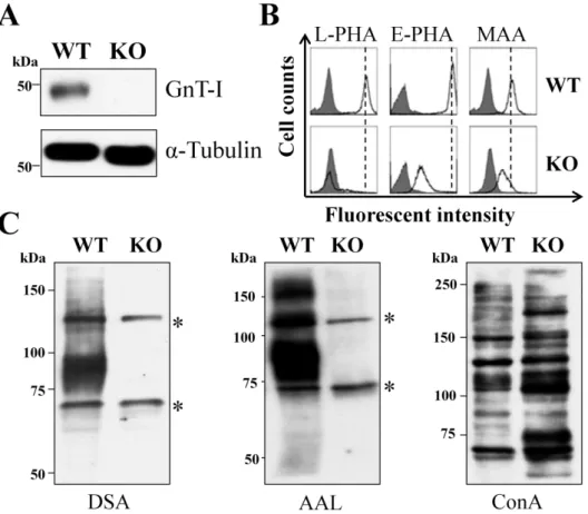
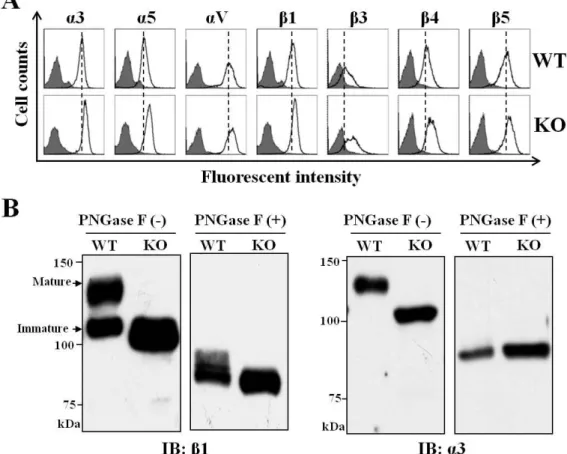
To distinguish the functions of only high-mannose N-glycans and mixed N-glycans including high-mannose, hybrid and complex types, we established GnT-I-KO cells using CRISPR/Cas9-based technology. As expected, the expression of GnT-I was not at detectable levels (Fig. 2A). Flow cytometric analysis and lectin blots using different kinds of lectins were performed to confirm the efficiency of GnT-I knockout in HeLa cells (Figs. 2B and C) and in MDA-MB-231 cells (data not shown). The reactive abilities of L-PHA, E-PHA, and MAA lectins preferentially recognized branched GlcNAc N-glycans, bisected N-glycans, and α2,3 sialylated N-glycans, respectively.
Those reactive abilities, however, were greatly suppressed in the KO cells compared with those in wild type (WT) cells (Fig. 2B). Lectin blots using DSA and AAL lectins preferentially recognized branched GlcNAc N-glycans and fucosylated N-glycans, respectively, which showed that the reactive abilities were almost totally blocked. The binding ability with ConA lectin, however, preferentially recognized high-mannose types of N-glycans that were augmented in the KO cells (Fig. 2C). These results clearly showed that the GnT-I gene knockout was successful.
Figure 2. Deficiency of complex types of N-glycans in GnT- I knockout cells.
(A) Equal amounts of cell lysates from WT and KO HeLa cells were immunoblotted by anti-GnT-I antibody, and α-tubulin served as a loading control. WT: wild type; KO:
knockout. (B) The WT and KO cells were collected and incubated with (bold line) or without (grey shadow) the indicated lectins: L-PHA, E-PHA and MAA, preferentially recognized branched GlcNAc N-glycans, bisected N-glycans, and α2,3 sialylated N-glycans, respectively, followed by incubation with Alexa Fluor 647 streptavidin subjected to flow cytometric analysis. (C) The same amounts of cell lysates from WT and KO cells were immunoblotted with the indicated lectins: DSA, AAL and ConA, which preferentially recognizes branched GlcNAc N-glycans, fucosylated N-glycans, and high-mannose types of N-glycans, respectively. Asterisks(*) indicate no specific bands.
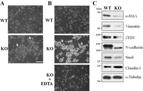
3.2 Deletion of GnT-I-induced cell aggregation and a MET-like phenotype
Next, we used these cell lines to compare the cell biological functions of GnT-I. First, during cell passages we noticed that the KO cells tended to form islands, which was not observed in the WT cells (Fig. 3A), suggesting that knockout of GnT-I might induce cell-cell adhesion. We performed a cell adhesion assay in the suspension. Both the WT and KO cells were incubated with rotation for 6 hours, as described in the MATERIALS AND METHODS section. Clearly, the KO cells tended to form many aggregates that resulted in larger sizes, while the WT cells remained as either single cells or smaller aggregates (Fig. 3B). The cell aggregates disappeared in the presence of EDTA (Fig. 3B, lower panel), suggesting the cell-cell adhesion had proceeded in a calcium-dependent manner. A similar phenomenon was also observed in MDA-MB-231 GnT-I-KO cells (data not shown). Of particular interest, the expression levels of mesenchymal markers such as α-SMA, vimentin, ZEB1, N-cadherin, and Snail were greatly suppressed in the KO cells, while epithelial markers such as claudin-1, which is a tight junction protein, were increased, compared with those in WT cells (Fig. 3C). It is noteworthy that the expression levels of E-cadherin in both WT and KO cells were undetectable. These results suggested that the deletion of GnT-I-induced cell aggregation and MET-like phenotypes.
Figure 3. Comparison of the cell-cell adhesion and expression levels of EMT markers between WT and KO cells.
(A) The WT and KO cells were cultured on normal culture dishes for 1 day, and were photographed by a phase-contrast microscope. (B) The WT and KO cells were detached from culture dishes and separated into single cells in 10% FBS culture media at 2 × 10⁵ cells/ml with or without EDTA, followed by a constant rotation (12 rpm) at 37 °C for 6 h. An aliquot of these cell suspensions was photographed using a phase-contrast microscope. (C) The same amounts of cell lysates from the WT and KO cells were immunoblotted with anti-α-SMA, vimentin, ZEB1, N-cadherin, Snail and claudin-1 antibodies, and α-tubulin was used as a loading control. Arrows indicate cell aggregation. Scale bar represents 50 μm.
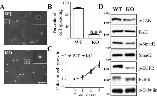
3.3 Deletion of GnT-I inhibited cell spreading and migration, but did not affect cell proliferation
As described above, the expression of GnT-I was important for cell-cell adhesion, and, therefore, we next explored whether GnT-I was also important for integrin-mediated cell-extracellular matrix (ECM) adhesion. By using a cell-spreading assay such as that described in the MATERIALS AND METHODS section, we found that the WT cells had already began to spread after replating the cells on FN-coated dishes for 30 min, while the KO cells continued to exhibit a rounded shape with white dots under phase-contrast photography (Fig. 4A and B). Unexpectedly, there were no significant changes in the proliferation of these two types of cells (Fig. 4C). However, we investigated some related cellular signaling pathways and found that p-Smad2, p-FAK, and p-EGFR, which correspond for TGF-β receptor-, integrin- and EGFR-mediated signaling, respectively, were decreased in the KO cells compared with those in the WT cells (Fig. 4C), which confirmed the notion that a complex type of N-glycans on these signal receptors plays an important role in cellular signaling.
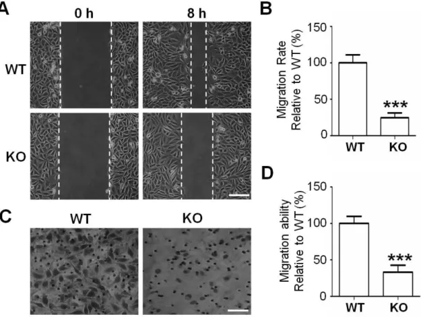
Cell migration is considered a crucial step in the metastatic cascade (42), and we examined the effects of GnT-I on cell migration by using wound healing and transwell assays as described in the MATERIALS AND METHODS section. The ability for wound closure was significantly suppressed in the KO cells compared with that in the WT cells (Figs. 5A and B). The integrin-mediated transwell ability was also greatly inhibited (Figs. 5C and D). Collectively, these data clearly showed that the expression of GnT-I is very important for integrin-mediated functions.
Figure 4. Effects of GnT-I deficiency on cell spreading, cell proliferation and cellular signaling.
(A) The WT and KO cells were detached, and then replated onto FN-coated plates as described in the MATERIALS AND METHODS section. After incubation for 30 min, cells were fixed and representative images were taken via a phase-contrast microscope.
The insertion is a magnified view of the area indicated by the smaller squared box.
Scale bar represents 50 μm. (B) The percentages of spread cells were statistically analyzed. ***, p<0.001. (C) The WT and KO cells were starved with serum-free DMEM for 24 h and then cultured in DMEM containing 10% FBS. The cell numbers were counted at the indicated times. Cell numbers were normalized to the group at 0 h.
Data are represented as the means ± S.E.M (n = 3). (D) The same amounts of cell lysates from the WT and KO cells were immunoblotted by anti-p-FAK, FAK, p-Smad2, Smad2, p-EGFR and EGFR antibodies. α-Tubulin was used as an internal control.
Figure 5. Comparison of cell migration between the WT and KO cells.
(A) These cells were grown to more than 90% confluency. The monolayers were incised with a pipette in each well. Photographs were taken using phase-contrast microscopy at 0 and 8 h. Scale bar represents 50 μm. (B) Quantification of cell migration rate relative to the WT (100%) was expressed as the means ±S.E.M from three independent experiments. (C) A transwell migration chamber was used to examine the migration capacity of WT and KO cells. Migrated cells were stained and photographed as described in the MATERIALS AND METHODS section. Scale bar represents 100 μm.
(D) Relative cell migration was expressed as the percentage of migrated cells compared with that of WT cells (100%) and results are representative of at least three independent experiments. ***, p<0.001.
3.4 The expression levels of integrins were up-regulated in the KO cells
Since integrin-mediated cell adhesion was inhibited in the KO cells, we next sought to determine whether the expression of integrin was also suppressed. Flow cytometric analysis and western blot were carried out to evaluate the expression levels of different integrins. As shown in Fig. 6A, the expression levels on the cell surface of integrins such as α3, α5, αV, β1, β3, β4, and β5 were increased in the KO cells, compared with their expression levels in the WT cells (Fig. 6A). Due to a lack of complex N-glycans on integrin α3 and β1 in the KO cells, the bands of those integrins were shifted on SDS-PAGE much faster than in the WT cells (Fig. 6B). In the WT cells, the so-called immature band of β1 integrin was similar to that in the KO cells, confirming that immature β1 integrin carries a high-mannose type of N-glycans. These differences were canceled via the removal of N-glycans using PNGase F. Treatment with PNGase F clearly showed enhanced expression levels of α3 and β1 integrin in the KO cells (Fig.
6B). These results suggested a negative feedback loop from integrin function to expression.
Figure 6. Effects of GnT-I deficiency on the expression levels of integrins and the effects of N-glycosylation on α3β1 integrin
(A) The WT and KO cells were collected and incubated with (bold line) or without (grey shadow) different types of integrins, α3, α5, αV, β1, β3, β4 and β5, followed by incubation with Alexa Fluor 647 conjugated with goat anti-mouse antibody, and then subjected to flow cytometry analysis. (B) The same amounts of cell lysates (20 μg) obtained from the WT and KO cells were treated with (+) or without (-) peptide:N-glycosidase F (PNGase F), and then immunoblotted with anti-β1integrin and anti-α3 integrin antibodies.
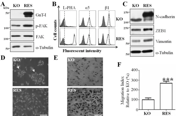
3.5 Restoration of GnT-I in KO cells rescued cell behaviors and induced an EMT-like phenotype
Given the observation that the KO cells exhibited aberrant cell behaviors, we restored GnT-I expression in the KO cells (RES) to examine whether it would rescue these phenotypes. The efficiencies of RES cells were confirmed by western blot by using anti-GnT-I antibody (Fig. 7A, upper panel) and flow cytometric analysis of L-PHA lectin (Fig. 7B, left panel). The integrin-mediated FAK phosphorylation (Fig. 7A, middle panel) was increased, while the expression levels of α5 integrin (Fig. 7B, middle panel) and β1 integrin (Fig. 7B, right panel) were decreased in the RES cells, further supporting the notion of a negative feedback loop from function to expression, as described above. Importantly, the expression levels of mesenchymal markers such as vimentin, ZEB1, and N-cadherin were increased in the RES cells, compared with those in the KO cells (Fig. 7C). Furthermore, these cell islands due to cell-cell adhesion, which could be observed in the KO cells, had disappeared in the RES cells (Fig. 7D).
The integrin-mediated cell migration ability was significantly increased in the RES cells (Fig. 7E and F). Taken together, these results clearly showed the importance of the GnT-I switching by the EMT process.
Figure 7. Forced expression of the GnT-I in KO cells rescued cell behaviors and induced EMT-like phenotypes.
(A) Equal amounts of cell lysates from the KO and restoration of GnT-I in the KO cells (RES cells) were immunoblotted by anti-GnT-I, anti-p-FAK and anti-FAK antibodies.
α-Tubulin was used as an internal control. (B) The KO and RES cells were collected and incubated with (bold line) or without (grey shadow) anti-GnT-I antibody, followed by incubation with Alexa Fluor 647 conjugated with goat anti-mouse antibody, and then subjected to flow cytometry analysis. (C) The expression levels of EMT markers such as N-cadherin, ZEB1 and vimentin were examined by western blotting. (D) Images were taken using a phase-contrast microscope to show representative cell morphologies of the KO and RES cells. Arrows indicate cell aggregation. Scale bar represents 50 μm.
(E) Photographs and (F) quantitative analysis of the transwell assays described in the MATERIALS AND METHODS section were employed to detect the cell migration abilities of the KO and RES cells. Scale bar represents 100 μm. ***, p<0.001
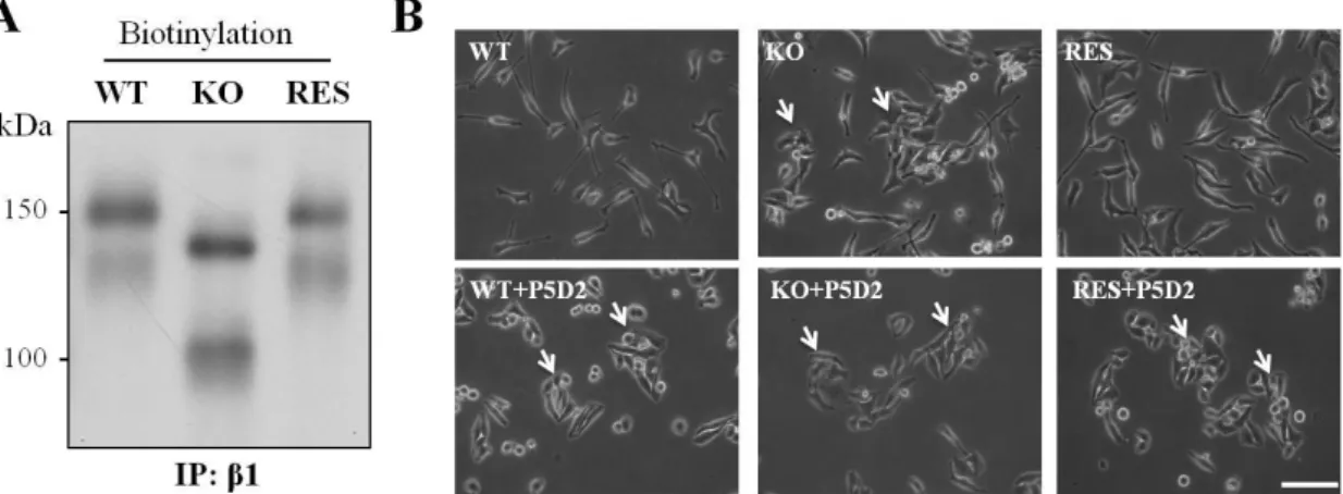
3.6 Influences of integrin and GnT-I on cell-cell adhesion
As described above, deletion of GnT-I induced integrin expression, which indicated that complex N-glycans are not essential for integrin expression. Here, we used biotinylation to further compare the expression levels of β1 integrin among the WT, KO and RES cells. Clearly, the expression levels of β1integrin and the associated α subunits on the cell surface were enhanced in KO cells, but restored to the levels of WT in the RES cells (Fig. 8A). The deletion of GnT-I enhanced the expression levels of integrins, but the integrin-mediated cell-ECM adhesion included FAK phosphorylation as well as cell adhesion and cell migration, all were down-regulated. We speculated as to whether the suppression of cell-ECM adhesion via blocking the process for complex-type N-glycans on integrins could induce cell-cell adhesion, as observed in the KO cells. To test the hypothesis, we added a functional blocking β1 integrin antibody (43), to attenuate cell-matrix adhesion in the cell culture. As shown in Fig. 8B, all three types of cells clearly exhibited cell aggregation in the presence of anti-β1 integrin antibody. These data might partly explain why cell aggregation occurred in the KO cells, and could further suggest that the modification of complex types of N-glycans on β1 integrin is very important for its ability to function.
Figure 8. Effects of GnT-I on integrin β1 maturation and the cell-cell adhesion induced by a blocking anti-β1 integrin antibody.
(A) The indicated cells were incubated with Sulfo-NHS-SS biotin for 1 h, and then the cell lysates were immunoprecipitated with anti-β1 integrin mAb (P5D2). The biotinylated proteins were detected as described in the MATERIALS AND METHODS section. (B) The indicated cells were replated onto 6-well culture dishes in normal culture media with or without a blocking anti-β1 integrin antibody (P5D2, 10 µg/ml).
After incubation for 16 h, phase-contrast photographs were taken. The arrows indicate cell aggregation. Scale bar represents 75 μm.
3.7 Deletion of GnT-I increased the drug-sensitivity
Since recent studies revealed the involvement of EMT on the efficacy of cancer drug responses (44, 45), we evaluated whether GnT-I deficiency affects drug sensitivities in HeLa cells. The three cells were incubated with cancer drugs such as Paclitaxel for 3 days (Fig. 9A) and Doxorubicin (Fig. 9B) for 2 days at indicated concentrations. Clearly, the KO cells were more sensitive to both drugs, compared with the WT and RES cells.
Furthermore, we monitored Doxorubicin cellular uptake using FACS. Consistently, more fluorescent intensities were observed in KO cells (Fig. 9C).
The ability of tumor cells to form colonies from a single cell is a well-known and important feature of metastatic potential (46). Cells that undergo EMT are also known to form more colonies (47), and, therefore, we evaluated the effects of GnT-I on colony formation in HeLa cells. As shown in Fig. 9D, the colony formation in soft agar was significantly suppressed in the KO cells, compared with the WT and the RES cells.
These data suggested that the expression of GnT-I greatly contributes to tumor survival and formation.
Figure 9. Loss of GnT-I chemosensitized the cells to cancer drugs.
The cells were cultured under normal culture media containing paclitaxel (A) and doxorubicin (B) at the indicated concentrations. Percentages of cell viability were measured via MTT assay. **, p<0.01 and ***, p<0.001 compared with paclitaxel or doxorubicin-treated WT cells, which was set as 100%. (C) After incubation with (bold line) or without (grey shadow) doxorubicin for 1 day, the indicated cells were collected and then subjected to flow cytometry via the FL2 (Phycoerythrin) channel as described in the MATERIALS AND METHODS section. (D) The indicated cells (1 × 10⁴ /per well) were cultured in soft-agar plates for 20 days, and then the colonies were stained with crystal violet and images were taken by light microscope. (E) The stained colonies were counted, and the quantitative data are presented as the means ± S.E.M from three independent experiments. Scale bar represents 5 mm. ***, p<0.001 compared to the WT group, which was set as 100%.
4. DISCUSSION
In the present study, we found that GnT-I works as a switch to activate EMT, based on the following observations: 1) deletion of GnT-I suppressed the expression of mesenchymal markers and cell-ECM adhesion, and it induced cell-cell adhesion; 2) loss of GnT-I decreased integrin-mediated cell migration; and, 3) restoration of GnT-I efficiently rescued the expression of mesenchymal markers and cell migration, and decreased cell-cell adhesion. In addition, the expression of GnT-I was important for cell survival against anticancer drugs.
Previous study has shown that mice lacking GnT-I died at mid-gestation (37).
Biochemical and morphological analyses of embryos have revealed GnT-I-null embryos that are developmentally retarded, particularly in neural tissue. The neural crest is known to be a transient embryonic cell population that migrates long distances through the vertebrate embryo during development, wherein the integrin family of cell adhesion receptors are required for proper cell migration, proliferation, survival, and differentiation. In fact, both major ECM molecules, laminin and fibronectin, have receptors that include α3β1 and α5β1 integrin and are abundantly expressed in neural crest migration pathways (48, 49). Furthermore, a loss of function of the murine α5 integrin by gene targeting posed a recessive embryonic lethal threat at mid-gestation, due to defects in the development of the cardiac neural crest, mesoderm formation, and to cell movement and function (50, 51). Our results clearly showed that GnT-I deficiency greatly suppresses integrin-mediated cellular signaling (FAK phosphorylation) as well as cell-ECM adhesion and migration, which may partly explain some of the important phenotypes shown in the GnT-I null embryos. We could speculate that the defect of integrin functions could be one of the main causes for the embryonic lethality at mid-gestation of GnT-I knockout mice. In fact, there is an increasing body of evidence implicating the importance of N-glycans on integrin function (52). For example, alterations in the oligosaccharide portion of integrins that
sialyltransferase (ST6GalI), differentiate in the regulation of cell malignant phenotypes such as in integrin-mediated cell migration and cell spreading. The expression of GnT-V stimulates α3β1 integrin-mediated cell migration, while the overexpression of GnT-III inhibits GnT-V-induced cell migration (19). Therefore, the lack of GnT-I might result in the loss of such dynamic and precise post-translational regulation, which plays a crucial role in embryo development. It is intriguing that GnT-I deficiency increases the expression levels of many types of integrins, although integrin functions were suppressed, while the restoration of GnT-I decreases integrin expression and enhances integrin-mediated cell adhesion and cellular signaling, which suggests the existence of a negative feedback loop from the function to the expression of integrins. Furthermore, these results support the notion that N-glycans closely regulate integrin function.
EMT is an important phenotype characterized by a loss of the cell-cell junction and the acquisition of cell migration, which plays crucial roles in not only physiological processes such as embryo implantation, embryogenesis and organ development, but also in tissue repair and pathological processes such as tissue fibrosis, tumor invasiveness, and metastasis (53). Different signaling pathways, for example, receptor tyrosine kinase, Notch, Wnt and TGF-β, are known to provide the necessary stimuli that modulate gene expression and triggers EMT and cell migration (54). The regulation of EMT by GnT-I might cross-link with those signaling pathways. Actually, glycosylation and those signaling mutually regulate, and affect cell behaviors. Inhibition of GnT-V-mediated N-glycosylation suppresses PI3K and MAPK signaling (55, 56). Inversely, PI3K/Akt (55) and ras-raf-ets2 (57) signaling pathways regulate GnT-V expression. In addition, core fucosylation is critical to the activation of TGF-β receptor type II (TβRII) and Smad2 signaling pathway (58) and high mannose type of TβRII decreases TGF-β-induced Smad2 activation (59), while TGF-β receptor-mediated signaling promotes GnT-V expression (60). Our data clearly showed that GnT-I deficiency
tissue was delayed in the GnT-I-null embryos. Consistently, the inhibition of complex types of N-glycans by SW efficiently inhibited cell invasion and EMT processes in esophageal carcinoma cells (30). In addition, the deletion of GnT-I suppressed the phosphorylation of both EGFR and Smad2, which further confirmed the switching function of GnT-I for EMT. Thus, high-mannose types of N-glycans might maintain MET-like phenotypes, while GnT-I initiates the EMT process.
Cell adhesion basically refers to cell attachment such as cell–cell adhesion and cell–matrix adhesion. Here, we also demonstrated how the deletion of GnT-I augmented cell-cell adhesion, which might be explained by the two following reasons. The first reason concerns a weakening of the cell-ECM adhesion in KO cells, as described above.
It is reasonable to speculate that the cell-cell adhesion and cell-ECM adhesion regulate each other in an opposing manner. Indeed, treatment with a blocking β1 integrin antibody (P5D2) greatly induced cell-cell adhesion in the WT and RES HeLa cells, which was observed in the KO cells without this treatment. Consistently, the knockout of β1 integrin resulted in cell-cell adhesion and in the suppression of FAK phosphorylation and cell migration (61). The second reason involves the possibility that high-mannose types of N-glycans might enhance cell-cell adhesion. E-cadherin is the core transmembrane glycoprotein of the adherens-junction, although the expression level of E-cadherin in HeLa cells is marginal. The modification of E-cadherin with complex N-glycans has been associated with the formation of dynamic and weak adherens-junctions, whereas E-cadherin modified either by high-mannose types or fewer N-glycans is known to promote the establishment of stable adherens junctions (62). E-cadherin was modified with N-glycans containing β1,6 branched and sialylation structures during acquisition of the malignant phenotype (63), while overexpression of GnT-III prolonged the turnover of E-cadherin on the cell surface due to a suppression of extensive branched complex N-glycan formation by the action of GnT-V , and then enhanced homophilic adhesion (15). Indeed, up-regulation of GnT-V expression was
and keratinocyte migration in GnT-V-transgenic mice (64). Therefore, smaller N-glycans such as high-mannose type or bisected N-glycans may promote cell-cell adhesion.
Here, it is also worth noting why GnT-I deficiency did not affect cell proliferation (Fig.
4B). Previous studies have shown that lost or reduced branched-complex N-glycans catalyzed by GnT-IV and GnT-V leads to a reduction in the retention time of glycoprotein receptors on the cell surface due to a weakening of their interactions with a galectin lattice (65, 66). Thus, it could be more plausible to speculate that deletion of GnT-I inhibits cell proliferation. However, our results showed there was no significant difference in cell growth between WT and KO cells, although the level of phosphorylated EGFR was decreased in the KO cells compared with that in the WT cells (Fig.4). In fact, recent studies have shown that cell proliferation of CHO cells was not affected by the genetic disruption of multiple α1,2-mannosidases to generate high-mannose types of N-glycans in HEK293 cells (67). Consistently, knockdown of GnT-I by shRNA did not alter cell proliferation rates in a human PC-3 prostate tumor cell line (38). These results indicated that complex types of N-glycans remodeled by glycosyltransferases on glycoprotein receptors may either positively or negatively regulate cellular signaling for cell proliferation. In fact, for instance, the lack of α1,6-fucosylation in TβRII reduced its binding with ligands, and subsequently suppressed intracellular signaling by phospho-Smad2, which suggests that α1,6-fucosylation positively regulates TGF-β downstream signaling (58). In contrast to TGF-β-mediated signaling, α1,6-fucosylation on an activin receptor, a member of the TGF-β superfamily, negatively regulates phospho-Smad2 signaling (68). Therefore, even the same types of N-glycans on different receptors may play distinctive roles in their cellular signaling. For example, cells expressing high levels of GnT-V increase migration (69) and EMT behaviors by up-regulating EGFR signaling (64). However,
Recently, inhibition of N-linked glycosylation has generated interest in its potential use as an anticancer agent (71, 72), since aberrant N-glycosylation is implicated in cancer growth and metastasis. In this study, we found that the KO cells were more sensitive to cancer drugs than WT and RES cells, and more drugs were detected in the KO cells.
The treatment with SW promoted cell apoptosis induced by paclitaxel (73). Inhibition of N-glycosylation treated with tunicamycin also greatly increased the sensitivity to Doxorubicin (74). These results indicate that the functions of some membrane glycoproteins such as ABC transporters and P-glycoprotein (75), which are involved in the export and import of drugs, may be regulated by N-glycans. In fact, N-glycosylation of ABC transporters has been associated with functional activity in rat hepatocytes (76) and the inhibition of protein glycosylation has enhanced the effect of chemotherapy by interfering with the localization and function of transporter proteins in several cancer cell lines (77). In addition, the decreased functions of integrin in KO cells might also suggest an increase in the sensitivity to drugs, since β1 integrin deficiency increased the inhibitory effects of AG1478, an EGFR tyrosine kinase inhibitor (61). The underlying mechanisms for drug sensitivity will require further study. In conclusion, our findings indicate that GnT-I plays a pivotal role in the regulation of key processes in cancer cells, such as EMT, cell adhesion, migration and chemoresistance. Thus, we believe that GnT-I is a promising cancer therapeutic target.
REFERENCES
1. Nieto, M. A., Huang, R. Y., Jackson, R. A., and Thiery, J. P. (2016) EMT: 2016.
Cell 166, 21-45
2. Lamouille, S., Xu, J., and Derynck, R. (2014) Molecular mechanisms of epithelial-mesenchymal transition. Nature reviews. Molecular cell biology 15, 178-196
3. Yang, J., and Weinberg, R. A. (2008) Epithelial-mesenchymal transition: at the crossroads of development and tumor metastasis. Developmental cell 14, 818-829
4. Li, Q., Hutchins, A. P., Chen, Y., Li, S., Shan, Y., Liao, B., Zheng, D., Shi, X., Li, Y., Chan, W. Y., Pan, G., Wei, S., Shu, X., and Pei, D. (2017) A sequential EMT-MET mechanism drives the differentiation of human embryonic stem cells towards hepatocytes. Nature communications 8, 15166
5. Jolly, M. K., Ware, K. E., Gilja, S., Somarelli, J. A., and Levine, H. (2017) EMT and MET: necessary or permissive for metastasis? Molecular oncology 11, 755-769
6. Thompson, E. W., and Haviv, I. (2011) The social aspects of EMT-MET plasticity. Nature medicine 17, 1048-1049
7. Adhikary, A., Chakraborty, S., Mazumdar, M., Ghosh, S., Mukherjee, S., Manna, A., Mohanty, S., Nakka, K. K., Joshi, S., De, A., Chattopadhyay, S., Sa, G., and Das, T. (2014) Inhibition of epithelial to mesenchymal transition by E-cadherin up-regulation via repression of slug transcription and inhibition of E-cadherin degradation: dual role of scaffold/matrix attachment region-binding protein 1 (SMAR1) in breast cancer cells. The Journal of biological chemistry 289, 25431-25444
8. Cai, D., Chen, S. C., Prasad, M., He, L., Wang, X., Choesmel-Cadamuro, V.,
Cell 157, 1146-1159
9. Chen, Q. K., Lee, K., Radisky, D. C., and Nelson, C. M. (2013) Extracellular matrix proteins regulate epithelial-mesenchymal transition in mammary epithelial cells. Differentiation; research in biological diversity 86, 126-132 10. Ganguly, K. K., Pal, S., Moulik, S., and Chatterjee, A. (2013) Integrins and
metastasis. Cell adhesion & migration 7, 251-261
11. Bates, R. C., Bellovin, D. I., Brown, C., Maynard, E., Wu, B., Kawakatsu, H., Sheppard, D., Oettgen, P., and Mercurio, A. M. (2005) Transcriptional activation of integrin β6 during the epithelial-mesenchymal transition defines a novel prognostic indicator of aggressive colon carcinoma. The Journal of clinical investigation 115, 339-347
12. Mori, S., Kodaira, M., Ito, A., Okazaki, M., Kawaguchi, N., Hamada, Y., Takada, Y., and Matsuura, N. (2015) Enhanced expression of Integrin αvβ3 induced by TGF-β is required for the enhancing effect of fibroblast growth factor 1 (FGF1) in TGF-β-induced epithelial-mesenchymal transition (EMT) in mammary epithelial cells. PloS one 10, e0137486
13. Li, X. L., Liu, L., Li, D. D., He, Y. P., Guo, L. H., Sun, L. P., Liu, L. N., Xu, H.
X., and Zhang, X. P. (2017) Integrin β4 promotes cell invasion and epithelial-mesenchymal transition through the modulation of Slug expression in hepatocellular carcinoma. Scientific reports 7, 40464
14. Li, X., Wang, X., Tan, Z., Chen, S., and Guan, F. (2016) Role of Glycans in cancer cells undergoing epithelial-mesenchymal transition. Frontiers in oncology 6, 33
15. Pinho, S. S., Figueiredo, J., Cabral, J., Carvalho, S., Dourado, J., Magalhaes, A., Gartner, F., Mendonfa, A. M., Isaji, T., Gu, J., Carneiro, F., Seruca, R., Taniguchi, N., and Reis, C. A. (2013) E-cadherin and adherens-junctions stability in gastric carcinoma: functional implications of glycosyltransferases involving N-glycan
et biophysica acta 1830, 2690-2700
16. Tu, C. F., Wu, M. Y., Lin, Y. C., Kannagi, R., and Yang, R. B. (2017) FUT8 promotes breast cancer cell invasiveness by remodeling TGF-β receptor core fucosylation. Breast cancer research 19, 111
17. Shao, K., Chen, Z. Y., Gautam, S., Deng, N. H., Zhou, Y., and Wu, X. Z. (2016) Posttranslational modification of E-cadherin by core fucosylation regulates Src activation and induces epithelial-mesenchymal transition-like process in lung cancer cells. Glycobiology 26, 142-154
18. Pochec, E., Litynska, A., Amoresano, A., and Casbarra, A. (2003) Glycosylation profile of integrin α3β1 changes with melanoma progression. Biochimica et biophysica acta 1643, 113-123
19. Zhao, Y., Nakagawa, T., Itoh, S., Inamori, K., Isaji, T., Kariya, Y., Kondo, A., Miyoshi, E., Miyazaki, K., Kawasaki, N., Taniguchi, N., and Gu, J. (2006) N-acetylglucosaminyltransferase III antagonizes the effect of N-acetylglucosaminyltransferase V on α3β1 integrin-mediated cell migration.
The Journal of biological chemistry 281, 32122-32130
20. Isaji, T., Gu, J., Nishiuchi, R., Zhao, Y., Takahashi, M., Miyoshi, E., Honke, K., Sekiguchi, K., and Taniguchi, N. (2004) Introduction of bisecting GlcNAc into integrin α5β1 reduces ligand binding and down-regulates cell adhesion and cell migration. The Journal of biological chemistry 279, 19747-19754
21. Taniguchi, N., and Kizuka, Y. (2015) Glycans and cancer: role of N-glycans in cancer biomarker, progression and metastasis, and therapeutics. Advances in cancer research 126, 11-51
22. Ho, W. L., Hsu, W. M., Huang, M. C., Kadomatsu, K., and Nakagawara, A.
(2016) Protein glycosylation in cancers and its potential therapeutic applications in neuroblastoma. Journal of hematology & oncology 9, 100
(2007) Comparison of biological activity among nonfucosylated therapeutic IgG1 antibodies with three different N-linked Fc oligosaccharides: the high-mannose, hybrid, and complex types. Glycobiology 17, 104-118
24. Porwoll, S., Loch, N., Kannicht, C., Nuck, R., Grunow, D., Reutter, W., and Tauber, R. (1998) Cell surface glycoproteins undergo postbiosynthetic modification of their N-glycans by stepwise demannosylation. The Journal of biological chemistry 273, 1075-1085
25. de Leoz, M. L., Young, L. J., An, H. J., Kronewitter, S. R., Kim, J., Miyamoto, S., Borowsky, A. D., Chew, H. K., and Lebrilla, C. B. (2011) High-mannose glycans are elevated during breast cancer progression. Molecular & cellular proteomics 10, M110 002717
26. Ozcan, S., Barkauskas, D. A., Renee Ruhaak, L., Torres, J., Cooke, C. L., An, H.
J., Hua, S., Williams, C. C., Dimapasoc, L. M., Han Kim, J., Camorlinga-Ponce, M., Rocke, D., Lebrilla, C. B., and Solnick, J. V. (2014) Serum glycan signatures of gastric cancer. Cancer prevention research 7, 226-235
27. Pochec, E., Bocian, K., Zabczynska, M., Korczak-Kowalska, G., and Litynska, A. (2015) Immunosuppressive drugs affect high-mannose/hybrid N-glycans on human allostimulated leukocytes. Analytical cellular pathology 2015, 324980 28. Chang, V. T., Crispin, M., Aricescu, A. R., Harvey, D. J., Nettleship, J. E.,
Fennelly, J. A., Yu, C., Boles, K. S., Evans, E. J., Stuart, D. I., Dwek, R. A., Jones, E. Y., Owens, R. J., and Davis, S. J. (2007) Glycoprotein structural genomics: solving the glycosylation problem. Structure 15, 267-273
29. Olden, K., Breton, P., Grzegorzewski, K., Yasuda, Y., Gause, B. L., Oredipe, O.
A., Newton, S. A., and White, S. L. (1991) The potential importance of swainsonine in therapy for cancers and immunology. Pharmacology &
therapeutics 50, 285-290
30. Ma, J., Wan, L., Li, J., Zhang, G., Tao, H., Li, X., Sun, D., and Hu, Y. (2018)
cells by targeting Twist1. Oncology research 26, 1207-1213
31. Shaheen, P. E., Stadler, W., Elson, P., Knox, J., Winquist, E., and Bukowski, R.
M. (2005) Phase II study of the efficacy and safety of oral GD0039 in patients with locally advanced or metastatic renal cell carcinoma. Investigational new drugs 23, 577-581
32. Helenius, A., and Aebi, M. (2001) Intracellular functions of N-linked glycans.
Science 291, 2364-2369
33. Stanley, P., Taniguchi, N., and Aebi, M. (2015) N-Glycans. In Essentials of Glycobiology (rd, Varki, A., Cummings, R. D., Esko, J. D., Stanley, P., Hart, G.
W., Aebi, M., Darvill, A. G., Kinoshita, T., Packer, N. H., Prestegard, J. H., Schnaar, R. L., and Seeberger, P. H., eds) pp. 99-111, Cold Spring Harbor (NY) 34. Fanata, W. I., Lee, K. H., Son, B. H., Yoo, J. Y., Harmoko, R., Ko, K. S.,
Ramasamy, N. K., Kim, K. H., Oh, D. B., Jung, H. S., Kim, J. Y., Lee, S. Y., and Lee, K. O. (2013) N-glycan maturation is crucial for cytokinin-mediated development and cellulose synthesis in Oryza sativa. The Plant journal : for cell and molecular biology 73, 966-979
35. He, X., Galpin, J. D., Tropak, M. B., Mahuran, D., Haselhorst, T., von Itzstein, M., Kolarich, D., Packer, N. H., Miao, Y., Jiang, L., Grabowski, G. A., Clarke, L.
A., and Kermode, A. R. (2012) Production of active human glucocerebrosidase in seeds of Arabidopsis thaliana complex-glycan-deficient (cgl) plants.
Glycobiology 22, 492-503
36. Strasser, R., Stadlmann, J., Schahs, M., Stiegler, G., Quendler, H., Mach, L., Glossl, J., Weterings, K., Pabst, M., and Steinkellner, H. (2008) Generation of glyco-engineered Nicotiana benthamiana for the production of monoclonal antibodies with a homogeneous human-like N-glycan structure. Plant biotechnology journal 6, 392-402
N-linked carbohydrates. Proceedings of the National Academy of Sciences of the United States of America 91, 728-732
38. Beheshti Zavareh, R., Sukhai, M. A., Hurren, R., Gronda, M., Wang, X., Simpson, C. D., Maclean, N., Zih, F., Ketela, T., Swallow, C. J., Moffat, J., Rose, D. R., Schachter, H., Schimmer, A. D., and Dennis, J. W. (2012) Suppression of cancer progression by MGAT1 shRNA knockdown. PloS one 7, e43721
39. Ran, F. A., Hsu, P. D., Wright, J., Agarwala, V., Scott, D. A., and Zhang, F. (2013) Genome engineering using the CRISPR-Cas9 system. Nature protocols 8, 2281-2308
40. Shalem, O., Sanjana, N. E., Hartenian, E., Shi, X., Scott, D. A., Mikkelson, T., Heckl, D., Ebert, B. L., Root, D. E., Doench, J. G., and Zhang, F. (2014) Genome-scale CRISPR-Cas9 knockout screening in human cells. Science 343, 84-87
41. Isaji, T., Sato, Y., Zhao, Y., Miyoshi, E., Wada, Y., Taniguchi, N., and Gu, J.
(2006) N-glycosylation of the β-propeller domain of the integrin α5 subunit is essential for α5β1 heterodimerization, expression on the cell surface, and its biological function. The Journal of biological chemistry 281, 33258-33267 42. Li, W., Ng, J. M., Wong, C. C., Ng, E. K. W., and Yu, J. (2018) Molecular
alterations of cancer cell and tumour microenvironment in metastatic gastric cancer. Oncogene 37, 4903-4920
43. Isaji, T., Sato, Y., Fukuda, T., and Gu, J. (2009) N-glycosylation of the I-like domain of β1 integrin is essential for β1 integrin expression and biological function: identification of the minimal N-glycosylation requirement for α5β1.
The Journal of biological chemistry 284, 12207-12216
44. Du, B., and Shim, J. S. (2016) Targeting Epithelial-Mesenchymal Transition (EMT) to Overcome Drug Resistance in Cancer. Molecules 21
45. Davis, F. M., Stewart, T. A., Thompson, E. W., and Monteith, G. R. (2014)