For Peer Review
Effects of mosapride on esophageal motor activity and esophagogastric junction distensibility in healthy volunteers
Journal: Journal of Gastroenterology Manuscript ID: JG-2013-06-OA-661 Manuscript Type: Original Article Date Submitted by the Author: 24-Jun-2013
Complete List of Authors: Fukazawa, Kousuke; Shimane University Faculty of Medicine, Second Department of Internal Medicine
Furuta, Kenji; Shimane University Faculty of Medicine, Second Department of Internal Medicine
Adachi, Kyoichi; Shimane Environment and Health Public Corporation, Health Center
Moritou, Yoshiya; Shimane University Faculty of Medicine, Second Department of Internal Medicine
Saito, Tsukasa; Shimane University Faculty of Medicine, Second Department of Internal Medicine
Kusunoki, Ryusaku; Shimane University Faculty of Medicine, Second Department of Internal Medicine
Uno, Goichi; Shimane University Faculty of Medicine, Second Department of Internal Medicine
Shimura, Shino; Shimane University Faculty of Medicine, Second Department of Internal Medicine
Aimi, Masahito; Shimane University Faculty of Medicine, Second Department of Internal Medicine
Ohara, Shunji; Shimane University Faculty of Medicine, Second Department of Internal Medicine
Ishihara, Shunji; Shimane University Faculty of Medicine, Second Department of Internal Medicine
Kinoshita, Yoshikazu; Shimane University Faculty of Medicine, Second Department of Internal Medicine
Category: Others
For Peer Review
Effects of mosapride on esophageal motor activity and esophagogastric junction distensibility in healthy volunteers
Running title: mosapride on EGJ compliance
Kousuke Fukazawa1, Kenji Furuta1, Kyoichi Adachi2, Yoshiya Moritou1, Tsukasa
Saito1, Ryusaku Kusunoki1, Goichi Uno1, Shino Shimura1, Masahito Aimi1, Shunji
Ohara1, Shunji Ishihara1, and Yoshikazu Kinoshita1
1
Second Department of Internal Medicine, Shimane University Faculty of Medicine
2
Health Center, Shimane Environment and Health Public Corporation
Correspondence to: Masahito Aimi, MD
Second Department of Internal Medicine, Shimane University Faculty of Medicine 89-1, Enya-cho, Izumo, Shimane, 693-8501, Japan
Phone: +81-853-20-2190 Fax: +81-853-20-2187 E-mail: [email protected] 5 6 7 8 9 10 11 12 13 14 15 16 17 18 19 20 21 22 23 24 25 26 27 28 29 30 31 32 33 34 35 36 37 38 39 40 41 42 43 44 45 46 47 48 49 50 51 52 53 54 55 56 57
For Peer Review
AbstractBackground: The effects of the prokinetic drug mosapride on esophageal motor
activity are variable at standard doses. In addition to esophageal motor activities, the distensibility of the esophagogastric junction (EGJ) is important for prevention of gastroesophageal reflux. However, the effect of mosapride on EGJ distensibility has not been reported. We investigated the effects of high-dose mosapride on esophageal motor activities and EGJ distensibility.
Methods: Nine healthy volunteers were enrolled in the study. Peristaltic esophageal
contraction and lower esophageal sphincter pressures before and after administration of 40 mg mosapride were examined by high resolution esophageal manometry. Esophageal distensibility was also investigated by intra-esophageal impedance planimetry
(EndoFLIP®).
Results: High-dose mosapride augmented peristaltic contractions, especially in the
distal esophageal segments (P<0.05). The mean resting lower esophageal sphincter pressure was elevated from 25.0 mmHg before administration to 28.9 mmHg after (P<0.05). In addition, mosapride significantly reduced EGJ distensibility (P<0.05).
Conclusions: Mosapride at 40 mg augmented esophageal motor activities and reduced
EGJ distensibility in healthy volunteers.
Keywords: distensibility, esophagogastric junction, manometry, mosapride
5 6 7 8 9 10 11 12 13 14 15 16 17 18 19 20 21 22 23 24 25 26 27 28 29 30 31 32 33 34 35 36 37 38 39 40 41 42 43 44 45 46 47 48 49 50 51 52 53 54 55 56 57
For Peer Review
IntroductionGastroesophageal reflux disease (GERD) is caused by the pathological reflux of gastric contents. Since acidic reflux is the main cause of GERD, inhibition of gastric acid secretion by administration of proton pump inhibitors (PPIs) is widely used as first-line therapy. However, approximately 30% of treated patients complain about reflux symptoms with PPI use and require additional treatment [1, 2]. As a second-line therapy, mosapride, a prokinetic agent activating the serotonin 5-HT 4 receptor, has been used in clinical practice, though reports concerning its beneficial effects for GERD are conflicting [3-6], as some have shown an augmenting action of mosapride on esophageal peristalsis and lower esophageal sphincter pressure [3-5], while others failed to find those activities [6]. These conflicting results may be derived from different dosages, with higher doses possibly necessary to gain beneficial therapeutic effects.
The esophago-gastric junction (EGJ) is an important structure to prevent
gastro-esophageal reflux. Patients with GERD show high distensibility of this area and the cross-sectional area (CSA) of EGJ during pressure distension is known to be larger than that in normal individuals [7]. Therefore, drugs that reduce EGJ distensibility and decrease the CSA of EGJ are expected to inhibit pathological gastroesophageal reflux.
Recently, endoluminal functional lumen-imaging probe (FLIP; EndoFLIP®, Crospon
Ltd, Galway, Ireland) has been demonstrated to be useful to evaluate the distensibility of upper and lower esophageal sphincter [7, 9-12]. In this study, the effect of high-dose mosapride on EGJ distensibility and esophageal motor function was investigated by using EndoFLIP system and high-resolution 36-channel manometry in healthy
volunteers to test the feasibility of the drug as a possible therapeutic agent for patients with GERD. 5 6 7 8 9 10 11 12 13 14 15 16 17 18 19 20 21 22 23 24 25 26 27 28 29 30 31 32 33 34 35 36 37 38 39 40 41 42 43 44 45 46 47 48 49 50 51 52 53 54 55 56 57
For Peer Review
MethodsNine normal healthy male volunteers (age 21-52 years old, mean 35.2 years) without any abdominal symptoms were enrolled in this study. After fasting for at least 8
hours, a high-resolution 36-channel manometry sensor catheter (ManoScan360; Sierra
Scientific Instruments, Los Angeles, CA) was transnasally introduced into the
esophagus and esophageal motor activity was measured in a supine position [8]. Next, the manometry catheter was withdrawn and an endoluminal functional lumen-imaging
probe (FLIP; EndoFLIP®, Crospon Ltd, Galway, Ireland) was transnasally inserted in
the same manner as the manometry catheter and positioned at the EGJ, then
distensibility of the EGJ was also measured in a supine position [7, 9-14]. After baseline measurements of esophageal motor activities and EGJ distensibility were obtained, mosapride was orally administered. The maximum serum concentration of mosapride is known to be reached at 2.7 hours after oral administration, and the maximal clinical dose of mosapride used in Japan is 40 mg in 2 divided doses with a 2-hour interval. Therefore, mosapride at 20 mg was orally administrated to the volunteers, followed by another 20 mg orally 2 hours later. One hour after the second dose, esophageal
manometry and EGJ distensibility measurements were repeated. Those values obtained before and after mosapride administration were then compared, and analyzed.
Esophageal motor function using high-resolution 36-channel manometry was evaluated according to a previously reported method [6, 15-17]. Based on the
manufacturer’s instructions, the transducers were calibrated before each measurement. The catheter was inserted transnasally, and then the measurements were performed in a supine position starting 5 minutes after insertion. Resting lower esophageal sphincter (LES) pressure was measured for at least 5 minutes. Esophageal body peristaltic
5 6 7 8 9 10 11 12 13 14 15 16 17 18 19 20 21 22 23 24 25 26 27 28 29 30 31 32 33 34 35 36 37 38 39 40 41 42 43 44 45 46 47 48 49 50 51 52 53 54 55 56 57
For Peer Review
contractions were measured after drinking 5 ml of water, which was repeated at 2-minute intervals until 5 complete contraction records were obtained. According to previous reports, esophageal body peristaltic contractions were separated into 3 segments (oral, middle, lower), as previously reported [18, 19] (Figure 1). We determined the maximum peristaltic contraction pressure in each segment and calculated the mean of 5 separate contractions in each subject.
The distensibility of EGJ was determined by using EndoFLIP system [9-14]. The EndoFLIP uses impedance planimetry with 16 electrodes to measure the CSA at 5-mm intervals inside a saline-filled bag (length 80 mm). With this system, the bag is attached to the tip of the catheter probe and can be filled with different volumes of fluid. In addition, the diameter and intra-bag pressure can be simultaneously assessed, and the CSA of EGJ and intra-bag distensibility pressure evaluated at the same time. The distension probe and pressure transducers were calibrated based on the manufacturer’s instructions. The pressure sensor was set to 0 before insertion of the catheter and then the deflated catheter was transnasally inserted. In our study protocol, the balloon was inflated to volumes of 20, 40, and 50 ml. Peristaltic waves were often visible and ignored, with data obtained as soon as a steady state of a combination of intra-bag pressure, minimum diameter (Dmin), and CSA was observed with real-time imaging.
We recorded diameter (mm), CSA (mm2), and distensibility indices (DI mm2/mmHg), as
previously reported [9, 13]. The EGJ distensibility index was based on the narrowest CSA in relation to the corresponding intra-bag pressure and calculated with the
following equation: [narrowest CSA/ (intra-bag pressure + intragastric pressure affects)]. EGJ distensibility was successfully measured in all 9 of the volunteers in this study.
A Wilcoxon signed rank test was used for statistical comparisons between the
5 6 7 8 9 10 11 12 13 14 15 16 17 18 19 20 21 22 23 24 25 26 27 28 29 30 31 32 33 34 35 36 37 38 39 40 41 42 43 44 45 46 47 48 49 50 51 52 53 54 55 56 57
For Peer Review
mosapride administered and control periods. All calculations were done with Stat View 5.0 software (Abacus Concepts Inc., Berkeley, CA, USA) for Macintosh. Differences at p<0.05 were considered to be statistically significant.
The study protocol was approved by the ethical committee of Shimane University Faculty of Medicine. Written informed consent was obtained from each of the enrolled volunteers. 5 6 7 8 9 10 11 12 13 14 15 16 17 18 19 20 21 22 23 24 25 26 27 28 29 30 31 32 33 34 35 36 37 38 39 40 41 42 43 44 45 46 47 48 49 50 51 52 53 54 55 56 57
For Peer Review
ResultsManometry data for 1 volunteer were not obtained because of mechanical trouble, thus those results from 8 volunteers were analyzed. The maximal contraction pressure was highest in the lower segment (segment 3) and lowest in the upper segment (segment 1). Mosapride administration tended to increase the peristaltic contraction pressure in segments 1 and 2 (upper and middle segments of esophagus), though the differences did not reach statistical significance. In the lower segment, mosapride given twice (40 mg total dose) resulted in a statistically significant increase in mean maximum contraction pressure (from 130.0 to 147.7 mmHg), as shown in Table 1. In addition, mean LES resting pressure in a supine position increased from 25.0 to 28.9 mmHg following administration of 40 mg of mosapride. Therefore, mosapride at a higher dose was considered to augment esophageal contractions, especially in the distal segments and EGJ areas.
The FLIP bag was configured into an hourglass shape when distended to straddle the EGJ, with the central constriction at the diaphragmatic hiatus during both the mosapride treated and non-treated periods. The diaphragmatic hiatus consistently fit to the least distensible locus with a minimal opening diameter (Figure 2). The hiatal diameter (D min) and CSA progressively increased with distending volume, as did the intra-bag pressure. During the mosapride administration period, intra-bag pressure was higher and the hiatal CSA was lower with the 40- and 50-ml intra-bag volumes (P<0.05), as shown in Table 2. At all distensible pressures, the extent of the EGJ opening during the mosapride treated period was smaller than that during the non-treated period (Figure 3). Using intra-bag pressure and hiatal CSA, an EGJ distensibility index (DI) was calculated for each distension volume (Table 2). This index was significantly and
5 6 7 8 9 10 11 12 13 14 15 16 17 18 19 20 21 22 23 24 25 26 27 28 29 30 31 32 33 34 35 36 37 38 39 40 41 42 43 44 45 46 47 48 49 50 51 52 53 54 55 56 57
For Peer Review
consistently lower during administration of mosapride at each distention volume, suggesting a reducing effect of mosapride on EGJ distensibility.
5 6 7 8 9 10 11 12 13 14 15 16 17 18 19 20 21 22 23 24 25 26 27 28 29 30 31 32 33 34 35 36 37 38 39 40 41 42 43 44 45 46 47 48 49 50 51 52 53 54 55 56 57
For Peer Review
DiscussionIn the present study, high-dose (40 mg) mosapride was found to not only augment peristaltic esophageal body contractions and LES pressure but also reduce EGJ distensibility. The anti-reflux mechanism of the esophagus is composed of 3 different factors. First, the high pressure zone at the LES prevents reflux of gastric contents. Indeed, free and stress-induced gastroesophageal refluxes are known to frequently occur in GERD patients with lowered LES pressure [20, 21]. Next, the limited range of EGJ distensibility reduces high volume fluid reflux from the stomach, whereas patients with GERD have been reported to have a higher amount of EGJ distensibility in studies using a FLIP or similar method [7, 22]. Finally, efficient esophageal body peristaltic contractions are important to clear refluxed gastric contents from esophagus [23-26]. These 3 factors composing the anti-reflux mechanism were investigated in the present study using 2 different techniques; high resolution manometry of the esophagus and a FLIP method.
We enrolled 9 normal volunteers without GERD to assess the feasibility of
mosapride as a possible drug to treat PPI-resistant GERD patients. Mosapride stimulates gastrointestinal contractions by activating the serotonin 5-HT 4 receptor and releasing acetylcholine from vagal efficient neurons [27]. With its administration, gastric
emptying has been reported to be accelerated [28]. Although lower doses of mosapride failed to augment esophageal motor functions [6], higher doses were reported to enhance esophageal contractions [29, 30]. In the present study, a high dose of 40 mg was shown to augment esophageal body peristaltic contractions and suggested to facilitate the esophageal clearance mechanism. In addition, we found that mosapride elevated resting LES pressure with possible efficient protection against stress-induced
5 6 7 8 9 10 11 12 13 14 15 16 17 18 19 20 21 22 23 24 25 26 27 28 29 30 31 32 33 34 35 36 37 38 39 40 41 42 43 44 45 46 47 48 49 50 51 52 53 54 55 56 57
For Peer Review
gastroesophageal reflux. In the present study, esophageal high-resolution manometry revealed the potential anti-GERD action of mosapride in normal individuals.
In addition to augmented esophageal contractions, mosapride was found to reduce EGJ distensibility. When a FLIP bag was serially inflated at the EGJ, the narrowest part of the hiatus gradually widened both before and during mosapride administration. The intra-bag pressure was higher with the same intra-bag volume during mosapride treatment, while the CSA of the narrowest hiatal locus was smaller. These changes in minimal diameter at the hiatal locus also showed a similar trend with those of the hiatal CSA. Together, our results indicated that intraluminal pressure-induced distension of the EGJ was reduced by mosapride administration, suggesting its effect to produce a stronger EGJ barrier against high volume fluid reflux.
There are some limitations in this study. First, the study subjects were normal individuals without GERD. In addition, we did not investigate the direct effects of mosapride on gastroesophageal reflux, because of the limited reflux present in the normal volunteers. The methods for dosage of mosapride in this study may be impossible for GERD patients in clinical practice, although our findings suggest the possible value of mosapride as a research target for potential use in PPI-resistant GERD patients. Therefore, further studies are recommended to demonstrate the efficacy of high-dose mosapride on GERD-related gastroesophageal reflux.
In summary, mosapride, a prokinetic drug featuring 5-HT4 stimulation, at a dose of 40 mg reduced EGJ distensibility, and also augmented esophageal body peristalsis and LES pressure.
5 6 7 8 9 10 11 12 13 14 15 16 17 18 19 20 21 22 23 24 25 26 27 28 29 30 31 32 33 34 35 36 37 38 39 40 41 42 43 44 45 46 47 48 49 50 51 52 53 54 55 56 57
For Peer Review
AcknowledgmentsKousuke Fukazawa, Kenji Furuta, Kyoichi Adachi, Yoshiya Moritou, Tsukasa Saito,
Ryusaku Kusunoki, Goichi Uno, Shino Shimura, Masahito Aimi, Shunji Ohara and
Shunji Ishihara performed the research; Kousuke Fukazawa, Kenji Furuta, Kyoichi
Adachi, Shunji Ishihara and Yoshikazu Kinoshita designed the research study; Kousuke
Fukazawa, Masahito Aimi, Yoshikazu Kinoshita contributed essential tool; Kousuke
Fukazawa, Kenji Furuta, Shino Shimura, Masahito Aimi, Shunji Ohara and Kyoichi
Adachi analyzed the data; Kousuke Fukazawa, Kenji Furuta, Kyoichi Adachi and
Yoshikazu Kinoshita wrote the paper.
This work was supported by a research fund of Shimane University Faculty of
Medicine.
Conflicts of interest
All authors declare that they have no conflicts of interests or financial interests.
5 6 7 8 9 10 11 12 13 14 15 16 17 18 19 20 21 22 23 24 25 26 27 28 29 30 31 32 33 34 35 36 37 38 39 40 41 42 43 44 45 46 47 48 49 50 51 52 53 54 55 56 57
For Peer Review
References1. Adachi K, Furuta K, Miwa H, Oshima T, Miki M, Komazawa Y, et al. A study on the efficacy of rebamipide for patients with proton pump inhibitor-refractory non-erosive reflux disease. Dig Dis Sci 2012; 57: 1609-17.
2. Fass R. Therapeutic options for refractory gastroesophageal reflux disease. J Gastroenterol Hepatol 2012; 27(Suppl 3): 3-7.
3. Hsu YC, Lin HJ. Addition of prokinetic therapy to a PPI in reflux diseases. Aliment Pharmacol Ther 2011; 33: 983-4.
4. Miwa H, Inoue K, Ashida K, Kogawa T, Nagahara A, Yoshida S, et al. Randomised clinical trial: efficacy of the addition of a prokinetic, mosapride citrate, to
omeprazole in the treatment of patients with non-erosive reflux disease - a
double-blind, placebo-controlled study. Aliment Pharmacol Ther 2011; 33: 323-32. 5. Madan K, Ahuja V, Kashyap PC, Sharma MP. Comparison of efficacy of
pantoprazole alone versus pantoprazole plus mosapride in therapy of
gastroesophageal reflux disease: a randomized trial. Dis Esophagus 2004; 17: 274-8. 6. Koshino K, Adachi K, Furuta K, Ohara S, Morita T, Nakata S, et al. Effects of
mosapride on esophageal functions and gastroesophageal reflux. J Gastroenterol Hepatol 2010; 25: 1066-71.
7. Kwiatek MA, Pandolfino JE, Hirano I, Kahrilas PJ. Esophagogastric junction distensibility assessed with an endoscopic functional luminal imaging probe (EndoFLIP). Gastrointest Endosc 2010;72: 272-8.
8. Bhatia SJ, Shah C. How to perform and interpret upper esophageal sphincter manometry. J Neurogastroenterol Motil 2013; 19: 99-103.
5 6 7 8 9 10 11 12 13 14 15 16 17 18 19 20 21 22 23 24 25 26 27 28 29 30 31 32 33 34 35 36 37 38 39 40 41 42 43 44 45 46 47 48 49 50 51 52 53 54 55 56 57
For Peer Review
9. Nathanson LK, Brunott N, Cavallucci D. Adult esophagogastric junction distensibility during general anesthesia assessed with an endoscopic functional luminal imaging probe (EndoFLIP®). Surg Endosc 2012; 26: 1051-5.
10. Rohof WO, Hirsch DP, Kessing BF, Boeckxstaens GE. Efficacy of treatment for patients with achalasia depends on the distensibility of the esophagogastric junction. Gastroenterology 2012; 143: 328-35.
11. Rieder E, Swanström LL, Perretta S, Lenglinger J, Riegler M, Dunst CM.
Intraoperative assessment of esophagogastric junction distensibility during per oral endoscopic myotomy (POEM) for esophageal motility disorders. Surg Endosc 2013; 27: 400-5.
12. Perretta S, Dallemagne B, McMahon B, D'Agostino J, Marescaux J. Improving functional esophageal surgery with a "smart" bougie: Endoflip. Surg Endosc 2011; 25: 3109.
13. Regan J, Walshe M, Rommel N, Tack J, McMahon BP. New measures of upper esophageal sphincter distensibility and opening patterns during swallowing in healthy subjects using EndoFLIP®. Neurogastroenterol Motil 2013; 25: e25-34. 14. Alqudah MM, Gregersen H, Drewes AM, McMahon BP. Evaluation of anal
sphincter resistance and distensibility in healthy controls using EndoFLIP©. Neurogastroenterol Motil 2012; 24: e591-9.
15. Yoshida K, Furuta K, Adachi K, Ohara S, Morita T, Tanimura T, et al. Effects of anti-hypertensive drugs on esophageal body contraction. World J Gastroenterol 2010; 16: 987-91.
16. Fox MR, Bredenoord AJ. Oesophageal high-resolution manometry: moving from research into clinical practice. Gut 2008; 57: 405-23.
5 6 7 8 9 10 11 12 13 14 15 16 17 18 19 20 21 22 23 24 25 26 27 28 29 30 31 32 33 34 35 36 37 38 39 40 41 42 43 44 45 46 47 48 49 50 51 52 53 54 55 56 57
For Peer Review
17. Salvador R, Dubecz A, Polomsky M, Gellerson O, Jones CE, Raymond DP, et al. A new era in esophageal diagnostics: the image-based paradigm of high-resolution manometry. J Am Coll Surg 2009; 208: 1035-44.
18. Clouse RE, Staiano A. Topography of the esophageal peristaltic pressure wave. Am J Physiol 1991; 261: G677-84.
19. Clouse RE, Staiano A. Topography of normal and high-amplitude esophageal peristalsis. Am J Physiol 1993; 265: G1098-1107.
20. Dodds WJ, Dent J, Hogan WJ, Helm JF, Hauser R, Patel GK, et al. Mechanisms of gastroesophageal reflux in patients with reflux esophagitis. N Engl J Med 1982; 307: 1547-52.
21. Dodds WJ. The pathogenesis of gastroesophageal reflux disease. AJR Am J Roentgenol 1988; 151: 49-56.
22. Pandolfino JE, Shi G, Trueworthy B, Kahrilas PJ. Esophagogastric junction opening during relaxation distinguishes nonhernia reflux patients, hernia patients, and normal subjects. Gastroenterology 2003; 125: 1018-24.
23. Ho SC, Chang CS, Wu CY, Chen GH. Ineffective esophageal motility is a primary motility disorder in gastroesophageal reflux disease. Dig Dis Sci. 2002; 47: 652-6. 24. Wong WM, Lai KC, Hui WM, Hu WH, Huang JQ, Wong NY, et al.
Pathophysiology of gastroesophageal reflux diseases in Chinese--role of transient lower esophageal sphincter relaxation and esophageal motor dysfunction. Am J Gastroenterol 2004; 99: 2088-93.
25. Chitkara DK, Fortunato C, Nurko S. Esophageal motor activity in children with gastro-esophageal reflux disease and esophagitis. J Pediatr Gastroenterol Nutr 2005; 40: 70-5. 5 6 7 8 9 10 11 12 13 14 15 16 17 18 19 20 21 22 23 24 25 26 27 28 29 30 31 32 33 34 35 36 37 38 39 40 41 42 43 44 45 46 47 48 49 50 51 52 53 54 55 56 57
For Peer Review
26. I Iwakiri K, Kawami N, Sano H, Tanaka Y, Umezawa M, Kotoyori M, et al. Mechanisms of excessive esophageal acid exposure in patients with reflux esophagitis. Dig Dis Sci 2009; 54: 1686-92.
27. Makimoto N, Sakurai-Yamashita Y, Furuichi A, Kawakami S, Enjoji A, Kanematsu T, et al. In vivo assessment of acceleration of motor activity associated with
acetylcholine release via 5-hydroxytriptamine. Jpn J Pharmacol 2002; 90: 28-35. 28. Kawamura E, Enomoto M, Kotani K, Hagihara A, Fujii H, Kobayashi S, et al. Effect
of mosapride citrate on gastric emptying in interferon-induced gastroparesis. Dig Dis Sci 2012; 57: 1510-6.
29. Ruth M, Finizia C, Cange L, Lundell L. The effect of mosapride on oesophageal motor function and acid reflux in patients with gastro-oesophageal reflux disease. Eur J Gastroenterol Hepatol 2003; 15: 1115-21.
30. Ruth M, Hamelin B, Röhss K, Lundell L. The effect of mosapride, a novel prokinetic, on acid reflux variables in patients with gastro-oesophageal reflux disease. Aliment Pharmacol Ther 1998; 12: 35-40.
5 6 7 8 9 10 11 12 13 14 15 16 17 18 19 20 21 22 23 24 25 26 27 28 29 30 31 32 33 34 35 36 37 38 39 40 41 42 43 44 45 46 47 48 49 50 51 52 53 54 55 56 57
For Peer Review
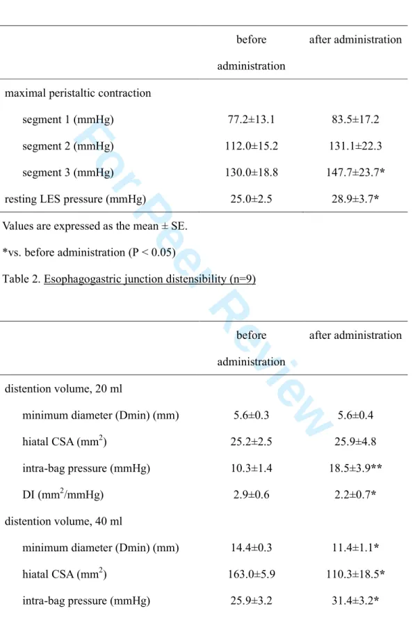
Table 1. Esophageal motor activity (n=8)
Values are expressed as the mean ± SE. *vs. before administration (P < 0.05)
Table 2. Esophagogastric junction distensibility (n=9) before administration
after administration
maximal peristaltic contraction
segment 1 (mmHg) 77.2±13.1 83.5±17.2
segment 2 (mmHg) 112.0±15.2 131.1±22.3
segment 3 (mmHg) 130.0±18.8 147.7±23.7*
resting LES pressure (mmHg) 25.0±2.5 28.9±3.7*
before administration
after administration
distention volume, 20 ml
minimum diameter (Dmin) (mm) 5.6±0.3 5.6±0.4
hiatal CSA (mm2) 25.2±2.5 25.9±4.8
intra-bag pressure (mmHg) 10.3±1.4 18.5±3.9**
DI (mm2/mmHg) 2.9±0.6 2.2±0.7*
distention volume, 40 ml
minimum diameter (Dmin) (mm) 14.4±0.3 11.4±1.1*
hiatal CSA (mm2) 163.0±5.9 110.3±18.5* intra-bag pressure (mmHg) 25.9±3.2 31.4±3.2* 5 6 7 8 9 10 11 12 13 14 15 16 17 18 19 20 21 22 23 24 25 26 27 28 29 30 31 32 33 34 35 36 37 38 39 40 41 42 43 44 45 46 47 48 49 50 51 52 53 54 55 56 57
For Peer Review
Values are expressed as the mean ± SE. *vs. before administration (P < 0.05) **vs. before administration (P < 0.01) DI: distensibility index
DI (mm2/mmHg) 7.1±0.9 4.1±0.8**
distention volume, 50 ml
minimum diameter (Dmin) (mm) 18.1±0.4 16.4±0.5**
hiatal CSA (mm2) 259.6±12.0 211.9±12.8** intra-bag pressure (mmHg) 33.1±2.3 37.1±3.7* DI (mm2/mmHg) 8.2±0.8 6.4±0.9** 5 6 7 8 9 10 11 12 13 14 15 16 17 18 19 20 21 22 23 24 25 26 27 28 29 30 31 32 33 34 35 36 37 38 39 40 41 42 43 44 45 46 47 48 49 50 51 52 53 54 55 56 57
For Peer Review
Figure legendsFig. 1.
Pressure tomography observed by high-resolution manometry in a representative case. Esophageal peristaltic contractions were divided into 3 segments by 2 troughs and the lower segment showed the strongest contractions. Esophageal peristaltic
contractions and esophageal resting pressure were higher in the mosapride-treated period (b) as compared to the non-treated period (a).
Fig. 2.
Shown is a representative set of volumetric FLIP distensions observed during mosapride treated (lower) and non-treated (upper) periods. Number of volume shows volume of inflated air. EGJ distensibility is illustrated as a cylinder of varying diameter with the corresponding intra-bag pressure. The hourglass shape of the EGJ was
narrowed at the hiatus (arrows). The EGJ became narrower with mosapride treatment.
Fig. 3.
EGJ distensibility with and without 40-mg mosapride administration. FLIP bag pressure (x-axis) and hiatal CSA (y-axis) were measured with the FLIP bag filled to 20 ml (triangles), 40 ml (squares), and 50 ml (circles) of fluid. Intra-bag pressures were consistently higher at similar hiatal CSA values with administration of mosapride, suggesting reduced EGJ distensibility.
5 6 7 8 9 10 11 12 13 14 15 16 17 18 19 20 21 22 23 24 25 26 27 28 29 30 31 32 33 34 35 36 37 38 39 40 41 42 43 44 45 46 47 48 49 50 51 52 53 54 55 56 57