東北薬科大学
審査学位論文(博士)
氏名(本籍) ワン ユーチン
王 玉琴(中国)
学位の種類 博士(薬科学)
学位記番号 博薬科第
3
号学位授与の日付 平成
27
年3
月17
日学位授与の要件 学位規則第4条1項該当
学位論文題名
α
1,6-Fucosyltransferase Is Required for Liver Regeneration and Chemical Induced
Hepatocarcinogenesis
論文審査委員
主査 教 授 井ノ口 仁 一
副査 教 授 細 野 雅 祐
副査 教 授 顧 建 国
α1,6-Fucosyltransferase Is Required for Liver Regeneration and Chemical Induced
Hepatocarcinogenesis
東北薬科大学大学院薬学研究科 王 玉琴
Contents
Summary……….…1
Part 1 Loss of α1,6-fucosyltransferase suppressed liver regeneration: implication of core fucose in the regulation of growth factor receptor-mediated cellular signaling 1.1 Introduction………..7
1.2 Materials and Methods………9
1.3 Results……….……….12
1.4 Discussion………... 19
Part 2 Loss of α1,6-fucosyltransferase inhibits chemical induced hepatocellular carcinoma and tumorigenesis by down-regulating several cell signaling pathways 2.1 Introduction……… 23
2.2 Materials and Methods……… ……… …… 25
2.3 Results……….……… 28
2.4 Discussion………...38
References………... 41
Acknowledgements………... 49
1
Summary
N-Linked glycosylation a common type of glycosidic bond. It is thought to be important for folding, stability, and vast degree of biological functions of glycoproteins. These different effects on glycoproteins mainly result from the different N-linked glycan structures determined by various glycosyltransferases. Among these, α1,6-fucosyltransferase (Fut8) is the only enzyme that catalyzes the transfer of a fucose from GDP-fucose to the innermost GlcNAc residue via α1,6-linkage to form core fucosylation in mammals. The enzymatic products, core fucosylated N-glycans, are widely distributed in a variety of glycoproteins and have been shown to play important roles in cell signaling. As examples, we previously showed that core fucosylation is crucial for the ligand binding affinity of transforming growth factor (TGF)-β1 receptor, epidermal growth factor (EGF) receptor, and integrin α3β1. Lacking the core fucose of these receptors led to a marked reduction in their ligand-binding ability and downstream signaling. Recently, our group found that a loss of core fucose on activin receptors resulted in an enhancement of the formation of activin receptor complexes, which constitutively activated intracellular signaling. These studies indicate that core fucosylation is able to negatively or positively affect signaling pathways through regulation of receptor binding ability, which could be important for appropriate signaling in vivo.
Part 1: Loss of α1,6-fucosyltransferase suppressed liver regeneration: implication of core fucose in the regulation of growth factor receptor-mediated cellular signaling
Liver regeneration after partial hepatectomy (PH) is a complicated process. At the cellular level,
it proceeds with the coordinated proliferation of all types of mature hepatic cells. Among these,
it has been generally accepted that the restoration of liver volume depends mainly on the
proliferation of hepatocytes. Molecularly, PH triggers multiple intracellular signaling cascades
(RAS/mitogen-activated protein kinase (MAPK) signaling, c-Met signaling, etc), leading to
great changes in the expression of genes associated with cell proliferation. The convergence of
these signaling pathways has been reportedly mediated via EGFR and hepatocyte growth factor
receptor (c-Met). Blocking the EGFR- or c-Met-mediated signaling pathway could cause a
severe delay of liver regeneration in mice. In addition to the expression level of EGFR and
c-Met proteins, it has been shown that the post-translational modification of these receptors
2
such as ubiquitination, phosphorylation, and glycosylation also plays a crucial role in the regulation of these signaling pathways.
Recently it was reported that core fucosylation on some glycoproteins, such as vitronectin, increased during liver regeneration after PH. However, the underlying mechanisms remain poorly understood. Here, we investigated the role of Fut8 in liver regeneration and showed for the first time that core fucosylation is physiologically associated with the liver regeneration.
Firstly, we detected the enzyme activities of Fut8 in the liver tissues at different time points after 70% PH. The Fut8 activities were increased in the first 4 days after operation, and returned to normal levels after liver mass is restored. These data indicated that the induction of Fut8 expression might be required for liver regeneration. To testify the hypothesis above, we performed a 70% PH on both Fut8
+/+and Fut8
-/-mice, and analyzed the restoration of their livers. Interestingly, the regeneration index calculated as an increase in liver-to-body weight ratio was significantly lower in Fut8
-/-mice than that in Fut8
+/+mice. Furthermore, a decrease in liver regeneration was also observed in the Fut8
+/-mice during the first 2 days.
Liver regeneration was achieved by the coordinated proliferation of all types of mature hepatic cells. Consistent with the results above, quantitative assessment of Ki67 by immunostaining revealed little difference between Fut8
-/-and Fut8
+/+mice without PH, while, the percentage of Ki67 positive versus TO-PRO-3 iodide positive cells in the livers of Fut8
-/-mice were markedly less than that in Fut8
+/+mice at day 2 after PH. These differences in cell proliferation were further reflected by the cell proliferation signaling. Overall, these data indicated that the delayed liver recovery in Fut8
-/-mice resulted from the lower cell proliferation.
It is known that two pathways for the synthesis of GDP-fucose in mammalian cells, the GDP-mannose-dependent de novo pathway and the free fucose-dependent salvage pathway.
And what is more, administration of oral L-fucose, an enhancement of the salvage pathway,
has been proven useful for correction of fucosylation defects in leukocyte adhesion deficiency
type II patients. To determine whether enhancing GDP-fucose salvage pathway could
complement the delayed liver regeneration of the Fut8
+/-mice as described above, we checked
the effects of L-fucose supplementation in the Fut8
+/-mice. Interestingly, an oral administration
of L-fucose significantly accelerated liver regeneration of the Fut8
+/-mice, but did not affect
sham mice. Consistently, in contrast to the little difference in the case of livers without 70% PH,
immunostaining with Ki67 showed the ratio of Ki67
+to TO-PRO-3 iodide
+cells in the livers
3
treated by PH were clearly increased after L-fucose administration. These results further suggest that Fut8 and its products are important for cell proliferation in liver regeneration.
The EGFR ligands and HGF are major mitogens for hepatocytes in the regenerating liver.
Lacking EGFR or c-Met in mice resulted in the liver regeneration abnormalities. To determine whether the delayed liver recovery in the Fut8
-/-mice is due to the impaired EGFR and/or c-Met signaling, we tested the expression levels of the key effectors in these signaling pathways. Although c-Met and EGFR associated signaling pathways were activated in both Fut8
+/+and Fut8
-/-mice 2 days post PH, the levels of phosphorylated c-Met and EGFR in Fut8
-/-mice were obviously lower than that in Fut8
+/+mice. These results indicated that loss of Fut8 impaired EGFR and c-Met associated signaling during liver regeneration.
To further corroborate the results above in vitro, we examined the downstream signaling cascades of EGF or HGF using the primary hepatocytes isolated from Fut8
+/+and Fut8
-/-mice.
Consistently, the treatments with EGF or HGF significantly increased the expression levels of phosphorylated ERK and AKT in the Fut8
+/+cells. However, these increases were greatly suppressed in the Fut8
-/-cells. The results above clearly demonstrated that the impaired regeneration in Fut8
-/-livers was due, at least mainly, to the down-regulated EGFR- and c-Met-mediated signaling in hepatocytes.
Overall, this study marks the first clear demonstration of the biological functions of Fut8 in the liver, suggesting that core fucosylation plays important roles in liver regenerating progression.
Part 2. Loss of α1,6-fucosyltransferase inhibits chemical induced hepatocellular carcinoma and tumorigenesis by down-regulating several cell signaling pathways
Hepatocellular carcinoma (HCC) is the third leading cause of cancer-related mortality worldwide, and hepatocarcinogenesis is a complicated process associated with the accumulation of pathological changes during the initiation, promotion, and progression of the disease. Identifying these changes may provide an avenue to develop a new generation of potential biomarkers, as well as therapeutic targets for HCC. It has been shown that altered expressions of growth factors such as EGF, TGF-α, TGF-β, and HGF, as well as their receptors during the development of HCC, influence the survival of cancerous cells by suppressing apoptosis and regulating the cell cycle.
It has been reported that the up-regulation of core fucosylation catalyzed by Fut8 has been
4
observed in pathological conditions such as HCC, and the fucosylated AFP (AFP-L3) is a reliable marker that can be used to distinguish patients with HCC from those with chronic hepatitis and liver cirrhosis. The HCC cases with high Fut8 expression is associated with the poor prognosis. These information prompted us to wonder what the pathological role of high Fut8 expression is in HCC progression and whether it could serve as a potential therapeutic target for liver cancer.
To explore the effects of high Fut8 expression and their molecular mechanisms in hepatocarcinogensis, here, the male wild-type (Fut8
+/+), hetero (Fut8
+/-) and knockout (Fut8
-/-) mice were used to establish the chemical induced HCC models by diethylnitrosamine (DEN) and pentobarbital (PB). The induction of HCC was significantly suppressed in Fut8
-/-mice, meanwhile, the expression of Fut8 was greatly increased in the liver tissues of Fut8
+/+mice during the process. Consistently, liver functions were destroyed in the Fut8
+/+mice and Fut8
+/-mice, but not Fut8
-/-mice.
DEN exerts carcinogenicity after being bioactivated by cytochrome P450 (CYP) enzymes in the liver. To check the early effects of DEN in Fut8
+/+and Fut8
-/-mice, semi-quantitative or quantitative PCR analysis of mRNAs encoding the CYP enzyme and Mgmt genes was performed at 2 h after DEN injection. There was no significant difference in the transcriptional levels of either CYP genes or DNA repair gene between Fut8
+/+and Fut8
-/-mice. DEN-induced tumor formation was further associated with substantial and marked induction of proinflammatory chemokines within the livers of mice. Quantitative PCR analysis of livers after DEN injection revealed increase in IL-6 and TNFα expression levels in both Fut8
+/+and Fut8
-/-mice, but there was no significant difference between the Fut8
+/+and Fut8
-/-mice.
Moreover, TUNEL assay for the livers showed the similar results. These data demonstrate that the expression of Fut8 may not affect the acute phase response to DEN.
Cell proliferation plays important roles in HCC process. In order to find the possible
mechanisms for the differences in tumorigenesis described above, we carried out Ki67
immunostaining for the frozen liver tissues of Fut8
+/+and Fut8
-/-mice. After chemical
induction, the Fut8
+/+mice showed a significant increase by more than ~6.5-fold in positive
immunostaining as compared with the untreated control. However, only a 2-fold increase was
observed in Fut8
-/-mice. The expression levels of cyclin mRNAs, including Cyclin B1, Cyclin
D1 and Cyclin E2, were also up-regulated in Fut8
+/+mice after DEN/PB treatment, and the
5
elevations in gene expression were attenuated by the ablation of Fut8. These results indicate that Fut8 may influence the progress of DEN/PB induced HCC by affecting the cell proliferation.
The effects of Fut8 expression on tumorigenesis were further confirmed by xenograft tumors formed by HepG2 cells in NOD/SCID mice. Knockout of Fut8 gene in the cells completely inhibited tumor formation. The HepG2 cell line was also used to reveal possible mechanisms of Fut8 for the regulatory roles. The responses to EGF and HGF were attenuated in the Fut8 knockout cells, although the expression levels of their receptors on the cell surface were similar.
Considering also our previously reported that core fucosylation was required for the binding of the EGF to EGFR, it is reasonable to conclude that lacking the core fucosylation on both receptors may decrease their biological functions in vitro or in vivo.
Taken together, it could be postulated that a loss of the Fut8 gene may affect the biological functions of some target membrane proteins and their subsequent downstream signaling, thereby inhibiting the hepatocarcinogenesis. These results suggest that the levels of core fucosylation are not only biomarkers, but also functional modulators in the liver. Thus, Fut8 might be a novel therapeutic target for HCC.
References
Wang Y., Fukuda T., Isaji T., Lu J., Gu W., Lee H., Ohkubo Y., Kamada Y., Taniguchi N., Miyoshi E. and Gu J.. Loss of α1,6-fucosyltransferase suppressed liver regeneration:
implication of core fucose in the regulation of growth factor receptor-mediated cellular
signaling. (2015) Scientific Reports 5, 8264 (DOI: 10.1038/srep08264)
6
Part 1
Loss of α1,6-fucosyltransferase suppressed liver regeneration:
implication of core fucose in the regulation of growth factor
receptor-mediated cellular signaling
7
1.1 Introduction
The adult liver has a remarkable capacity to regenerate, which makes it possible to use partial livers from living donors for transplantation. However, certain hepatic conditions, including cirrhosis, steatosis, and conditions due to old age, also have impaired liver regeneration that results in increased morbidity and mortality in response to liver transplantation (1). Therefore, in the past decade, numerous studies have been focused on dissecting the molecular mechanisms underlying liver regeneration.
Seventy percent partial hepatectomy (PH) is the most common technique that is used to study the regeneration of liver. Namely, it describes a surgical procedure which removes 70% of liver mass in rodents (rats and mice). Due to the multi-lobed structure of the rodent liver, three of the five liver lobes (representing 70% of its liver mass) can be removed. The residual lobes enlarge and reconstitute the original size of the liver within 2 weeks (2, 3). Regeneration after PH is a complicated process. At the cellular level, it proceeds with the coordinated proliferation of all types of mature hepatic cells. Among these, it has been generally accepted that the restoration of liver volume depends mainly on the proliferation of hepatocytes (4). This is not only because hepatocytes account for about 80% of liver weight and 70% of all liver cells, but also they are the first cells to enter into DNA synthesis and produce mitogenic signals for other hepatic cells (4, 5). Molecularly, PH triggers multiple intracellular signaling cascades (RAS/mitogen-activated protein kinase (MAPK) signaling, c-Met signaling, etc), leading to great changes in the expression of genes associated with cell proliferation (1, 6). The convergence of these signaling pathways has been reportedly mediated via epidermal growth factor receptor (EGFR) and hepatocyte growth factor receptor (c-Met) (4). Blocking the EGFR- or c-Met-mediated signaling pathway could cause a severe delay of liver regeneration.
In addition to the expression level of EGFR and c-Met proteins, it has been shown that the post-translational modification of these receptors such as ubiquitination, phosphorylation, and glycosylation also plays a crucial role in the regulation of these signaling pathways (7, 8).
Fucosylation is one type of glycosylation. It describes the attachment of a fucose residue to
N-glycans, O-glycans, and glycolipid catalyzed by a family of enzymes called
fucosyltransferases (Futs) (9). Among these, α1,6-fucosyltransferase (Fut8) is the only enzyme
that catalyzes the transfer of a fucose from GDP-fucose to the innermost GlcNAc residue via
8
α1,6-linkage to form core fucosylation in mammals as shown in Figure 1c. The enzymatic products, core fucosylated N-glycans, are widely distributed in a variety of glycoproteins and have been shown to play important roles in cell signaling. As examples, we previously showed that core fucosylation is crucial for the ligand binding affinity of TGF-β1 receptor (10), EGF receptor (11), and integrin α3β1 (12). Lacking the core fucose of these receptors led to a marked reduction in their ligand-binding ability and downstream signaling. Recently, our group found that a loss of core fucose on activin receptors resulted in an enhancement of the formation of activin receptor complexes, which constitutively activated intracellular signaling (13). These studies indicate that core fucosylation is able to negatively or positively affect signaling pathways through regulation of receptor binding ability.
Abnormal expression of Fut8 has been pathologically correlated with diverse carcinomas
including liver (14), ovarian (15), lung (16) and colorectal cancers (17). Recently it was
reported that core fucosylation on some glycoproteins, such as vitronectin, increased during
liver regeneration after PH (18). However, the underlying mechanisms remain poorly
understood. Here, we investigated the role of Fut8 in liver regeneration and showed for the first
time that core fucosylation is physiologically associated with the liver regeneration. In
particular, we show that the liver regeneration was significantly inhibited in Fut8 deficient
(Fut8
-/-) and Fut8 hetero (Fut8
+/-) mice as compared to wild type (Fut8
+/+) mice. It is intriguing
that this effect could be attenuated by L-fucose supplementation in the Fut8
+/-mice. Moreover,
intracellular signaling analysis using primary hepatocytes isolated from Fut8
+/+and Fut8
-/-mice
clearly demonstrated that Fut8 is important for the initiation of hepatocyte proliferation. Taken
together, our data here provide novel insight for the function of core fucosylation in liver
regeneration.
9
1.2 Materials and Methods
1.2.1 Mice
The Fut8-deficient mice line used for these studies has been described previously (10, 19).
Male mice on an ICR background at 6 to 8 weeks of age were used for the experiments in the present study, comparing Fut8
-/-animals with Fut8
+/+littermates. Mice were housed in a temperature-controlled room with a 12-h dark/12-h light cycle. Food and water were provided ad libitum. The present study was approved by the Institutional Animal Care and Use Committee of Tohoku Pharmaceutical University, Japan.
1.2.2 70% partial hepatectomy
All experiments were carried out in accordance with relevant guidelines and regulations. For liver regeneration studies, 7- to 8- week-old mice were anesthetized with pentobarbital sodium and subjected to mid-ventral laparotomy with a two-third liver resection, as previously described (20, 21). The left and median liver lobes were surgically resected without injuring the remaining liver tissue. The removed parts represented the resting liver. At least three mice from each group were euthanized at each analysis time point. For L-fucose (Nacalai tesque Inc.) supplementation, 6-week-Fut8
+/-mice were orally administrated with L-fucose (4 g/L in water) for 12 days prior to partial hepatectomy (PH), and then the livers were harvested at 48h after operation.
1.2.3 Immunostainings
The hepatic lobules were assessed based on 10 m frozen sections. Proliferative cells in the liver were detected through immunostaining with a monoclonal antibody recognizing Ki67 (Abcam), and examined with Olympus confocal laser scanning microscope (Olympus).
1.2.4 Cell culture
Primary hepatocytes of 8-week old mice were isolated using the standard method of in situ
collagenase (Gibco) perfusion and digestion of liver with low-speed centrifugation (50×g, 1
min), as previously reported (22, 23). Isolated cells were plated on collagen type I-coated
dishes in Dulbecco’s modified Eagle’s medium (DMEM) with 10% (v/v) fetal bovine serum
10
(FBS), 100 IU/mL penicillin, and 100 g/ml streptomycin. Hepatocytes were incubated for 6 h at 37 ˚C in a humidified atmosphere with 95% air and 5% CO
2, allowing for cell attachment to the plate. The medium was then changed, which involved replacement by 0.1% FBS contained DMEM with or without EGF or HGF for stimulation at indicated times.
1.2.5 Western blotting analyses
Total protein was isolated from frozen liver tissue and cultured cells with TBS (20 mM Tris, 150 mM NaCl, PH 7.4) containing 1% triton X-100. Protein concentration was measured using a bicinchoninic acid protein assay kit (Thermo Scientific). Equal protein samples were separated by SDS-PAGE and then transferred onto nitrocellulose or polyvinylidinedifluoride (Millipore) membranes. After blocking with 5% skim milk, the membranes were incubated with specific antibodies against the indicated antibodies at 4 ˚C overnight, followed by incubation with horseradish peroxidase-conjugated secondary antibody. Immunoreactivity was visualized by HRP substrate peroxide solution (Millipore). The related antibodies that are used included ERK1 (BD), phospho-ERK, phospho-AKT, AKT, phospho-Met (Tyr1234/5), c-Met, phospho-EGFR (Tyr1068), EGFR, rabbit IgG (Cell Signaling) and mouse IgG (Sigma).
1.2.6 Enzyme activity assays for Fut8
Frozen liver tissues were homogenized in TBS containing 1% protease inhibitor cocktail (Nacalai tesque Inc.). After centrifugation at 900 g for 10 min, the supernatant was collected for enzyme activity assays. Each sample containing 800 g of total protein was centrifuged at 105,000 g for 1 h, then the pellet was resuspended in 0.1 M MES-NaOH (PH 7.0) for reactions.
Equal amounts of protein were used in Fut8 activity assays. The specific activities of Fut8 were determined using a substrate, 4-(2-pyridylamino)-butylamine (PABA)-labeled GlcNAcβ1-2Manα1-6(GlcNAcβ1-2Manα1-3)Manβ1-4GlcNAcβ1-4GlcNAc-Asn
(GnGn-Asn-PABA). Each assay used 2 mM of acceptor substrate and 2 mM GDP-L-fucose as
a donor (in 10 l of total reaction solution). The reactions were terminated by boiling after 2h
of incubation at 37 ˚C, and the reaction mixtures were centrifuged at 10,000 g for 10 min. The
result supernatants were applied to high-performance liquid chromatography (HPLC) equipped
with a TSK-gel, ODS-80TM column (4.6×150mm) in order to separate and quantitate the
products. Elution was performed isocratically at 55 ˚C using a 20 mM acetate buffer (pH 4.0)
11
containing 0.15% butanol. The column eluate was monitored for fluorescence using a detector operating at excitation and emission wavelengths of 320 and 400 nm, respectively. The activities of endogenous Fut8 were measured by HPLC, expressed as the pmol of fucose transferred/h/mg of proteins (24).
1.2.7 Statistical analysis
Results are given as the mean ± standard error of the mean (SEM). The data were analyzed
using Prism 5.0 software (GraphPad Software Inc.). Comparisons were carried out using
2-tailed Mann-Whitney tests and/or a Tukey’s Multiple Comparison test. A P value of less than
0.05 was considered significant.
12
1.3 Results
1.3.1 70% PH induced the expression of Fut8
It has been reported that lacking N-acetylglucosaminyltransferase III suppressed the liver tumor progression and liver regeneration in mice, indicating the importance of glycosylation in liver (25). In the present study, we investigated the roles of Fut8 in liver regeneration. Firstly, we detected the enzyme activities of Fut8 in the liver tissues at different time points after 70%
PH. As shown in Figure 1a and 1b, the Fut8 activities were increased in the first 4 days after operation, and returned to normal levels after liver mass is restored. On the other hand, the expression levels of L-fucosidase after PH were not changed confirmed by RT-PCR (data not shown). These data indicated that the induction of Fut8 expression might be required for liver regeneration.
Figure1. The activities of Fut8 were increased after 70% partial hepatectomy (PH).
The liver tissues were harvested for the determination of enzyme activities at indicated times as described in
“Methods”. (a) A representative elution pattern on HPLC for Fut8 activities in Fut8+/+ mouse with (lower
13
panel) or without (upper panel) PH. S: substrate; P: product. (b) The quantitative assay for enzyme activities in Fut8+/+ mice after PH. *, P<0.05, compared to the group without PH (sham), which was set as 1, n=3. (c) Reaction for synthesis of α1,6-fucose.
1.3.2 Loss of Fut8 inhibited recovery of liver mass after a two-third liver resection
To testify the hypothesis above, we performed a 70% PH on both Fut8
+/+and Fut8
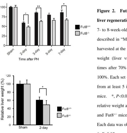
-/-mice, and analyzed the restoration of their livers. Interestingly, the regeneration index calculated as an increase in liver-to-body weight ratio was significantly lower in Fut8
-/-mice than that in Fut8
+/+mice (Figure 2a). Furthermore, a decrease in liver regeneration was also observed in the Fut8
+/-mice during the first 2 days (Figure 2b). The results above indicated that the liver regeneration was inhibited in Fut8
-/-mice as compared to Fut8
+/+mice.
Figure 2. Fut8 expression was required for liver regeneration after PH.
7- to 8-week-old mice were surgically resected as described in “Methods”, and then the livers were harvested at the indicated times. (a) Relative liver weight (liver vs whole body) at the indicated times after 70% PH. The sham group was set as 100%. Each set of the reported data was obtained from at least 5 individuals of Fut8+/+ and Fut8−/−
mice. *, P<0.05; **, P<0.01. (b) Comparison of relative weight at 2 days after PH between Fut8+/+
and Fut8+/- mice (C57BL/6 genetic background).
Each data was obtained from at least 8 individuals.
*, P<0.05, compared with the Fut8+/+ mice.
Liver regeneration was achieved by the coordinated proliferation of all types of mature hepatic
cells (2). Consistent with the results above, quantitative assessment of Ki67 by immunostaining
revealed little difference between Fut8
-/-and Fut8
+/+mice without PH, while, the percentage of
Ki67 positive versus TO-PRO-3 iodide positive cells in the livers of Fut8
-/-mice were
14
markedly less than that in Fut8
+/+mice at day 2 after PH (Figure 3a and 3b). These differences in cell proliferation were further reflected by the cell proliferation signaling. As shown in figure 3c, the phosphorylation levels of ERK were remarkably lower in the Fut8
-/-mice as compared with Fut8
+/+mice, although the MAPK signaling pathways were activated by PH in both Fut8
+/+and Fut8
-/-mice. Overall, these data indicated that the delayed liver recovery in Fut8
-/-mice resulted from the lower cell proliferation.
Figure 3. Cell proliferation was suppressed in the livers of Fut8−/− mice.
(a) Immunostaining for liver tissues (10 m frozen section) of Fut8+/+ and Fut8−/− mice using anti-Ki67 antibody (200×field). The positive cells of the immunostaining were labeled with the green spots (left panel), and the nuclei were labeled by TO-PRO-3 iodide (red spots, middle panel). (b) The quantitative data were obtained from at least 3 mice in each group. **, P<0.01. (c) Equal protein of liver lysates at day 2 after PH were separated by 10% SDS-PAGE and blotted with anti-phospho-ERK and anti-ERK1 antibodies. The quantitative data were obtained from 3 mice in each group. *, P<0.05, **, P<0.01.
15
1.3.3 L-fucose administration in Fut8
+/-mice attenuated the inhibitory effect in cell proliferation as described above.
GDP-fucose is the donor for fucosyltransferases. It is known that two pathways for the synthesis of GDP-fucose in mammalian cells, the GDP-mannose-dependent de novo pathway and the free fucose-dependent salvage pathway (26). And what is more, administration of oral L-fucose, an enhancement of the salvage pathway, has been proven useful for correction of fucosylation defects in leukocyte adhesion deficiency type II (LAD II) patients (27). To determine whether enhancing GDP-fucose salvage pathway could complement the delayed liver regeneration of the Fut8
+/-mice as described above, we checked the effects of L-fucose supplementation in the Fut8
+/-mice. Interestingly, an oral administration of L-fucose significantly accelerated liver regeneration of the Fut8
+/-mice, but did not affect sham mice (Figure 4a). Consistently, in contrast to the little difference in the case of livers without 70%
PH, immunostaining with Ki67 showed the ratio of Ki67
+to TO-PRO-3 iodide
+cells in the livers treated by PH were clearly increased after L-fucose administration (Figure 4b and 4c).
Moreover, as shown in figure 4d and 4e, the phosphorylation levels of ERK and EGFR were
induced in Fut8
+/-mice after PH. Furthermore, the L-fucose administration up-regulated their
phosphorylation levels, although there was no significant difference between the mice treated
with or without L-fucose by statistical analysis. These results further suggest that Fut8 and its
products are important for cell proliferation in liver regeneration.
16
Figure 4. L-fucose supplementation attenuated the decreased regeneration of Fut8+/- mice.
(a) Relative liver weight (liver vs whole body) at 2-day after PH in Fut8+/- mice with or without administration of L-fucose. Prior to operation, 6-week old Fut8+/- mice were administrated with L-fucose at 4g/L in water for 12 days, and then the livers were harvested at 48 hours after PH. The sham group without L-fucose treatment was set as 100%. *, P<0.05, compared with the mice without L-fucose treatment (n >10 mice). (b) Immunostaining for liver tissues using anti-Ki67 antibody (200×field). (c) The quantitative data were obtained from at least 3 mice in each group, *, P<0.05. (d and e) Equal protein of liver lysates at day 2 after PH were separated by SDS-PAGE (10% for pERK/ERK1, 7% for pEGFR/EGFR) and blotted with anti-phospho-ERK and anti-ERK1 antibodies. The quantitative data were obtained from 3 mice in each group.
1.3.4 The intracellular signaling was inhibited in the Fut8
-/-primary hepatocytes upon stimulation with EGF or HGF
The EGFR ligands and HGF are major mitogens for hepatocytes in the regenerating liver.
Lacking EGFR or c-Met in mice resulted in the liver regeneration abnormalities (28, 29). To
determine whether the delayed liver recovery in the Fut8
-/-mice is due to the impaired EGFR
and/or c-Met signaling, we tested the expression levels of the key effectors in these signaling
pathways. As shown in Figure 5a and b, although c-Met and EGFR associated signaling
17
pathways were activated in both Fut8
+/+and Fut8
-/-mice 2 days post PH, the levels of phosphorylated c-Met (Tyr1234/5) and EGFR (Tyr1068) in Fut8
-/-mice were obviously lower than that in Fut8
+/+mice. These results indicated that loss of Fut8 impaired EGFR and c-Met associated signaling during liver regeneration.
To further corroborate the results above in vitro, we examined the downstream signaling cascades of EGF or HGF using the primary hepatocytes isolated from Fut8
+/+and Fut8
-/-mice.
Consistently, the treatments with EGF or HGF significantly increased the expression levels of phosphorylated ERK and AKT in the Fut8
+/+cells. However, these increases were greatly suppressed in the Fut8
-/-cells (Figure 5d and e). The results above clearly demonstrated that the impaired regeneration in Fut8
-/-livers was due, at least mainly, to the down-regulated EGFR- and c-Met-mediated signalings in hepatocytes.
Figure 5. Intracellular signaling was suppressed in Fut8-/- mice upon either PH or EGF and HGF stimulation.
8-week-old Fut8+/+ or Fut8-/- mice were surgically resected as described in “Methods”, and then the livers were harvested at 2 days. The liver homogenates were separated by 7% SDS-PAGE and blotted with
18
anti-EGFR and anti-phospho-EGFR antibodies (a), and anti-c-Met and anti-phospho-c-Met antibodies (b).
The quantitative data were obtained from 3 mice in each group. *, P<0.05, **, P<0.01. The primary hepatocytes isolated from 8-week old Fut8+/+ and Fut8-/- mice were cultured in DMEM containing with 10%
FBS for 12 h, and then cultured under DMEM containing with 0.1% FBS for 24 hours. After the starvation, these cells were stimulated with or without EGF at indicated concentrations for 5 min (c and d), or HGF at indicated concentrations for 10 min (e). The cell lysates were immunoblotted with anti-pEGFR and anti-EGFR, anti-pAKT and anti-AKT antibodies, anti-pERK and anti-ERK1 antibodies. The quantitative data were obtained from at least 3 independent experiments, *, P<0.05, **, P<0.0 1.
19
1.4 Discussion
In the present study, we used a well-established regeneration model, to investigate the functions of Fut8 in liver regeneration, and found the following: i) The expression of Fut8 was markedly up-regulated during the regenerating process in the Fut8
+/+mice; ii) the liver regeneration was greatly inhibited in Fut8
-/-mice compared to Fut8
+/+mice; iii) L-fucose supplementation could reverse the delayed regeneration in Fut8
+/-mice; and, iv) the responses to growth factors such as EGF and HGF, were decreased in Fut8 deficient hepatocytes compared to wild-type hepatocytes. Overall, this study marks the first clear demonstration of the biological functions of Fut8 in the liver, suggesting that core fucosylation plays important roles in liver regenerating progression as shown in Fig.6.
Figure 6. Proposed molecular mechanisms for the delayed liver regeneration in Fut8-/- mice. It is well known that the 70% partial hepatectomy could activate several cell proliferation associated signaling pathways including Ras/MAPK signaling, c-Met signaling, and Akt/mammalian targets of rapamycin (mTOR) signaling, which up-regulate the cell proliferation, and consequently lead to the restoration of liver. Loss of core fucosylation on growth factor receptors such as EGFR and c-Met may alter their conformation and impair their ligand binding, thereby inhibiting their downstream signalings, and ultimately suppressing the cell proliferation. Overall, a loss of Fut8 gene results in a decrease in liver regeneration.
Liver regeneration after PH is a complicated process with the coordinated proliferation of all
types of mature hepatic cells, which involves numerous molecules and signaling pathways (1, 2,
6, 30). Among these, the EGFR-mediated signaling has been reported to be critical for liver
regeneration (29). Lacking EGFR in hepatocytes increased the mouse mortality rate after PH,
and delayed the hepatocyte proliferation (28), although little effect was observed on liver
function. We have previously shown that core fucosylation on EGFR is required for its binding
20
to EGF and downstream signaling in embryonic fibroblast cells (11). Therefore, it is reasonable to consider that the delayed liver recovery of Fut8
-/-mice could be attributed, at least mainly, to the loss of the core fucosylation on the EGFR protein (Figure 5c and d). In agreement with this hypothesis, we found here that knockout of Fut8 led to an inhibition of the EGFR-mediated signaling cascade both in vivo and in vitro.
In addition to EGFR, c-Met has also been shown to play an irreplaceable role in liver regeneration. c-Met gene deficient or suppressed by shRNAs significantly inhibited the proliferation of hepatocytes after PH (28, 31, 32). In the present study, we found that knockout of Fut8 also attenuated the response to an HGF stimulus in primary hepatocytes (Figure 5e).
Since c-Met is also a core fucosylated protein which had been confirmed by using human cell lines (data not shown), one possibility for this attenuated response is that like EGFR, the core fucosylation on c-Met may be necessary for its ligand binding and downstream signaling as well. Obviously, we could not exclude other possibilities. Recently, Tobias Speicher et al.
reported that theβ1-integrin knockout or knockdown in mice inhibited liver regeneration by impairing the ligand-induced phosphorylation of EGFR and c-Met, as well as their downstream signalings (33). Considering also that α3β1 integrins were highly modified by Fut8 and loss of core fucosylation could result in the malfunction of β1-integrin (12), Fut8 may also affect the c-Met-mediated signaling in the liver regeneration by regulating the core fucosylation status of β1-integrin. Further investigation is required to confirm the hypotheses above.
Increasing evidence indicated the importance of core fucosylation in protein-protein interaction,
and we proposed here that Fut8 may affect the liver regeneration through modulating some
associated receptor-ligand bindings. However, the mechanistic roles of Fut8 underlying the
protein-protein interaction remain poorly understood. Recently, two research teams determined
the complex structures of glycosylated FcγRIIIa and human core fucosylated or afucosylated
Fc of IgG (34, 35). Interestingly, the crystal structures indicated that core fucose depletion
increased the incidence of the active conformation of the Tyr-296 of Fc, and thereby
accelerated the formation of the high-affinity complex with its receptor. These findings clearly
explained why the lack of a core fucose on IgG could greatly enhance antibody-dependent
cell-mediated cytotoxicity as previously reported (36, 37). From a more general viewpoint,
these studies provide direct evidence for the mechanistic roles of Fut8 in different biological
processes, where the attachment of core fucose leads to an alteration of glycoprotein
21
conformation, which determines its protein dynamics coupled with the selection of protein-protein interactions and complex formation, and consequently affects the intracellular signaling pathways.
The excellent results of liver transplantation have led to an increasing number of patients on
the waiting list, while the number of liver donors remains stable (1, 38). Studies on potential
hepatoprotective factors in liver injury may contribute to increasing the success ratio of liver
transplantation. Here, we showed that liver regeneration is significantly inhibited in Fut8
-/-mice. Moreover, L-fucose administration could partially complement the delayed liver recover
in Fut8
+/-mice. Clearly, there is a need to further elucidate the underlying mechanisms and
confirm these observations of core fucosylation in human. However, the current study provides
important evidence for the effect of L-fucose supplementation on liver regeneration and
indicates the important role of Fut8 in liver regeneration.
22
Part 2
Loss of α1,6-fucosyltransferase inhibits chemical induced hepatocellular carcinoma and tumorigenesis by down-regulating
several cell signaling pathways
23
2.1 Introduction
N-Linked glycosylation a common type of glycosidic bond. It has been shown to be important for folding, stability, and vast degree of biological functions of glycoproteins. These different effects on glycoproteins mainly result from the different N-linked glycan structures determined by various glycosyltransferases (9). Among these, α1,6-Fucosyltransferase (Fut8) is the only enzyme that catalyzes the transfer of a fucose from GDP-fucose to the innermost GlcNAc residue via α1,6-linkage in mammals. The resulting core fucosylated N-glycans are widely distributed in a variety of glycoproteins. It has been reported that core fucosylation is crucial for the ligand binding affinity of transforming growth factor (TGF)-β1 receptor (10), epidermal growth factor (EGF) receptor (11), and integrin α3β1 (12). Lacking the core fucose led to a marked reduction in ligand-binding ability and downstream signaling. Recently, we found that loss of core fucose on activin receptors also resulted in an enhancement of the formation of activin receptor complexes, which constitutively activated intracellular signaling (13). Those studies clearly suggest that Fut8 plays important roles in cell signal transduction.
Hepatocellular carcinoma (HCC) is the third leading cause of cancer-related mortality
worldwide (39), and hepatocarcinogenesis is a complicated process associated with the
accumulation of pathological changes during the initiation, promotion, and progression of the
disease (40, 41). Identifying these changes may provide an avenue to develop a new generation
of potential biomarkers, as well as therapeutic targets for HCC. It has been shown that altered
expressions of growth factors such as EGF, TGF-α, TGF-β, and hepatocyte growth factor
(HGF), as well as their receptors during the development of HCC, influence the survival of
cancerous cells by suppressing apoptosis and regulating the cell cycle (41-44). Inhibitors
targeting those receptors mediated signaling pathways have been successfully developed and
used to improve the survival of patients with HCC (42). However, the therapeutic resistance
also develops (45, 46). Recently, an inhibition of N-linked glycosylation had been reported as a
novel therapeutic strategy for the treatment of gliomas and other malignant tumors (47). In
contrast, the role of N-glycosylation in HCC is largely unknown. It has been shown that Fut8
expression is up-regulated in both liver and serum during the process of hepatocarcinogenesis
(14, 48), and the fucosylated alpha fetoprotein (AFP-L3) is a reliable marker that can be used
to distinguish patients with HCC from those with chronic hepatitis and liver cirrhosis (49-51).
24
The HCC cases with high Fut8 expression is associated with the poor prognosis (52). These information prompted us to wonder what the pathological role of high Fut8 expression is in HCC progression and whether it could serve as a potential therapeutic target for liver cancer.
To address these questions, here, the typical HCC model was established in the male wild-type
(Fut8
+/+), hetero (Fut8
+/-) and knockout (Fut8
-/-) mice by administration of diethylnitrosamine
(DEN), which induces hepatocyte DNA damage, in conjunction with phenobarbital (PB), a
well-known promoter of liver carcinogenesis (53-55). DEN and PB effectively induced the
tumor formation in Fut8
+/+mice, while the tumor incidence was almost completely inhibited in
Fut8
-/-mice. The inhibitory effects of Fut8 were further confirmed by using human hepatoma
cell line, HepG2. Knockout of Fut8 gene abolished tumorigenesis of the xenograft tumors
formed by HepG2 cells in NOD/SCID mice. These results clearly demonstrate the importance
of Fut8 in hepatocarcinogenesis and implicate Fut8 as a novel therapeutic approach for HCC
treatment.
25
2.2 Materials and Methods
2.2.1 Chemical HCC induction
Six-week-old Fut8
-/-, Fut8
+/-and Fut8
+/+male mice were divided into two groups. The experimental group (n >10) received a single intraperitoneal injection of DEN (100 mg per kg, Sigma-Aldrich, St. Louis, MO, USA) and was provided 0.05% phenobarbital (Tokyo chemical, Japan) in water after 4 weeks, and the serum and livers were harvested after 40 weeks.
Meanwhile, the control group received the same operation without agents. A blood chemical analysis was performed by using a Vet Scan VS2 automated analyzer (ABAXIS, Union City, CA, USA) with a mammalian liver profile multi-rotor.
2.2.2 Liver histology and immunostaining
The hepatic lobules were assessed based on 4 m hematoxylin and eosin-stained paraffin sections. Masson's trichrome staining was performed to show collagen fiber deposition.
Immunostaining for P. squarrosa lectin (PhoSL), which specifically recognizes core fucose (56), was used to detect changes in core fucosylation during HCC induction. An in situ cell-death kit (Roche, Laval, Canada) was used for TUNEL staining with frozen sections (10
m), according to the manufacturer’s instructions. Proliferative cells in the liver were detected through immunostaining with a monoclonal antibody recognizing Ki67 (Abcam, Cambridge, MA, USA), and the liver sinusoidal endothelial cells were labelled with anti-CD31 antibody (BD Bioscience, San Jose, CA, USA).
2.2.3 Quantitative and semi-quantitative real-time PCR
Total RNA from livers was extracted with TRI Reagent (MRC Inc., Cincinnati, OH, USA).
Complementary DNA synthesis was carried out with PrimeScript
TMRT reagent Kit (TaKaRa,
Japan) according to the manufacturer’s instructions. Quantitative real-time PCR reactions were
performed with SYBR Premix Ex Taq
TMⅡ kit (TaKaRa), and semi-quantitative PCR actions
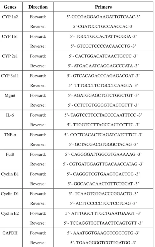
were carried out by using TaKaRa Ex Taq. Primers were listed in Table 1. Values of qPCR
reactions were quantified using the comparative CT method, and normalized to GAPDH
mRNA.
26
Table 1: Primer sequences used for qRT-PCR and RT-PCR
Genes Direction Primers
CYP 1a2 Forward:
Reverse:
5’-CCCGAGGAGAAGATTGTCAAC-3’
5’-CGATCCCTGCCAACCAC-3’
CYP 1b1 Forward:
Reverse:
5’- TGCCTGCCACTATTACGGA -3’
5’- GTCCCTCCCCACAACCTG -3’
CYP 2e1 Forward:
Reverse:
5’- CACTGGACATCAACTGCCC -3’
5’- ATGAGAATCAGGAGCCCATA -3’
CYP 3a11 Forward:
Reverse:
5’- GTCACAGACCCAGAGACGAT -3’
5’- TTTGCCTTCTGCCTCAAGTA -3’
Mgmt Forward:
Reverse:
5’- AGATGGAGCTGTCTGGCTGT -3’
5’- CCTCTGTGGGGTCAGTGTTT -3’
IL-6 Forward:
Reverse:
5’- TAGTCCTTCCTACCCCAATTTCC -3’
5’- TTGGTCCTTAGCCACTCCTTC -3’
TNF-α Forward:
Reverse:
5’- CCCTCACACTCAGATCATCTTCT -3’
5’- GCTACGACGTGGGCTACAG -3’
Fut8 Forward:
Reverse:
5’- CAGGGGATTGGCGTGAAAAAG -3’
5’- CGTGATGGAGTTGACAACCATAG -3’
Cyclin B1 Forward:
Reverse:
5’- CAGGGTCGTGAAGTGACTGG -3’
5’- GGCACACAACTGTTCTGCAT -3’
Cyclin D1 Forward:
Reverse:
5’- TCAAGTGTGACCCGGACTG -3’
5’- ACTTCCCCCTCCTCCTCAG -3’
Cyclin E2 Forward:
Reverse:
5’- ATTTGGCTTTGCTGAATGAAGT -3’
5’- TCCAGGTTGTTAACTTCAGTGTT -3’
GAPDH Forward:
Reverse:
5’- AAATGGTGAAGGTCGGTGTG -3’
5’- TGAAGGGGTCGTTGATGG -3’
2.2.4 Cell culture and transfection
Human hepatoma HepG2 cells were grown in Dulbecco’s modified Eagle’s medium (DMEM)
with 10% (v/v) fetal bovine serum (FBS), 100 IU/mL penicillin, and 100 g/ml streptomycin.
27
The Fut8 knockout CompoZr Zinc Finger Nuclease (ZFN) plasmids (Sigma) and GFP control plasmid were transfected into the cell lines via a TransIT-2020 Transfection Kit (Mirus Bio, Madison, WI, USA), and stable cell lines were selected via mushroom Pholiota Squarrosa lectin (PhoSL) staining by BD FACSAria II. The cells were incubated for 6 h at 37 ˚C in a humidified atmosphere with 95% air and 5% CO
2, allowing for cell attachment to the plate.
The medium was then changed, which involved replacement by 0.1% FBS contained DMEM with or without EGF or HGF for stimulation at indicated times.
2.2.5 Genomic PCR to detect Fut8
The removal of the Fut8 gene in HepG2 cell was confirmed by PCR, ZFN Primer F; 5'-
TGTCAGGTGAAGTGAAGGACA -3' and Primer R; 5'-
ATCAGATTCTTACCCAATAACTGG-3'.
2.2.6 Western blotting and lectin blot analyses Shown in Chapter 1.2.5
2.2.7 Immunoprecipitation and cell surface biotinylation
The cell lysate (500 g total protein) was incubated with 10 l FG streptavidin beads (Tamagawa Seiki, Japan) containing 2 g PhoSL for 2 hours at 4 ˚C, followed by washing 3 times with lysis buffer. The immunoprecipitate was dissolved in a 20l SDS-PAGE sample solution. For cell surface biotinylation, cells were incubated with 0.2 mg/ml sulfosuccinimidobiotin (Thermo Scientific) on ice for 2 hours, and then were solubilized in lysis buffer. The biotinylated proteins were precipitated with 50 l streptavidin agarose beads (Millipore, 2h, 4 ˚C), and visualized by Western blot analysis.
2.2.8 Established tumors originating from HepG2 cells
Six-week-old male NOD/SCID mice were purchased from Charles River Laboratories, INC.
The mice were inoculated subcutaneously with 5×10
6HepG2 WT and Fut8 KO cells into the
dorsal flanks, and the tumor tissues were harvested after 4 weeks.
28
2.3 Results
2.3.1 Loss of the Fut8 gene inhibited DEN/PB-induced tumor formation
To assess the effects of Fut8 on the development of HCC, 6-week-old Fut8
+/+, Fut8
+/-and Fut8
-/-male mice were exposed to the carcinogen DEN and liver tumor promoter PB. The absence of Fut8 significantly decreased HCC formation in response to DEN and PB when compared with Fut8
+/+mice. Interestingly, the Fut8
+/-mice also showed a lower incidence of tumors, although there was no significant difference compared with the Fut8
+/+mice (Figure 1b). Morphologic examination revealed that livers of Fut8
+/+model mice showed multiple large, vascularized nodules at the 40
thweek, while this phenomenon was rarely observed in Fut8
-/-mice (Figure 1a). Consistently, Masson's trichrome staining showed much more collagen deposition in DEN/PB-treated Fut8
+/+group as compared with the control and Fut8
-/-mice.
Meanwhile, in agreement with the tumor incidence, a significant increase in liver/body weight
ratios after DEN/PB treatment was observed for the Fut8
+/+model mice, but not the Fut8
-/-mice (Figure 1c). The ratio for Fut8
+/-mice showed a decreased tendency when compared with
that of Fut8
+/+, albeit not as compared with that of Fut8
-/-mice (Figure 1c). Furthermore, the
serum biochemical analysis for alanine aminotransferase (ALT), alkaline phosphatase (ALP),
and bile acids (BA), showed that DEN and PB induction destroyed liver functions of both
Fut8
+/+mice and Fut8
+/-mice, but not the Fut8
-/-mice (Figure 1d). These results strongly
suggest that the expression of Fut8 plays an important role in pathological functions.
29
Figure 1. Hepatocarcinogenesis was inhibited in Fut8−/− mice.
(a) The DEN/PB treated Fut8+/+ mice showed multiple large, vascularized nodules at the 40th week compared with the control group, while this phenomenon was rarely observed in Fut8−/− mice (upper panel, bar=1cm).
Sections (4 μm thick) from formalin-fixed, paraffin-embedded liver lobe of control and DEN/PB-treated Fut8+/+ and Fut8−/− male mice were used for histologic analysis by H&E (middle panel, bar=1 mm) and Masson's trichrome staining (lower panel, bar=1 mm). (b) Tumor incidence in different groups after DEN/PB-treatment (n>10). (c) Relative liver weight in different groups after 40 weeks. *, P<0.05, compared to Fut8+/+ control mice. (d) The expression levels of ALT, ALP and BA in the serum were analyzed by using a Vet Scan VS2 automated analyzer as described in “Materials and Methods”. *, P<0.05, compared to Fut8+/+
control mice.
30
2.3.2 Chemical treatment induced the expression of Fut8
The expression of Fut8 and its products are elevated in both liver and serum during the process
of human hepatocarcinogenesis (14). The increase in core fucosylated N-glycans has been also
observed in DEN-induced HCC rat models (57). To confirm whether it is also the case in
mice, here, we tested the activities of Fut8 in Fut8
+/+mice. As shown in Figure 2a and 2b, the
Fut8 enzyme activities were remarkably enhanced in DEN/PB treatment models as compared
with the control. The mRNA expression of Fut8 also showed a dual phase elevation during the
process of hepatocarcinogenesis, which reached a plateau after 36 weeks (Figure 2c). The
underlying molecular mechanism for the dual phase reaction is unclear. Immunostaining
analysis using PhoSL which specifically recognized the core fucose showed that the increases
in Fut8 expression and enzyme activities were accompanied by the enhancement of its
enzymatic products (Figure 2d). And these core fucose did not localize in the liver sinusoidal
endothelial cells, which were stained by anti-CD31 antibody (Figure 2e). Thus, we believed
that it might mainly localize in hepatocytes. Taken together, these results indicate that Fut8 is
expressed at lower level under physiological conditions in the liver, which could be
up-regulated after stimulus, and thereby affect the subsequent pathological processes.
31
Figure 2. The expression of Fut8 and its products were induced after DEN/PB treatment.
(a) The relative activities of Fut8, as determined by HPLC, as described in “Chapter 1.2.6”. The red arrows show the products of Fut8 after the reaction. (b) The quantitative assay for enzyme activities in Fut8+/+ mice with or without the DEN/PB treatment. *, P<0.05, compared to the control group without the treatment. (c) The expression of Fut8 mRNA showed a dual phase increase during the DEN/PB induced tumorigenesis. *, P<0.05, compared to the control group without the treatment (n=3-4). (d) PhoSL immunostaining for
paraffin-embedded sections (4 μm thick) of liver tissues. The immunostaining for Fut8−/− mice was used as a negative control. (e) The frozen liver sections (10 μm thick) of Fut8+/+ mice treated with DEN/PB were co-immunostained with PhosSL (green) and anti-CD31 antibody (red), which is a marker for the sinusoidal endothelial cells. The nuclei were labeled by TO-PRO-3 iodide (blue).
32
2.3.3 Comparison of the acute phase responses for DEN injection between Fut8
+/+and Fut8
-/-mice
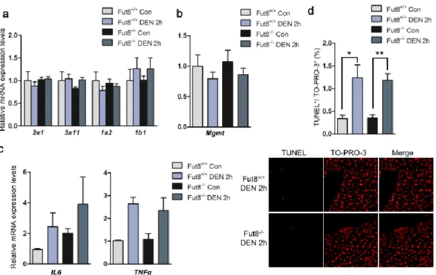
DEN itself does not exert carcinogenicity. It needs to be bioactivated by cytochrome P450 (CYP) enzymes in the liver (58). For instance, CYP2e1-deficient mice showed lower tumor incidence and multiplicity compared with wild-type mice for DEN-induced hepatocarcinogenesis (59). After activated, DEN resulted in DNA-adducts which forming through an alkylation mechanism, and the alkylated DNA-adducts can be removed by a DNA repair gene O
6-methylguanine-DNA methyltransferase (Mgmt) (60). To check the early effects of DEN in Fut8
+/+and Fut8
-/-mice, semi-quantitative or quantitative PCR analysis of mRNAs encoding the CYP enzyme (Figure 3a) and Mgmt (Figure 3b) genes was performed at 2 h after DEN injection. There was no significant difference in the transcriptional levels of either CYP genes or DNA repair gene between Fut8
+/+and Fut8
-/-mice. DEN-induced tumor formation was further associated with substantial and marked induction of proinflammatory chemokines within the livers of mice. After DEN injection, IL-6 and TNF-α were induced, resulting in the cytokine-driven compensatory proliferation (61). Quantitative PCR analysis of livers after DEN injection revealed an increase in IL-6 expression in both Fut8
+/+and Fut8
-/-mice, although the expression level in Fut8
+/+control mice was lower than that in Fut8
-/-control ones.
The mRNA expression levels of TNFα were also induced after DEN injection, but there was no
significant difference between the Fut8
+/+and Fut8
-/-mice (Figure 3c). Moreover, TUNEL
assay for the livers showed the similar results (Figure 3d). These data demonstrate that the
expression of Fut8 may not affect the acute phase response to DEN.
33
Figure 3. No big difference in the acute response was observed between the Fut8+/+ and Fut8-/- mice after DEN injection.
(a) Semi-quantitative PCR analysis of cytochrome P450 enzymes after DEN injection (n=3-5). (b) Quantitative PCR analysis of the DNA repairing gene (Mgmt) after DEN injection (n=3-5). (c) The mRNA expression levels of IL-6 and TNFα in the liver were detected by quantitative PCR after DEN injection at 2 hours (n=3-5). (d) TUNEL analysis was performed using the frozen liver tissues (10μm thick, green), the nuclei were labeled by TO-PRO-3 iodide (blue). The quantitative data were obtained from at least 3 mice. **, P<0.01, *, P<0.05, compared to the control mice.
2.3.4 Cell proliferation induced by DEN/PB treatment is inhibited in the Fut8
-/-mice
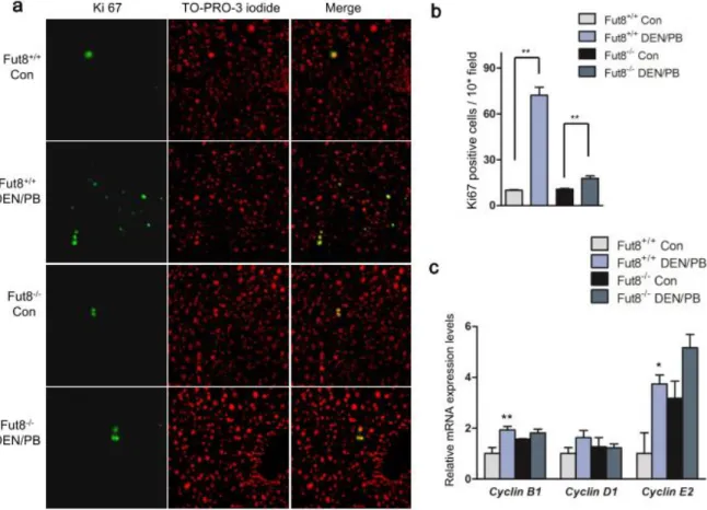
Cell proliferation plays important roles in HCC process (41, 62). In order to find the possible
mechanisms for the differences in tumorigenesis described above, we carried out Ki67
immunostaining for the frozen liver tissues of Fut8
+/+and Fut8
-/-mice. Without DEN/PB
treatment, both Fut8
+/+and Fut8
-/-mice showed few positive cells in the liver, and there was no
significant difference between the control groups. After chemical induction, the Fut8
+/+mice
showed a significant increase by more than ~6.5-fold in positive immunostaining as compared
with the untreated control. However, only a 2-fold increase was observed in Fut8
-/-mice
(Figure 4a and 4b). To further confirm the difference in cell proliferation after DEN/PB
34
induction between Fut8
+/+and Fut8
-/-mice, qPCR analysis of mRNAs encoded by cell cycle control genes was performed. The expression levels of cyclin mRNAs, including Cyclin B1, Cyclin D1 and Cyclin E2, were up-regulated in Fut8
+/+mice after DEN/PB treatment, and the elevations in gene expression were attenuated by the ablation of Fut8 (Figure 4c). These results indicate that Fut8 may influence the progress of DEN/PB induced HCC by affecting the cell proliferation.
Figure 4. Knockout of Fut8 led to a suppression of cell proliferation.
(a) Immunostaining for liver tissues of Fut8+/+ and Fut8−/− mice using anti-Ki67 antibody (40×field). The positive cells of the immunostaining were labeled with the green spots (left panel), and the nuclei were labeled by TO-PRO-3 iodide (red spots, middle panel). (b) The quantitative data were obtained from at least 3 mice in each group (10×field). **, P<0.01, compared to the Fut8+/+ mice. (c) qPCR Analysis of cell cycle control genes-encoded mRNAs. **, P<0.01, *, P<0.05, compared to the control mice. n=3-4.