総 説
循環系の基礎と臨床
(6)動脈硬化―病因と慢性腎臓病の関与
東京女子医科大学東医療センター内科 オ ガ ワ テ ツ ヤ小川 哲也
(受理 平成 29 年 11 月 7 日)Circulatory System: Basic and Clinical Research
(6) Arteriosclerosis: Pathogenesis and Involvement in Chronic Kidney Disease
Tetsuya OGAWA
Department of Medicine, Tokyo Women s Medical University Medical Center East
Arteriosclerosis is usually classified into atherosclerosis and arteriosclerosis. Nephrosclerosis and renal ar-tery stenosis are important regarding arteriosclerosis in the kidney. Nephrosclerosis advances because of hyper-tension and aging. Glomerulosclerosis includes two types of abnormalities that are complete opposites, namely ischemia-related sclerosing lesions and sclerosing lesions with glomerular hypertension. Furthermore, it is thera-peutically important to decide which of these is the primary pathological condition. Moreover, 90 % of renal ar-tery stenosis cases are atherosclerotic. Activation of the renin-angiotensin-aldosterone system by hypoperfusion of the stenotic kidneys causes renal contraction and renal fibrosis in the long term. Renal artery stenosis worsens the prognosis because of the occurrence of cardiovascular events.
Arteriosclerosis in chronic kidney disease (CKD) is a promoting/aggravating factor of CKD itself. The pro-gression of arterial wall sclerosis of the aorta is a characteristic finding of CKD. For diagnosing arteriosclerosis, it is common to perform carotid ultrasonography, imaging diagnostic modalities such as computed tomography/ magnetic resonance imaging, structure evaluation of the vessels, and vascular function testing of the cardio-ankle vascular index, pulse-wave velocity, and ankle-brachial index. Recently, the relationship between intestinal bacte-ria and arteriosclerosis has garnered attention. Trimethylamine N-oxide (TMAO) in blood has been reported to promote arteriosclerosis, and TMAO is produced by the metabolism of trimethylamine in the liver, which is a degradation product of dietary choline by intestinal bacteria.
Key Words: atherosclerosis, arteriosclerosis, nephrosclerosis, renal artery stenosis, intestinal bacteria
はじめに
動脈硬化は発症部位からアテローム性(粥状)動
脈硬化症(atherosclerosis),メンケベルグ型中膜硬
化症(Moncheberg s medial sclerosis),細小動脈硬化
症(arteriolosclerosis)に分類される
1).これらは,相
互に関連しながら動脈硬化病変を形成している.た
だし,メンケブルグ型中膜硬化症は筋型動脈に出現
する頻度が少なく石灰化沈着として捉えられること
が多いためメンケブルグ型中膜石灰化と表現される
こともある.通常,動脈硬化はアテローム型動脈硬
:小川哲也 〒116―8567 東京都荒川区西尾久 2―1―10 東京女子医科大学東医療センター内科 Email: [email protected] doi: 10.24488/jtwmu.87.6_151Copyright Ⓒ 2017 Society of Tokyo Women s Medical University
! # $ 東女医大誌 第 87 巻 第 6 号 頁 151∼159 平成 29 年12月 " # %
Fig. 1 Histological features of benign nephrosclerosis
a) Arteriolar hyaline change in benign nephrosclerosis (arrow, PAS staining, original magnification, ×400) b) Intimal thickening of the interlobular arteries in be-nign nephrosclerosis
(PAS staining, original magnification, ×400)
c) Global glomerular sclerosis in benign nephrosclerosis (PAS staining, original magnification, ×400)
a
a
b
b
c
c
化と細動脈硬化症に分けられている
2)が,慢性腎臓
病,特に透析患者ではメンケブルグ型中膜石灰化を
来すことが多い
3).本稿では,腎血管および腎疾患を
中心とした動脈硬化の病理,動脈硬化の臨床像およ
び診断,動脈硬化の関連因子のトピックスについて
概説する.
動脈硬化の病理
1.正常な動脈構造と機能
一般に動脈は内膜,中膜,外膜の 3 層構造を有し
ており,それぞれ層と層との間には内弾性板と外弾
性板が存在する.内膜は 1 層の内皮細胞と内皮細胞
の基底膜で構成されている.中膜には多数の弾性線
維や平滑筋が含まれ,外膜は線維組織で構成されて
いる.3 層の構造の特徴および血管のサイズから弾
性動脈,筋型動脈,小動脈および細動脈に分類され
る.弾性動脈は弾性線維が豊富で弾力性に富み,大
動脈,肺動脈など心臓に近く心臓のポンプ作用の補
助的なポンプ作用を果たす.筋型動脈,小動脈およ
び細動脈では中膜の平滑筋が発達しており,この平
滑筋が拡張と収縮をすることにより血流を調整す
る
4).冠動脈および腎動脈は筋型動脈に分類される.
小動脈と細動脈は抵抗血管と呼ばれ臓器の血圧を減
少させると同時に全身血圧にも影響を与える.小動
脈は平滑筋層が最大 8 層程度だが,細動脈は 1∼2
層とされている.腎内動脈では,葉間動脈が筋型動
脈,細い小葉間動脈と輸入細動脈は細動脈に相当す
る
5).
2.細小動脈硬化症(arteriolosclerosis)
組織 像 は,①hyaline arteriolosclerosis/arteriolar
hyalinosis(硝子化),②intimal
thickening,③hyper-plastic
arteriolosclerosis,④急性で重症な障害時(ac-ceralated hypertension)に出現する組織像,に分類
される
1).
①Hyaline arteriolosclerosis/arteriolar hyalinosis
(硝子化)
PAS 染色強陽性,HE 染色で好酸性均一な物質で
内皮下から中膜にかけて存在し,免疫蛍光法で C3,
C1q,IgM が病変に沈着することがある(Fig. 1-a).
硝子化は内皮細胞障害により内皮の透過性が高ま
り,その結果内皮下の貯留した血清の浸出物と思わ
れている
5).糖尿病,高血圧
6),加齢
7),肥満,高尿酸
血症
8)などで出現する.
②Intimal thickening
平滑筋の増殖,線維化,内弾性板(弾性線維束)の
重複による病変である(Fig. 1-b).内弾性板の重複は
長期間の高血圧状態を反映している
9).
③Hyperplastic arteriolosclerosis
平滑筋とその周辺の基底膜様物質が層状に増殖す
ることにより内腔が狭窄する病変であり,onion skin
lesion と呼ばれている
5).
④急 性 で 重 症 な 障 害 時(acceralated
hyperten-sion)に出現する組織像
内膜の粘液腫状の変化(滲出性変化),内皮下浮腫,
内皮細胞の脱落や腫大,壊死,フィブリノイドや破
砕赤血球の出現,血栓性微小血管症(thrombotic
microangiopathy:TMA)などが観察される
5).
3.アテローム型動脈硬化
粥状硬化とは主として動脈の内膜に限局性に起こ
る変化で,脂質,産生ムコ多糖類,血液とそれに由
来する物質,線維性結合識の増殖と石灰沈着などの
種々の組み合わせからなる病変で,中膜の変化を伴
う.内皮細胞に機能障害や傷害がもたらされると,
接着分子の発現,白血球の遊走・接着,内皮細胞層
の透過性亢進が起こり,単球由来のマクロファージ
や T リンパ球などの炎症細胞の浸潤が増加する
10).
血漿成分には平滑筋細胞遊走および増殖因子が含ま
れており平滑筋細胞の遊走および増殖をもたらし,
内膜の細胞線維性肥厚巣が形成される.
腎疾患と動脈硬化
1.腎硬化症
腎硬化症は,糖尿病性腎症,慢性糸球体腎炎に次
ぐ 3 番めに多い新規透析導入の原疾患であり,高血
圧や高齢化により進展し経年的に増加傾向であるた
め腎疾患において重要な疾患である.弓状動脈およ
び小葉間動脈を含む小動脈や輸入細動脈の血管構築
の変化により起こる.圧負荷,容量負荷による血圧
上昇による内皮細胞障害によって弾性線維,膠原線
維,筋線維芽細胞の増加,増殖を来し小動脈の内膜
肥厚をもたらす.また輸入細動脈では内皮の透過性
が亢進し血漿成分が内皮下に浸み込み硝子化をもた
らす.内膜肥厚により内腔が狭小化すると末梢では
間質の線維化とともに炎症性単核球細胞の浸潤,尿
細管の萎縮を認め糸球体硬化に進展する.よって高
血圧性腎硬化症に特徴的な病理所見は,全節性硬化,
細動脈硝子化,小葉間動脈,弓状動脈の血管内膜肥
厚,間質線維化・尿細管萎縮と考えられる
11)(Fig. 1
a-c).
腎内小細動脈硬化症は最終的には自身の灌流する
糸球体の硬化病変を引き起こすが,糸球体硬化症病
変には虚血に関連した硬化病変と糸球体高血圧に関
連した硬化病変の全く逆の 2 種類の糸球体血行動態
異常が存在することが示唆されている
12).輸入細動
脈にみられる硝子化病変が高度で内腔の狭小化を伴
う場合には虚血性障害,硝子化病変が軽度である場
合には輸入細動脈の収縮障害や輸出細動脈の過剰収
縮による糸球体高血圧をもたらしている可能性が報
告されている(Fig. 2)
13).この 2 種類の病態が,1
個の腎臓に同時に存在しており病態によって分布の
割合が異なっている.糸球体高血圧主体の病態にお
いては厳格な降圧と renin-angiotensin:RA 系阻害
薬使用による輸出細動脈の拡張による糸球体内圧の
低下が重要であるが,虚血主体の病態においては過
度な降圧や RA 系阻害薬の使用は腎障害の進展を促
進させるおそれがあると考えられる.臨床において,
糸球体高血圧主体であるか虚血が主体であるかを見
極めることが治療上,非常に重要となる.蛋白尿の
有無が鑑別に有用である可能性が考えられる.すな
わち,蛋白尿陰性の病態では,虚血の病態が主体で
あると考える必要がある.特に高齢者では,高度腎
内小細動脈病変を有する高齢者が少なくないため,
蛋白尿陰性で血圧コントロールが良好にもかかわら
ず腎機能が悪化する症例では,虚血の病態を念頭に
おいて降圧目標の上方への設定,RA 系阻害薬の減
量の中止,カルシウム(Ca)拮抗薬への変更などを
考慮する必要がある
13).
腎硬化症には,拡張期血圧が 120∼130 mmHg 以
上が持続し,腎機能障害の急速な進行および未治療
な状態が持続すると心不全,高血圧脳症,脳出血ま
で発展する予後不良な病態があり,これを加速型―悪
性高血圧とし,上記の病態を良性腎硬化症として区
別している
14).
悪性高血圧の病態の機序としては,急激な血圧上
昇により輸入細動脈が障害され自動調整機序が破綻
することで高い圧が糸球体に直接伝播することで糸
球体障害を来す.糸球体高血圧により管腔内にヒア
リン様物質の充満を伴い糸球体硬化をもたらす.一
方で,輸入細動脈は内皮障害に伴い内皮下の浮腫や
増殖性内膜炎,フィブリノイド壊死がみられ,時間
の経過と共に同心円状の線維性内膜増殖(onion-skin
lesion)を伴う.また,内皮障害に起因すると思われ
る TMA を合併すると内腔狭窄を助長して灌流糸
球体の虚血性障害と尿細管間質の虚血性障害をもた
らす.治療としては,著明な血圧上昇の病態に RA
系の亢進の関与が示唆されており,RA 系阻害薬の
治療効果が期待される.悪性高血圧は,すでに高血
圧罹患期間が長く細動脈のリモデリングが進行して
いる症例が多いため,急速な降圧による臓器虚血を
予 防 す る た め 降 圧 目 標 は 拡 張 期 血 圧 100∼110
Fig. 2 Mechanism of glomerularsclerosis related to hypertension, including two types of
abnormalities
Glomerulosclerosis includes two types of abnormalities that are complete opposites, namely sclerosing lesions with glomerular hypertension and ischemia-related sclerosing lesions. Furthermore, the distribution in a single kidney varies depending on the patho-logical condition. (Adapted from reference 13)
Aging
Disorder of lipid metabolism Smoking, Hyperuricemia
Afferent arteriole sclerosis
Dilatation of afferent arteriole Stenosis of the afferent arteriole
Glomerular hypertension
Glomerular ischemia
Proteinuria Tubulointerstitial
lesion
Glomerulosclerosis
Hypertension
Kohagura K et al: Benign nephrosclerosis and malignant nephrosclerosis. Jpn J Nephrol 58 (2): 85-91, 2016 (in Japanese)
mmHg にとどめることが推奨されている
13).
2.腎動脈狭窄症
腎動脈狭窄症は,腎動脈内腔の狭窄を認める病態
であり,その原因として 90 %が動脈硬化性(粥状硬
化性 atherosclerotic)であり
15)大動脈壁に生じたプ
ラークが腎動脈に進展して狭窄することが多く腎動
脈本幹の中枢側に頻発する.その他の原因として線
維筋性異形成(fibromuscular dysplasia:FMD),大
動脈解離,大動脈炎症候群,血栓塞栓症などがある.
腎動脈狭窄は狭窄側の腎の低灌流をもたらし,その
結果としてレニン・アンジオテンシン・アルドス
テ ロ ン 系(renin-angiotensin-aldosterone system:
RAAS)の賦活化により,長期的には腎萎縮および腎
線維化をもたらす
16).腎動脈狭窄によって生じる高
血圧症が腎血管性高血圧症であるが,血圧が高くな
くても慢性の腎虚血により虚血性腎症をもたらし,
無症候性であっても心血管イベントの発生により予
後を悪化させる.腎動脈狭窄症が潜在的に存在して
いる場合に突然の原因不明の肺水腫・うっ血性心不
全,原因不明の腎機能低下,冠動脈多枝病変,難治
性不安定狭心症を来すことがあり,これらの病態で
は腎動脈狭窄の存在を積極的に疑う必要がある
15).
診断は腎動脈のドップラー法の臨床的有用性が高
く,最大収縮期血流速度(peak systolic velocity)が
180∼200 cm/sec 以上で,かつ腎動脈―大動脈血流速
度比(renal-aortic ratio)が 3.5 以上である場合に有
意な腎動脈狭窄と診断する
17).治療は,腎血管性高血
圧を有する場合には降圧療法が有効である.ただし,
両側腎動脈狭窄の場合には RAAS 阻害薬は腎機能
悪化を来す場合がある.腎動脈狭窄に対する血行再
建法としては腎動脈ステントが第一選択となる.特
に腎動脈狭窄による神経液性因子の変化で生じる
cardiac disturbance syndrome と呼ばれている心不
全・肺水腫例が積極的適応である
18).
3.慢 性 腎 臓 病(chronic kidney disease:CKD)
と動脈硬化
CKD における動脈硬化には,粥状硬化,腎不全に
比較的特徴的といわれた中膜硬化,高血圧性動脈硬
化指標としての細動脈硬化があげられる.動脈硬化
は,それ自体が CKD の進展・増悪因子となってい
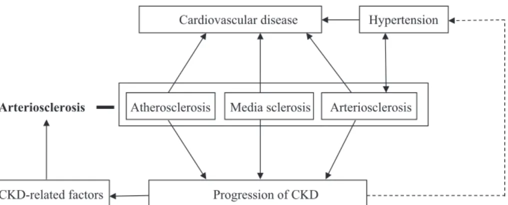
Fig. 3 Arteriosclerosis and chronic kidney disease
Arteriosclerosis in chronic kidney disease (CKD) is a promoting/aggravating factor of CKD itself, and CKD-related factors also promote arteriosclerosis. Arteriosclerosis causes cardio-vascular diseases and progression of CKD. (Adapted from reference 19)
Cardiovascular disease Hypertension
Arteriosclerosis Atherosclerosis Media sclerosis Arteriosclerosis
CKD-related factors Progression of CKD
Nakayama M et al: Epidemiology and clinical significance of nephrosclerosis. Jpn J Nephrol 58 (2): 92-96, 2016 (in Japanese)
Fig. 4
a) Aortic arch calcification by three-dimensional (3-D) analysis of computed tomography in a hemodialysis patient.
b) Calcification of the inner surface of the aortic arch as shown in the 3-D computed tomog-raphy analysis in a hemodialysis patient.
a
a
b
b
ることから,CKD と動脈硬化は相互に増悪因子とな
り病態の悪循環を形成している(Fig. 3)
19).末期腎不
全症例では透析導入時に冠動脈病変が高頻度に存在
するという報告からも CKD は粥状動脈の促進因子
となっている可能性が高いと考えられる.特に,
CKD 患者では,動脈硬化危険因子として古典的な危
険因子だけでなく,CKD に関連した危険因子の存在
により動脈硬化が進展していると考えられる
19).
しかし,新規透析患者の基礎疾患として粥状動脈硬
化の腎疾患は少なく必ずしも粥状硬化を CKD の動
脈硬化の普遍的な基本病態と捉えることはできな
い
20).
大動脈の動脈壁硬化の進行は CKD の特徴的な所
見とされ,さらに,動脈硬化の進行に伴い末梢動脈
疾患(peripheral arterial disease:PAD)も CKD で
頻度が高い.また,血管中膜の石灰化は CKD 患者の
血管硬度上昇の重要な要因となっている
21).CKD
stage5D の末期腎不全患者において,血管石灰化が
顕著である
22)(Fig. 4).血管石灰化の病理学的特徴
は,動脈内膜の粥状硬化巣に起こる石灰化と動脈中
膜の平滑筋層に起こるメンケブルグ型石灰化の 2 種
類がある
23).血管石灰化には Table 1 に示すような
因子が関連しているが,特に Ca とリン(P)代謝の
異常が重要である
24).メンケブルグ型石灰化では,
Ca・P 代謝異常により平滑筋細胞が骨芽細胞様に
変化することが関係していると報告されている
25).
高 P 血症は,ナトリウム依存性リン酸トランスポー
ターである Pit-1 を介して直接的に血管障害を引き
Fig. 5 The Kaplan-Meier event-free survival analysis of cardiovascular events according
to aortic arch calcification (AoAC) grade in the hemodialysis patients
The Kaplan-Meier analysis revealed that the incidence of cardiovascular events was sig-nificantly higher in the patients with higher AoAC grade (grades 2 and 3) than in those with grade 0 or 1.
p
Table 1 Risk factors of vascular calcification
Clinical Age
Duration of dialysis Kidney function/uremia Diabetes
Known coronary artery disease Abnormal bone
Biochemical Hyperphosphatemia Hypertension
High parathyroid hormone Low fetuin A
Increased aldosterone Oxidative stress Low pyrophosphate
Decreased matrix Gla protein
Decreased bone morphogenic protein 7 Medications Calcium-containing phosphate bindings
High-dose vitamin D
Coumadin (decreased active MGP)
起こしている可能性があるが,過剰な Ca と P 負荷
が血管石灰化にとって最も重要な因子である
26).血
中ビタミン D 値は早期の CKD ですでに低下を認め
ており,25 ヒドロキシビタミン D(25(OH)D)低
値は死亡率を高めている
27).一方,25(OH)D は
RAAS を抑制し動物モデルでの血管石灰化を抑制
することが報告されている
28).fibroblast growth
fac-tor(FGF)-23 は骨芽細胞で産生される因子であり P
およびビタミン D の調整を行っているが,この調整
は,FGF-23 と FGF 受容体および老化遺伝子である
Klotho と結合することにより作用が発揮される.
CKD では FGF-23 は高値,Klotho は低値を示してお
り,動物実験および臨床研究では FGF-23 および
klotho の異常が血管石灰化に密接に関与している
と報告されている
29).近年,冠動脈や大動脈の血管石
灰化が,CKD 患者の発症・進展に寄与していること
が報告されている
30).血管石灰化は,動脈の弾力性を
低下させ,後負荷の増大による左室肥大を惹起し
31),
さらに左室拡張能の低下にも寄与する
32)ため心血管
疾患(cardiovascular disease:CVD)の発症に関与
すると考えられている.さらに,大動脈弓部の血管
石灰化が総死亡および心血管死亡の予後予測因子で
あり
33),さらに大動脈弓部血管石灰化の程度が強い
ほど心血管イベントが多い
34)(Fig. 5).
動脈硬化の診断
動脈硬化の診断として頸動脈エコー検査,com-puted tomography(CT)・magnetic resonance
im-aging(MRI)などの画像診断,血管機能検査が非侵
襲的な検査として用いられてきている.
頸動脈エコー法は,早期動脈硬化の指標として有
用であり心疾患のサロゲートマーカーとして脳梗
塞・心筋梗塞のハイリスク群の層別化に広く用いら
れている
35).また,頸動脈肥厚度(intima media
thick-ness:IMT)をサロゲートマーカーとして用いて,
酸化ストレス,慢性炎症反応,プラークの破綻と血
栓形成の病態に遺伝的な素因が深く関与することが
報告されている
36).さらに心血管イベントとの関連
についても IMT がハイリスク群の層別化に有用で
あり,我々も,維持透析患者においても IMT 高値群
において脳血管障害が有 意 に 高 い こ と を 報 告 し
た
37).
動脈硬化症を動脈瘤や閉塞性動脈硬化症などの動
脈の拡張と閉塞をきたす病態として認識することも
可能であり,その診断には CT や MRI が広く用いら
れている.大動脈瘤は大動脈壁が脆弱化し,正常径
の 1.5 倍以上に拡張した状態であり胸部大動脈瘤は
4.5 cm 以上,腹部大動脈瘤は 3 cm 以上と定義され
る
38).動脈硬化では紡錘状の真性瘤が多いが,囊状瘤
では急速に増大する危険があり注意が必要である.
胸部大動脈瘤は,先天性心疾患,結合織異常および
弁疾患の割合が多い上行大動脈瘤と,動脈硬化の割
合が多い弓部および胸腹部大動脈瘤とがある.腹部
大動脈瘤は,大半が腎動脈分岐部以下に発生し,動
脈硬化に伴う腸骨動脈末梢の動脈硬化による血液の
反射波(反跳波)も影響している.閉塞性動脈硬化
症(arteriosclerosis obliterans:ASO)の画像診断と
して超音波検査は狭窄部の性状や壁の石灰化の程度
を簡便に観察可能である.MDCT(multi
detector-row CT)は大動脈から下肢動脈まで短時間に詳細な
壁 性 状 の 評 価 が 可 能 で あ る.MR angiography
(MRA)は造影 MRA と非造影 MRA があるが,腎障
害がある場合には MRI の造影剤であるガドリニウ
ム造影剤による腎性全身性線維症(nephrogenic
sys-temic fibrosis)の発生が報告されている.非造影
MRA と し て time-of-flight 法 と 新 た な fresh blood
imaging(FBI)法が血流評価として用いられてい
る
39).
動脈硬化症の診断には血管の構造評価と共に,画
像診断による血管機能検査が用いられており,心臓
足 首 血 管 指 数(cardio-ankle vascular index:
CAVI),脈 波 伝 搬 速 度(pulse wave velocity:
PWV),足関節上腕血圧比(ankle-brachial pressure
index:ABI)が繁用されている.
CAVI は,血圧に依存しない動脈の硬さ(arterial
stiffnesss)を測定する指数であり高血圧,糖尿病,
脂質異常,メタボリックシンドローム,肥満患者,
喫煙,腎不全で上昇している
40).PWV は,血液が左
室から大動脈に押し出された際に発生する波動が血
管壁を伝わる速度を示し,心血管事故の予測因子の
動脈硬化指数として用いられることが多い
41).動脈
硬化の進展に伴い下肢の動脈の閉塞性病変を生じる
が,この末梢動脈疾患(peripheral arterial disease:
PAD)の診断に ABI が用いられる.ABI は足関節血
圧と比較的動脈硬化の起こりにくい上腕動脈の血圧
の比で表し,1.0∼1.4 は正常値,0.9∼0.99 は境界領
域,0.9 以下で有意な閉塞性病変の存在を疑う.1.4
以上は,下肢動脈の石灰化が強く偽の上昇と判断さ
れる.
動脈硬化と腸内細菌
近年,腸内細菌が注目されているが動脈硬化との
関連においても注目されている.卵・牛肉・チー
ズ・エ ビ な ど に 含 ま れ る ホ ス フ ァ チ ジ ル コ リ ン
(phosphatidylcholine:PC)が消化管で代謝されコ
リン(choline)となり,さらに腸内細菌で代謝され
てトリメチルアミン(trimethlamin:TMA)となる.
TMA は肝臓で代謝されてトリメチルアミン N オ
キシド(trimethylamine N-oxide:TMAO)となる
が,この TMAO の血中濃度が高いほど心血管イベ
ントの発生が高いことが臨床研究で示された
42).さ
らに,動物実験においても動脈硬化モデルマウスで
あるアポリポ蛋白 E 遺伝子欠損マウス(apoE-KO
マウス)への PC 投与で,普通食投与より TMAO
の血中濃度が上昇し動脈硬化が悪化するが,抗生物
質 で 腸 内 細 菌 を 除 去 す る と PC 投 与 で の 血 中
TMAO の増加は抑制され動脈硬化の悪化を抑制す
ることが示されている.TMAO の動脈硬化促進の
作用機序として,マクロファージの泡沫化の増加,
コレステロールの逆転送系の抑制,血小板機能の亢
進が関与していることが示されている
43)44).泡沫化マ
クロファージが集積すると,細胞外脂質の集積と相
まって次第にアテローム(粥腫)が形成される.現
在のところ Pseudomonas や Candida が TMA 産生
に関わることが明らかになっているが
45),腸内細菌
層のうちどの菌種が主として TMA 産生に働いて
いるのか不明である.
謝 辞 病理写真の選定に関してご指導頂きました昭和大学 医学部顕微解剖学 本田一穂教授に深謝申し上げます. 開示すべき利益相反状態はない. 文 献1)Jannette JC, D Agati VD, Olson JL et al: Renal disease caused by hypertension. In Heptinstall s Pa-thology of the Kidney 7th ed, pp849―896, Lippincott Williams & Wilkins, Philadelphia (2014)
2)細動脈硝子化.「糖尿病性腎症と高血圧腎硬化症の
病理診断への手引き」(和田隆志,湯澤由紀夫監),
pp54―60,東京医学社,東京(2015)
3)London GM, Guerin AP, Marchais SJ et al: Arte-rial media calcification in end-stage renal disease : impact on all-cause and cardiovascular mortality. Nephrol Dial Transplant 18: 1731―1740, 2003 4)Schiffrin EL : Vascular remodeling in
hyperten-sion: mechanisms and treatment. Hypertension 59: 367―374, 2012
5)上杉憲子:動脈硬化の原理.日腎会誌 58(2):97― 103,2016
6)Ninomiya T, Kubo M, Doi Y et al: Prehyperten-sion increases the risk for renal arteriosclerosis in autopsies: the Hisayama study. J Am Soc Nephrol
18: 2135―2142, 2007
7)Rule AD, Amer H, Cornell LD et al: The associa-tion between age and nephrosclerosis on renal bi-opsy among healthy adults. Ann Intern Med 152 : 561―567, 2010
8)Kohagura K, Kochi M, Miyagi T et al: An associa-tion between uric acid levels and renal arteriolopa-thy in chronic kidney disease: a biopsy-based study. Hypertens Res 36: 43―49, 2013
9)Kubo M, Kiyohara Y, Kato I et al: Risk factors for renal glomerular and vascular changes in an autopsy-based population survey : the Hisayama study. Kidney Int 63: 1508―1515, 2003
10)稲葉真由美,杉岡憲一,上田真貴子:動脈硬化症: 診断と治療の進歩 II.促進因子 病因と病態 2. 病理面 か ら:冠 動 脈 を 中 心 に.日 内 会 誌 102: 297―304,2013 11)古市賢吾,和田隆志:腎硬化症の定義,臨床と病理. 日腎会誌 58(2):80―84,2016
12)Hill GS, Heudes D, Jacquot C et al: Morphometric evidence for impairment of renal autoregulation in advanced essential hypertension. Kidney Int 69 : 823―831, 2006 13)古波蔵健太郎,大屋祐輔:良性腎硬化症と悪性腎硬 化症.日腎会誌 58(2):85―91,2016 14)特殊条件下高血圧.「高血圧治療ガイドライン 2014」 (日本高血圧学会治療ガイドライン作成委員会編), pp108―114,ライフサイエンス出版,東京(2014) 15)Safian RD, Textor SC : Renal-artery stenosis. N
Engl J Med 344 (6): 431―442, 2001
16)Caps MT, Zierler RE, Polissar NL et al: Risk of atrophy in kidneys with atherosclerotic renal ar-tery stenosis. Kidney Int 53: 735―742, 1998
17)Olin JW, Piedmonte MR, Young JR et al: The util-ity of duplex ultrasound scanning of the renal arter-ies for diagnosing significant renal artery stenosis. Ann Intern Med 122: 833―838, 1995
18)Hirsch AT, Haskal ZJ, Hertzer NR et al: ACC / AHA 2005 guidelines for the management of pa-tients with peripheral arterial disease ( lower ex-tremity, renal mesenteric, and abdominal aortic). J Am Coll Cardiol 47: 1239―1312, 2006
19)中山昌明,田中健一,旭 浩一:CKD における動脈 硬化とその危険因子.日腎会誌 58(2):92―96, 2016
20)Drüeke TB, Massy ZA: Atherosclerosis in CKD:
differences from the general population. Nat Rev Nephrol 6: 723―735, 2010
21)Briet M, Boutouyrie P, Laurent S et al: Arterial stiffness and pulse pressure in CKD and ESRD. Kidney Int 82: 388―400, 2012
22)Goodman WG, London G, Amann K et al: Vascu-lar calcification in chronic kidney disease. Am J Kidney Dis 43: 572―579, 2004
23)London GM, Guérin AP, Marchais SJ et al: Arte-rial media calcification in end-stage renal disease : impact on all-cause and cardiovascular mortality. Nephrol Dial Transplant 18: 1731―1740, 2003 24)Jono S, McKee MD, Murry CE et al: Phosphate
regulation of vascular smooth muscle cell calcifica-tion. Circ Res 87: E10―E17, 2000
25)Moe SM, Chen NX : Pathophysiology of vascular calcification in chronic kidney disease. Circ Res 95: 560―567, 2004
26)Nitta K : Vascular calcification in patients with chronic kidney disease. Ther Apher Dial 15 (6): 513― 521, 2011
27)Barreto DV, Barreto FC, Liabeuf S et al: Vitamin D affects survival independently of vascular calcifi-cation in chronic kidney disease. Clin J Am Soc Nephrol 4: 1128―1135, 2009
28)Mathew S, Lund RJ, Chaudhary LR et al: Vitamin D receptor activators can protect against vascular calcification. J Am Soc Nephrol 19: 1509―1519, 2008 29)Lanske B, Razzaque MS: Mineral metabolism and
aging: the fibroblast growth factor 23 enigma. Curr Opin Nephrol Hypertens 16: 311―318, 2007
30)Cannata-Andía JB, Rodríguez-García M,
Carri-llo-López N et al: Vascular calcifications :
patho-genesis, management, and impact on clinical out-comes. J Am Soc Nephrol 17 (12 Suppl 3): S267―S 273, 2006
31)Guérin AP, London GM, Marchais SJ et al: Arte-rial stiffening and vascular calcifications in end-stage renal disease. Nephrol Dial Transplant 15 : 1014―1021, 2000
32)Fujiu A, Ogawa T, Matsuda N et al: Aortic arch calcification and arterial stiffness are independent factors for diastolic left ventricular dysfunction in chronic hemodialysis patients. Circ J 72: 1768―1772, 2008
33)Ogawa T, Ishida H, Akamatsu M et al: Progres-sion of aortic arch calcification and all-cause and cardiovascular mortality in chronic hemodialysis patients. Int Urol Nephrol 42: 187―194, 2010 34)Inoue T, Ogawa T, Ishida H et al: Aortic arch
cal-cification evaluated on chest X-ray is a strong inde-pendent predictor of cardiovascular events in chronic hemodialysis patients. Heart Vessels 27 (2): 135―142, 2012
35)O Leary DH, Polak JF, Kronmal RA et al: Carotid-artery intima and media thickness as risk factor for myocardial infarction and stroke in older adults: Cardiovascular Health Study Collaborative Research Group. N Engl J Med 340: 14―22, 1999 36)Katakami N, Saito M, Kaneto H et al: Combined
polymor-循環系の基礎と臨床―掲載予定― 執筆者 所属 テーマ 掲載号 澤田達男 病理学(第一) 1.脳の微小循環 87(1・2) 槍澤大樹 輸血・細胞プロセシング科 2.血管新生 87(1・2) 森本 聡 高血圧・内分泌内科 3.高血圧 87(3) 瀧田守親 薬理学 4.転移 87(4) 江 太一 解剖学・発生生物学 5.リンパ管発生 87(5) 小川哲也 東医療センター内科 6.動脈硬化(透析も含めた腎血管) 87(6)
phisms on the progression of carotid atherosclero-sis in Japanese type 2 diabetes. Atheroscleroatherosclero-sis 207 (1): 29―31, 2009
37)Sato M, Ogawa T, Sugimoto H et al: Relation of carotid intima-media thickness and silent cerebral infarction to cardiovascular events and all-cause mortality in chronic hemodialysis patients. Intern Med 51 (16): 2111―2117, 2012
38)高本眞一,石丸 新,上田裕一ほか:循環器病の診 断と治療に関するガイドライン(2004-2005 年度合 同研究班報告) 大動脈瘤・大動脈解離診療ガイド ライン(2006 年改訂版).Circ J 70(suppl IV): 1569―1646,2006
39)岡田宗正,松永尚文:動脈硬化症:診断と治療の進 歩 トピックス III.診断の進歩 3.CT(三次元再 構成画像),MRI.日内会誌 102:325―334,2013 40)Shirai K, Hiruta N, Song M et al: Cardio-ankle
vas-cular index (CAVI) as a novel indicator of arterial stiffness: theory, evidence and perspectives. J Athe-roscler Thromb 18 (11): 924―938, 2011
41)Vlachopoulos C, Aznaouridis K, Stefanadis C : Prediction of cardiovascular events and all-cause mortality with arterial stiffness : a systematic re-view and meta-analysis. J Am Coll Cardiol 55 (13): 1318―1327, 2010
42)Wang Z, Klipfell E, Bennett BJ et al: Gut flora me-tabolism of phosphatidylcholine promotes cardio-vascular disease. Nature 472: 57―63, 2011
43)Tang WH, Wang Z, Levison BS et al: Intestinal microbial metabolism of phosphatidylcholine and cardiovascular risk. N Engl J Med 368: 1575―1584, 2013
44)Zhu W, Gregory JC, Org E et al: Gut microbial me-tabolite TMAO enhances platelet hyperreactivity and thrombosis risk. Cell 165: 111―124, 2016 45)Liffourrena AS, Salvano MA, Lucchesi GI:
Pseu-domonas putida A ATCC 12633 oxidizes trimethyl-amine aerobically via two different pathways. Arch Microbiol 192 (6): 471―476, 2010