緒 言
特 発 性 肺 線 維 症(idiopathic pulmonary fibrosis:
IPF)において,呼吸困難が強い症例1),一定観察期間 における FVC や%DLco の漸減する症例2〜4),6 分間歩 行試験で desaturation をきたす症例5)や歩行距離が短い 症例6),網状病変および蜂巣肺が広範に広がっている症 例7)などが予後不良であることが報告されてきた.近年,
Cottin ら8)や Mejía ら9)により報告された,気腫を合併し
た特発性肺線維症(combined pulmonary fibrosis and emphysema:CPFE)は,予後不良とされている.一 方で,予後に差はなく,むしろ気腫合併例では年間の肺 活量および肺拡散能変化率が IPF のみの患者に比べて 軽度であるとする報告もみられ10),その予後ならびに予 後不良因子に関しては,一定の見解が得られていない.
そこで今回,気腫を合併した IPF を CPFE とし,その 臨床的特徴ならびに予後不良因子を検討した.さらに CPFE の発癌部位に関して,病理組織学的特徴を検討し た.
対象および方法
2003 年 4 月から 2009 年 10 月までに東邦大学医療セ ンター大森病院呼吸器内科に入院した気腫性病変と両肺 に蜂巣肺を呈する CPFE 患者(E 群,clinical IPF with emphysema:pathological IPF with emphysema=29 例:6 例)35 例を対象に,同時期に入院した気腫非合併 IPF 患者(F 群,clinical IPF:pathological IPF=48 例:
●原 著
気腫合併特発性肺線維症の臨床病理学的特徴
伊藤 貴文a 杉野 圭史a 坂本 晋a 黒崎 敦子b 植草 利公c 本間 栄a
要旨:気腫を合併した特発性肺線維症(idiopathic pulmonary fibrosis:IPF)を combined pulmonary fibro- sis and emphysema(CPFE)として,その臨床的特徴ならびに予後因子を明らかにし,次に CPFE の発癌 部位に関して病理組織学的特徴を明らかにすることを目的とした.過去 6 年間に,東邦大学医療センター 大森病院にて胸部 HRCT 検査を行った,気腫性病変と両肺に蜂巣肺を呈する CPFE 患者(E 群)35 例を対 象に,同時期に入院した気腫非合併 IPF 患者(F 群)57 例と比較し,臨床的特徴を検討した.次に原発性 肺癌合併のために外科的肺切除術を施行され,病理組織学的に E 群と診断された 6 例を対象に,気腫ある いは線維化と発癌の関連性について,F 群,肺気腫群と対比し病理組織学的にその特徴を検討した.患者背 景では E 群で有意に喫煙指数が高く,原発性肺癌の合併も多かった.予後は E 群で有意に不良で,予後不 良因子として肺気腫,原発性肺癌,肺高血圧症,composite physiologic index 増加ならびに急性増悪などが 挙げられた.また肺癌合併例を除いた両群においても,予後は E 群で有意に不良で,肺気腫の合併が単独 の予後不良因子であった.さらに E 群において,肺癌合併の有無別の予後に差は認めず,予後不良因子は,
急性あるいは慢性増悪であった.病理組織学的に原発性肺癌合併 E 群の発癌部位は気腫あるいは線維化病 変を伴った気腫に隣接しており,この線維化病変は線維芽細胞巣,肺胞虚脱および胸膜下病変が目立たず構 造破壊が顕著で,F 群にみられる線維化とは異なるものであった.E 群では F 群に比して原発性肺癌の合併 率が高いものの,肺癌合併の有無にかかわらず予後不良であった.病理組織学的には,E 群の発癌母地とし て F 群とは異なる線維化を伴った気腫の存在が重要であることが示唆された.
キーワード:気腫合併特発性肺線維症,特発性肺線維症,肺気腫,原発性肺癌
Combined pulmonary fibrosis and emphysema,Idiopathic pulmonary fibrosis,
Pulmonary emphysema,Primary lung cancer
連絡先:杉野 圭史
〒143‑8541 東京都大田区大森西 6‑11‑1
a東邦大学医療センター大森病院呼吸器内科
b虎の門病院放射線診断科
c 独立行政法人労働者健康福祉機構関東労災病院病理診
断科
(E-mail: [email protected])
(Received 12 Sep 2011/Accepted 30 Nov 2011)
9 例)57 例と比較し,はじめに臨床的特徴ならびに予後 不良因子を検討した.なお,気腫の診断は,HRCT 上 で壁のない,あるいは 1 mm 未満の壁厚を有する低吸収 領域が,上葉を中心に散在性に 25%以上分布するもの と定義した11).IPF の診断は,外科的肺生検あるいは American Thoracic Society/European Respiratory Soci- ety(ATS/ERS)の international consensus statement12)
および厚生労働省びまん性肺疾患研究班第 4 次診断基 準13)に準じた.画像所見の評価は,両側の肺葉ごとに honeycombing(HC)と ground glass opacity(GGO)
の程度を数値化し検討した14).また判定は放射線科医1人,
呼吸器内科医 2 人の同意によった.呼吸機能検査は CHEST 社製の Chestac-33 を使用し,拡散能は 1 回呼吸 法で測定した.推定収縮期肺動脈圧(estimated systolic pulmonary arterial pressure:esPAP)の測定は,室内 気吸入下で経胸壁心臓超音波を用いて行われ,安静時の esPAPが35 mmHgを超えるものを肺高血圧(pulmonary arterial hypertension:PH)と診断した15).また,com- posite physiologic index(CPI)は,[91−(0.65×%pre- dicted DLco)−(0.53×%predicted FVC)+(0.34×%pre- dicted FEV1.0)]により算出した.
次に原発性肺癌合併のために外科的肺切除術を施行さ れたE群6例について,F群(
n
=5)および肺気腫群(n
=5)を対照として,切除肺を用いて気腫あるいは線維化と発 癌の関連性について,hematoxylin and eosin(HE)染 色および Elastica van Gieson(EVG)染色を行い,光顕 で病理組織学的に比較検討した.なお,肺癌周囲の肺組 織におけるそれぞれの病理組織学的所見に関しては,以 下のように半定量的評価を行った.
(1)線維芽細胞巣,扁平上皮化生:200 倍 5 視野中 3 個以上の線維芽細胞巣あるいは扁平上皮化生を認める場 合を 3+,2 個を 2+,1 個を 1+,0 個を陰性(−)と した.
(2)平滑筋増生,II 型肺胞上皮細胞増生,腺様化生:
100 倍 5 視野中 4〜5 視野で線維化病変内に平滑筋増生,
II 型肺胞上皮細胞増生あるいは腺様化生を認める場合を 3+,2〜3 視野を 2+,1 視野を 1+,0 個を陰性(−)
とした.
(3)弾性線維増生:100 倍 1 視野あたり線維化病変内 の80%以上を占める場合を3+,50%以上80%未満を2+,
50%未満を 1+,増生のない場合を陰性(−)とした.
(4)肺胞構造の破壊,拡張:ルーペ像で明らかな肺胞 構造の破壊,拡張を認める場合を 3+,4 倍で認める場 合を 2+,10 倍で認める場合を 1+,0 個を陰性(−)
とした.
統計学的解析として,2 群間の比較には,Mann-Whit- ney
U
検定およびχ2検定を用い,予後因子ならびに危険因子の解析には Cox 比例ハザード回帰分析,ロジス ティック解析を用いた.また,肺癌合併の予測因子の解 析における smoking index,%TLC,%FVC の最適カッ トオフ値については,receiver operating characteristic
(ROC)線を作成することにより求めた.生存率は Ka- plan-Meier 法で算出し,その有意差検定には log rank test を用いた.成績は平均値±標準偏差で示し,有意差 水準を 5%未満とした.なお統計解析ソフトは Stat View(version 5.0,SAS Institute, Cary,NC,USA)
を使用した.
本研究は東邦大学医療センター大森病院倫理委員会で 承認されている(承認番号 22-36)。
結 果
Table 1 に示すように,患者背景では,E 群は男性で 多く,喫煙歴,smoking index,肺癌合併率はともに E 群で有意に高かった.また,肺癌組織型の内訳としては,
E 群では,扁平上皮癌(38.9%)が最多で,次いで小細 胞癌(22.2%),腺癌(16.7%)であった.一方,F 群では,
扁平上皮癌(12.5%),腺癌(12.5%),小細胞癌(25.0%)
で両群間に有意差は認められなかった.6 分間歩行試験 では,歩行距離(272±119 m vs. 279±134 m,p=NS),
Lowest SpO2(87.2±4.7% vs. 88.3±5.1%,
p
=NS),Δ
SpO2(8.1±3.7% vs. 7.3±4.6%,p
=NS),Borg scale(2.59±2.12 vs. 2.58±2.14,
p
=NS)ともに両群間で有意 差は認めなかった.画像所見では,HC score(9.3±3.0 vs. 11.4±3.9,p=0.008),GGO score(7.0±4.1 vs. 8.9±4.3,
p
=0.04)ともに F 群において有意に高かった.呼吸機 能検査では FVC,%FVC,FEV1.0,%TLC は E 群で有 意に高く,FEV1.0%,%DLco/VA は E 群で有意に低かっ た.さらに Fig. 1 に示すように,CPFE および IPF の 診断後の生存期間中央値は,E 群で 18ヶ月,F 群で 25ヶ月と F 群に比して E 群で有意に生存期間が短かっ た(p
=0.007).死因としては,急性増悪 19 例,肺癌 13 例,呼吸不全の進行 10 例,細菌性肺炎 4 例,心不全 3 例,その他,心筋梗塞 1 例,気胸 1 例,胃癌 1 例であった.
そこで予後不良因子を明らかにするためCox比例ハザー ド回帰分析を行ったところ,単変量解析では,年齢
(Hazard Ratio(HR)=1.001,
p
=0.96),性別(HR=1.750,p
=0.18),smoking index(HR=1.000,p
=0.23),気腫 合併(HR=2.202,p=0.008),原発性肺癌合併(HR=3.254,
p
=0.0001),MMRC scale(HR=1.821,p
=0.0001),esPAP(HR=1.057,
p
=0.0003),6MWD(HR=0.997,p
=0.02),CPI(HR=1.023,p
=0.02),FVC(HR=1.015,p
=0.93),%FVC(HR=0.995,p
=0.46),%DLco(HR=0.976,
p=0.007),急性増悪(HR=2.730, p=0.0007)
であった.次に多変量解析を行ったところ,原発性肺癌
合併,esPAP 上昇,CPI 増加,急性増悪が有意な予後 不良因子であった(Table 2).そこで最も強い予後不良 因子である原発性肺癌合併に影響する因子として多変量 ロジスティック解析を行ったところ,肺気腫,smoking index>775 が有意な危険因子であった(Table 3).さ らに原発性肺癌合併例を除いた67例(E群:F群=17例:
50 例)で上記同様に検討を行ったが,E 群は F 群に比 して有意に予後不良で,その予後不良因子は肺気腫合併 が単独因子として挙げられた(Table 4).次に,Table 5 に示すように,E 群(
n
=35 例)の原発性肺癌合併群(
n
=18 例),非合併群(n
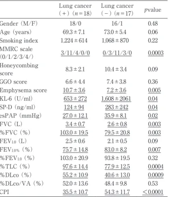
=17 例)を比較検討したところ,原発性肺癌合併群では肺気腫スコアが非合併群に比して 有意に高値であり,非合併群では MMRC scale,血清 SP-D 値,esPAP が原発性肺癌合併群に比して有意に高 値であった.呼吸機能検査では,原発性肺癌合併群で FVC,%FVC,%TLC,%DLco が有意に高く,一方,
FEV1.0%,CPI が有意に低かった.また両群間の予後に 差は認められず(生存期間中央値,肺癌合併群:非合併 群=18.8ヶ月:17.7ヶ月),その予後不良因子は急性ある Table 1 Comparison of baseline patient characteristics between Groups E and F
Group E (n=35) Group F (n=57) p value
Gender (M/F) 34/1 43/14 0.007
Age (years) 71.5 ± 6.5 74.1 ± 6.2 0.03
Smoking history (Current/Former/Never) 11/23/1 8/39/10 0.02
Smoking index 1,148±742 699±610 0.003
Dust exposure 12/26 (46.2%) 17/54 (31.5%) 0.30
Duration from onset of symptoms to hospital visiting (days) 144±255 303±378 0.04
MMRC scale (0/1/2/3/4) 3/14/15/3/0 8/27/16/4/2 0.30
Disease severity of IPF by JRS classification (I/II/III/IV) 16/5/9/5 30/5/12/10 0.76
Complication of lung cancer 18 (51.4%) 7 (12.3%) <0.0001
Acute exacerbation 7 (20.0%) 12 (21.1%) 0.99
Histological type of lung cancer (AD/SCC/SCLC/others) 3/7/4/4 2/1/2/3 0.58
FVC (L) 3.0±0.8 2.1±0.7 <0.0001
%FVC (%) 91.6±23.2 73.0±18.2 <0.0001
FEV1.0 (L) 2.3±0.6 1.8±0.5 0.0003
FEV1.0% (%) 79.1±12.5 86.4±7.4 0.0008
%FEV1.0 (%) 98.5±20.5 90.1±19.7 0.053
%TLC (%) 88.0±16.7 71.4±14.9 <0.0001
%Dlco (%) 48.1±13.9 51.0±18.5 0.44
%DLco/VA (%) 50.4±12.1 69.9±19.5 <0.0001
CPI 44.6±14.6 49.8±14.6 0.10
MMRC, Modified British Medical Research Council; JRS, Japanese Respiratory Society; AD, adenocarcinoma; SCC, squamous cell carcinoma; SCLC, small cell lung cancer; CPI,composite physiological index.
Fig. 1 The Kaplan-Meier survival curve in Groups E and F patients. Survival time was significantly lower in Group E patients than in those of Group F (p=
0.007).
Table 2 Prognostic factors (Groups E and F, n=92):
Multivariate Cox Proportion Hazard Regression Analysis
HR 95% CI p value Age (years) 1.010 0.927‑1.100 0.82 Male gender 0.930 0.265‑3.265 0.91 Complication of emphysema 2.288 0.952‑5.498 0.06 Complication of lung cancer 10.996 3.979‑30.387 <0.0001
MMRC scale 1.393 0.852‑2.278 0.18
esPAP (mmHg) 1.047 1.012‑1.084 0.009
6MWD (m) 1.002 0.998‑1.005 0.34
%DLco (%) 1.037 0.974‑1.104 0.25
CPI 1.093 1.009‑1.184 0.03
Acute exacerbation 2.581 1.016‑6.557 0.04 MMRC, Modified British Medical Research Council; esPAP, es- timated systolic pulmonary arterial pressure; 6MWD, 6-min walking distance; CPI, composite physiologic index.
Table 4 Prognostic factors (Groups E and F without lung cancer, n=67): Multivariate Cox Proportion Hazard Regression Analysis
HR 95% CI p value Age (years) 1.039 0.926‑1.166 0.51 Male gender 0.896 0.208‑3.851 0.88 Complication of emphysema 3.659 1.033‑12.964 0.04
MMRC scale 1.716 0.927‑3.178 0.08
esPAP (mmHg) 1.058 0.995‑1.125 0.07
6MWD (m) 1.001 0.997‑1.006 0.64
%FVC (%) 1.000 0.961‑1.040 0.99
%DLco (%) 1.034 0.938‑1.140 0.50
FEV1.0%(%) 1.073 0.983‑1.172 0.11
CPI 1.069 0.918‑1.246 0.39
Acute exacerbation 2.503 0.825‑7.589 0.10 MMRC, Modified British Medical Research Council; esPAP, es- timated systolic pulmonary arterial pressure; 6MWD, 6-min walking distance; CPI, composite physiological index.
Table 3 Predictive factors of complication of lung cancer (Groups E and F, n=92): Multivariate Logis- tic Analysis
OR 95% CI p value Complication of emphysema 4.031 1.258‑12.916 0.02 Smoking index>775 4.261 1.197‑15.169 0.03
%TLC>81.1% 5.530 0.902‑33.919 0.06
%FVC>76.5% 1.227 0.181‑8.322 0.83
Table 5 Comparison of baseline patient characteris- tics between 2 groups with or without lung cancer in CPFE
Lung cancer
(+) (n=18) Lung cancer
(−)(n=17) pvalue
Gender (M/F) 18/0 16/1 0.48
Age (years) 69.3±7.1 73.0±5.4 0.06 Smoking index 1,224±614 1,068±870 0.22 MMRC scale
(0/1/2/3/4/) 3/11/4/0/0 0/3/11/3/0 0.0003 Honeycombing
score 8.3±2.1 10.4±3.4 0.09
GGO score 6.6±4.4 7.4±3.8 0.36
Emphysema score 10.7±3.6 7.2±3.6 0.005 KL-6 (U/ml) 653±272 1,608±2061 0.04
SP-D (ng/ml) 124±94 283±242 0.04
esPAP (mmHg) 27.0±12.1 35.9±8.1 0.02
FVC (L) 3.4±0.7 2.6±0.8 0.003
%FVC (%) 103.0±19.5 79.5±20.8 0.003 FEV1.0 (L) 2.5±0.6 2.1±0.5 0.09 FEV1.0% (%) 75.7±14.8 83.0±8.2 0.007
%FEV1.0 (%) 103.0±20.9 93.8±19.5 0.32
%TLC (%) 97.6±14.4 77.9±12.5 0.0004
%DLco (%) 55.2±10.9 40.6±13.0 0.0009
%DLco/VA (%) 52.0±13.6 48.4±9.8 0.53
CPI 35.5±10.7 54.3±11.7 <0.0001
GGO, ground glass opacity; CPI, composite physiological index.
Table 6 HRCT findings and semiquantitative analysis of histological findings in CPFE, IPF, and PE
Case Sub- types
Localization of lung cancer
(HRCT images)
Background lung structure adjacent to lung
cancer
(HRCT images)
Subtypes of lung cancer
Fibroblas- tic focus
Smooth muscle prolifera- tion
Elastosis Hyperplas- tic type II AECs
Bronchi-
olization Squamous metaplasia
Alveolar destruction
and enlarge-
ment
1 CPFE LUL (S1+2) E SCC + ++ + ++ ++ + +++
2 CPFE LUL (S3) E SCC + + ++ + ++ + ++
3 CPFE LLL (S6-8) F AD − − + + ++ − +++
4 CPFE RLL (S9) E+F SCC − + ++ +++ ++ +++ ++
5 CPFE LLL (S9) E+F SCLC − + + ++ +++ ++ +++
6 CPFE RLL (S8) F SCLC + + + ++ ++ − ++
7 IPF RLL (S6) F AD + ++ +++ ++ ++ − −
8 IPF LLL (S6) F AD ++ +++ +++ +++ +++ + +
9 IPF RUL (S2) F SCLC ++ +++ +++ ++ ++ − −
10 IPF LLL (S9) F SCC ++ ++ +++ ++ +++ − −
11 IPF RLL (S8) F AD +++ +++ +++ ++ ++ + −
12 PE RUL (S2) E AD − − − ++ + − +++
13 PE LUL (S3) E AD − − − − ++ − +++
14 PE LUL (S1+2) E SCC − − − − + − +++
15 PE RUL (S6) E SCC − − − + ++ ++ +++
16 PE RUL (S3) E SCLC − + − + ++ + +++
Histological scoring: −, negative; +, weak; ++, moderate; +++, intense. CPFE, combined pulmonary fibrosis and emphysema; IPF, idiopathic pulmonary fibrosis; PE, pulmonary emphysema; LUL, left upper lobe; LLL, left lower lobe; RUL, right upper lobe; RLL, right lower lobe; E, emphysematous lesion; F, fibrotic lesion; AD, adenocarcinoma; SCC, squamous cell carcinoma; SCLC, small cell lung can- cer; IP, interstitial pneumonia; AECs, alveolar epithelial cells.
いは慢性増悪であった(HR=3.044,
p
=0.01).次に発癌母地に関する画像病理組織学的検討(Table 6)
において,F 群(case 7〜11)では,画像上,全例で癌 は線維化病変に隣接して認められた.この線維化病変は,
病理組織学的に胸膜下,小葉辺縁性に分布しており,健 常部と隣接していた.一部で気腔が単純化しており,蜂 巣肺を呈していた.線維化内には肺胞虚脱および平滑筋 増生が目立ち,ところどころ線維芽細胞巣もみられた.
それらの蜂巣肺病変に隣接,または一部浸潤して癌の増 生を認めた(Fig. 2A).次に肺気腫群(case 12〜16)で は,画像上,全例で癌は気腫性病変に隣接して認められ た.病理組織学的には,胸膜側から中枢側にかけて,肺 胞の破壊,拡張が認められたが,IPF や fibrotic NSIP
でみられるような線維化は存在せず,一部の症例で限局 的あるいは非常に不規則な肺胞虚脱を伴わない線維化を 認めるのみであった.それらの気腫性病変に隣接して癌 の増生を認めた(Fig. 2B).一方,E 群(case 1〜6)では,
画像上,線維化病変,気腫性病変ならびに両者の混在部 に隣接する症例がそれぞれ 2 例ずつに認められた.病理 組織学的に癌は気腫あるいは線維化病変を伴った気腫に 隣接して存在し(Fig. 2C),この線維化病変は線維芽細 胞巣,肺胞虚脱および胸膜下病変が目立たず構造破壊が 顕著で,F 群や肺気腫群でみられる線維化とは異なるも のであった(Fig. 2D).
Fig. 2 (A) Lung cancer associated with idiopathic pulmonary fibrosis. Patchy interstitial fibrosis with archi- tectural distortion such as frequent honeycomb change shows a striking subpleural and perilobular distri- bution, alternating with areas of normal lung. Marked smooth muscle proliferation and collapse of alveoli, along with fibroblastic foci, are also present in dense collagen fibrosis. Squamous cell carcinoma of lung
(arrows) invade adjacent to honeycomb lesions (Elastica van Gieson stain, scale bar: 250 μm). (B) Lung cancer associated with pulmonary emphysema. Alveolar destruction and enlargement is seen in lung pa- renchyma. The local and irregular fibrosis without collapse of alveoli are present. Lung cancer, histological- ly squamous cell carcinoma (arrows), is located next to the emphysematous lesions (Elastica van Gieson stain, scale bar: 1 mm). (C) Lung cancer associated with combined pulmonary fibrosis and emphysema.
Small-cell carcinoma of lung (arrows) is located next to emphysematous lesions with or without fibrosis
(Elastica van Gieson stain, scale bar: 1 mm). (D) Microscopic high-power view of the fibrosis correspond- ing to the circular mark in (C) shows a marked architectural distortion and less significant fibroblastic foci and a collapse of alveoli (Elastica van Gieson stain, scale bar; 250 μm).
考 察
CPFE は気腫を伴う肺線維症として欧米に先駆けて厚 生省特定疾患研究班で,特発性間質性肺炎慢性型 B 群 としてすでにその概念が記載されていたものの16),いま だ一定の診断基準は定められていない.このようにその 概念は古くから認識されていたが,2005 年に Cottin ら8)
が画像学的に CPFE という概念を提唱したのを契機に その存在が再度注目されるようになった.彼らの報告で は,CPFE の臨床的特徴として,①重喫煙者の男性に多 い,② HRCT で上葉優位の気腫性病変(小葉中心性あ るいは傍隔壁性)と下葉優位の線維化病変(主に usual interstitial pneumonia:UIP pattern)がみられる,③ 1 秒率や%肺活量の異常は軽度であるが高度の拡散能低下 がみられ,労作時の desaturation が顕著である,④肺 高血圧症の合併が予後不良因子となる,というもので あった.今回の検討でも,E 群で 35 例中 34 例に喫煙歴 があり,smoking index も平均 1,148 と非常に高値であっ た.さらに原発性肺癌の高い合併率(51.4%)が挙げら れるが,諸家17)18)の報告でも,CPFE において原発性肺 癌の合併が約 50%で認められ,肺癌の組織型としては 扁平上皮癌が最多であった.これらの臨床的特徴からも,
CPFE の原因として喫煙が強く関連していると考えられ た.しかしながら,その予後ならびに予後不良因子に関 しては一定の見解が得られていない.近年,Mejía ら9)は,
CPFE 患者は,IPF 単独患者に比べて死亡率が高く,さ らに肺高血圧が予後不良因子の一つであったと報告して いる.一方,Akagi ら10)は,その予後に差はなく,気腫 合併例では年間の肺活量および肺拡散能変化率が IPF のみの患者に比べて軽度であると指摘している.また Kurashima ら19)の報告では,むしろ IPF 単独患者のほ うが予後不良であったとしており,これは直接死因にお いて IPF 単独患者で急性増悪の頻度が有意に高かった ことなどが影響しているのではないかと考えられた.最 近,Todd ら20)も間質性肺炎単独例で予後不良であった と報告しており,さらに CPFE 群では,気腫スコアが 高いほど予後良好であったとしている.このように予後 に関する見解が報告により異なる理由として,間質性肺 炎および肺気腫の重症度や画像スコアならびに病変分布 の偏り,間質性肺炎の中に IPF 以外の fibrotic NSIP や その他の慢性線維化型間質性肺炎が混在している可能性 などが影響しているのではないかと推測された.そこで 間質性肺炎を IPF に限定した今回の検討では,E 群で は F 群に比して有意に予後不良であり,肺気腫,原発 性肺癌,肺高血圧症,CPI 増加ならびに急性増悪などが 有意な予後不良因子で,特に原発性肺癌合併で最も強い 関連が認められた.さらに原発性肺癌合併例を除いた症
例においても,E群はF群に比して有意に予後不良であっ たことから,肺癌合併の有無にかかわらず両群間には予 後に差があり,多変量 Cox 比例ハザード解析の結果,
肺気腫合併が単独の予後不良因子であった.Schmidt ら21)は,321 例の IPF 患者のうち,約半数に HRCT 上,
肺気腫を合併していたとしており,その中で中等度〜高 度の気腫を合併している CPFE 患者において,12ヶ月 後に FEV1.0が 10%減少した症例では,死亡率が 3.7 倍 に上昇し,CPFE 患者において最も信頼できる予後因子 としている.今回,さらに E 群において原発性肺癌合 併群,非合併群に分けて比較検討したところ,原発性肺 癌合併群では非合併群に比して気腫スコアが有意に高 か っ た. ま た 呼 吸 機 能 検 査 で は,FVC,%FVC,%
TLC が有意に高く,FEV1.0%が有意に低かった.したがっ て,CPFE の中でも気腫の影響がより強い症例において,
原発性肺癌を合併する可能性が高いことが示唆された.
これらのことから,CPFE 患者の中でも特に気腫スコア 10 前後の気腫性変化が強い症例においては,原発性肺 癌の合併に注意が必要である.
次に原発性肺癌の発生母地としての CPFE の病理組 織学的特徴に注目したところ,肺癌は気腫あるいは線維 化病変を伴った気腫に隣接して発生しており,この線維 化病変は線維芽細胞巣,肺胞虚脱および胸膜下病変が目 立たず構造破壊が顕著で,気腫非合併 IPF にみられる 線維化とは異なるものであった.元来,肺気腫は肺胞構 造の破壊を伴い,明らかな線維化がみられないものと定 義されているが,肺気腫と肺線維症との関係について,
TGF-
β,TNF- α,PDGF- β,IL-13 のトランスジェニッ
クマウスで肺の線維化病変に加えて気腫性病変も生じる ことが報告されており22),肺の炎症や損傷に対する修復 機転の過程において気腫性病変と線維化病変の両者が生 じる可能性が示唆されている.また最近では,airspace enlargement with fibrosis(AEF)を肺気腫と間質性肺 炎の接点にあるものととらえる研究がされており,この AEF の頻度は喫煙量とともに増加することが示されて いる23).さらに Rogliani ら24)は,免疫組織学的染色によ りマトリックスメタロプロテアーゼ(MMP)の陽性率 を比較したところ,間質の線維芽細胞において,肺気腫 単 独 症 例 に 比 べ て CPFE 症 例 で MMP-2,MMP-7,MMP-9,membrane type 1-metalloproteinase の陽性率 が高くなっていることを報告している.しばしば肺気腫 にみられる線維化と CPFE の線維化を,形態学的に HE 染色のみで鑑別することは困難な事が多く,このような 免疫組織学的手法により,これら線維化の質的相違点が 明らかになれば,大変有用なものになりうると考えられ た.
以上,CPFE 患者では原発性肺癌の合併率が高いもの
の,肺癌合併の有無にかかわらず予後不良で,肺気腫合 併が単独の予後不良因子であった.また,病理組織学的 には,CPFE における発癌母地として気腫非合併 IPF とは異なる線維化を伴った気腫の存在が重要であること が示唆された.今後は,この発癌母地に浸潤する炎症細 胞や線維化・リモデリングに関連した増殖因子の局在・
分布を検討し,さらに発癌部における p53 蛋白および Ki-67 labeling index からみた増殖能についても評価する ことによって,気腫と線維化の形成機序,さらには発癌 との関連性を明らかにしたい.
謝辞:病理診断に関して貴重な助言を賜った東邦大学医療 センター大森病院病理科の密田亜希博士に深謝する.
引用文献
1)Nishiyama O, Taniguchi H, Kondoh Y, et al. A sim- ple assessment of dyspnea as a prognostic indicator in idiopathic pulmonary fibrosis. Eur Respir J 2010;
36: 1067‑72.
2)Flaherty KR, Mumford JA, Murray S, et al. Prognos- tic implications of physiologic and radiographic changes in idiopathic interstitial pneumonia. Am J Respir Crit Care Med 2003; 168: 543‑8.
3)Latsi PI, du Bois RM, Nicholson AG, et al. Fibrotic idiopathic interstitial pneumonia. The prognostic value of longitudinal functional trends. Am J Respir Crit Care Med 2003; 168: 531‑7.
4)Zappala CJ, Latsi PI, Nicholson AG, et al. Marginal de- cline in FVC is associated with a poor outcome in idio- pathic pulmonary fibrosis. Eur Respir J 2010; 35: 830‑6.
5)Lederer DJ, Arcasoy SM, Wilt JS, et al.:Six-minute- walk distance predicts waiting list survival in idio- pathic pulmonary fibrosis. Am J Respir Crit Care Med 2006; 174: 659‑64.
6)Flaherty KR, Andrei AC, Murray S, et al. Idiopathic pulmonary fibrosis: prognostic value of changes in physiology and six-minute-walk test. Am J Respir Crit Care Med 2006; 174: 803‑9.
7)Sumikawa H, Johkoh T, Colby TV, et al. Computed tomography findings in pathological usual intersti- tial pneumonia:relationship to survival. Am J Respir Crit Care Med 2008; 177: 433‑9.
8)Cottin V, Nunes H, Brillet PY, et al. Combined pul- monary fibrosis and emphysema: a distinct under- recognised entity. Eur Respir J 2005; 26: 586‑593.
9)Mejía M, Carrillo G, Rojas-Serrano J, et al. Idiopathic pulmonary fibrosis and emphysema: decreased sur- vivial associated with severe pulmonary arterial hy- pertension. Chest 2009; 136: 10‑5.
10)Akagi T, Matsumoto T, Harada T, et al. Coexistent
emphysema delays the decrease of vital capacity in idiopathic pulmonary fibrosis. Respiratory Medicine 2009; 103: 1209‑15.
11)Goddard PR, Nicholson EM, Laszlo G, et al. Comput- ed tomography in pulmonary emphysema. Clin Ra- diol 1982; 33: 379‑87.
12)American Thoracic Society; European Respiratory Society. Idiopathic pulmonary fibrosis: diagnosis and treatment: international consensus statement.
Am J Respir Crit Care Med 2000; 161: 646‑64.
13)日本呼吸器学会びまん性肺疾患診断・治療ガイドラ イン作成委員会.特発性間質性肺炎診断と治療の手 引き.東京 : 南江堂. 2004.
14)Kazerooni EA, Martinez FJ, Flint A, et al. Thin-sec- tion CT obtained at 10-mm increments versus limit- ed three-level thin-section CT for idiopathic pulmo- nary fibrosis. Am J Roentgenol 1997; 169: 977‑83.
15)Nadrous HF, Pellikka PA, Krowka MJ, et al. Pulmo- nary hypertension in patients with idiopathic pul- monary fibrosis. Chest 2005; 128: 2393‑9.
16)本間行彦,斎木茂樹,土井修他,他.特発性間質性 肺炎(IIP)の臨床的診断基準 第 3 次改定案.厚生 省特定疾患びまん性肺疾患調査研究班 平成 3 年度 報告書.1992; 20‑32.
17)小谷敬太,村田和子,吉田祥二.肺気腫と特発性肺 線維症の併存症例における胸部 CT の評価.断層映 像研究会雑誌 2004; 31: 25‑9.
18)Kitaguchi Y, Fujimoto K, Hanaoka M, et al. Clinical characteristics of combined pulmonary fibrosis and emphysema. Respirology 2010; 15: 265‑71.
19)Kurashima K, Takayanagi N, Tsuchiya N, et al. The effect of emphysema on lung function and survival in patients with idiopathic pulmonary fibrosis. Res- pirology 2010; 15: 843‑8.
20)Todd NW, Jeudy J, Lavania S, et al. Centrilobular emphysema combined with pulmonary fibrosis re- sults in improved survival. Fibrogenesis Tissue Respir 2011; 4: 6.
21)Schmidt SL, Nambiar AM, Tayob N, et al. Pulmo- nary function measures predict mortality different- ly in IPF versus combined pulmonary fibrosis and emphysema. Eur Respir J 2011; 38: 176‑83.
22)Gauldie J, Kolb M, Ask K, et al. Smad3 signaling in- volved in pulmonary fibrosis and emphysema. Proc Am Thorac Soc 2006; 8: 696‑702.
23)Kawabata Y, Hoshi E, Murai K, et al: Smoking-relat- ed changes in the background lung of specimens re- sected for lung cancer: a semiquantitative study with correlation to postoperative course. Histopa- thology 2008; 53: 707‑14.
24)Rogliani P, Mura M, Mattia P, et al. HRCT and histo- pathological evaluation of fibrosis and tissue de-
struction in IPF associated with pulmonary emphy- sema. Respir Med 2008; 102: 1753‑61.
Abstract
Clinicopathological characteristics of patients with combined pulmonary fibrosis and emphysema Takafumi Ito a, Keishi Sugino a, Susumu Sakamoto a, Atsuko Kurosaki b,
Toshimasa Uekusa c and Sakae Homma a
a Department of Respiratory Medicine, Toho University Omori Medical Center
b Department of Diagnostic Radiology, Toranomon Hospital
c Department of Pathology, Labor Health and Welfare Organization Kanto Rosai Hospital
The aim of this study was to clarify the clinicopathological characteristics and the prognostic factors of com- bined pulmonary fibrosis and emphysema (CPFE), which is defined as pulmonary emphysema associated with id- iopathic pulmonary fibrosis (IPF). Of 92 IPF patients admitted to our hospital during the past 6 years, 35 were di- agnosed as CPFE (Group E) and 57 as IPF without emphysema (Group F). We retrospectively compared the clinical features between these 2 groups. Furthermore, we investigated the histopathological localization and characteristic background parenchymal architecture of lung cancer in 6 patients with CPFE who had undergone surgical lung resection for complications of lung cancer. Lastly, we compared the histopathological differences in background lung structures of the origins of the lung cancers in the following 3 groups: Group E, Group F, and pul- monary emphysema. The smoking index values and the incidence of lung cancer were higher in patients in Group E than in Group F, but the survival times were significantly lower in Group E patients. The prognostic factors in- cluded emphysema, lung cancer, pulmonary arterial hypertension, increased composite physiological index, and acute exacerbation. Histopathologically, the localization of lung cancer in Group E patients was adjacent to emphy- sematous lesions with or without associated fibrosis, which showed a more marked architectural distortion with less significant fibroblastic foci, collapse of alveoli, and subpleural fibrosis than in Group F patients. In conclusion, this study demonstrated that the incidence of lung cancer was extremely high in patients with CPFE. However, the prognosis was unfavorable regardless of the presence of lung cancer, with the chief adverse prognostic factor being complications of pulmonary emphysema. Emphysematous lesions with fibrosis in Group E that are different from those seen in Group F may be background lung structures characteristic of the carcinogenesis of lung can- cer.