UNCORRECTED
PROOF
Contents lists available at ScienceDirectJournal of Affective Disorders
journal homepage: www.elsevier.comElectroconvulsive shock restores the decreased coverage of brain blood vessels by
astrocytic endfeet and ameliorates depressive-like behavior
Ilhamuddin A. Azis
a, b, c, Sadayuki Hashioka
a, ⁎, Keiko Tsuchie
a, Tsuyoshi Miyaoka
a, Rostia A. Abdullah
a,
Erlyn Limoa
b, Ryosuke Arauchi
a, Ken Inoue
d, Shoko Miura
a, Muneto Izuhara
a, Misako Kanayama
a,
Koji Otsuki
a, Michiharu Nagahama
a, Kiminori Kawano
a, Tomoko Araki
a, Maiko Hayashida
a, Rei Wake
a,
Arata Oh-Nishi
a, Andi J. Tanra
b, Jun Horiguchi
a, Masatoshi Inagaki
aaDepartment of Psychiatry, Shimane University, 89-1 Enya-cho, Izumo 693-8501, Japan
bDepartment of Psychiatry, Hasanuddin University, Jl. Perintis Kemerdekaan Km. 10, Makassar 90245, South Sulawesi, Indonesia cDepartment of Biochemistry, Hasanuddin University, Jl. Perintis Kemerdekaan Km. 10, Makassar 90245, South Sulawesi, Indonesia dHealth Service Center, Kochi University, Akebono-cho, Kochi 780-8520, Japan
A R T I C L E I N F O A B S T R A C T
Background: Although growing evidence indicates that ECT affects astrocytes, the exact mechanisms of the
ther-apeutic effect of ECT are still unknown. Astrocytic endfeet express the water channel aquaporin (AQP) 4 abun-dantly and ensheath brain blood vessels to form gliovascular units. It has been shown that the coverage of blood vessels by AQP4-immunostained endfeet is decreased in the prefrontal cortex (PFC) of patients with major de-pression. This study was made to determine whether ECT restores the astrocytic coverage of blood vessels with amelioration of depressive symptoms.
Methods: After electroconvulsive shock (ECS) administration to rats, the forced swimming test (FST) and Y-maze
test were performed. Subsequently, immunofluorescence analysis was conducted to measure the coverage of blood vessels by astrocytic endfeet in the PFC and hippocampus by using the endothelial cell marker lectin and anti-AQP4 antibody. We also performed Western blot to examine the effects of ECS on the hippocampal expres-sion of AQP4 and the tight junction molecule claudin-5.
Results: Gunn rats showed learned helplessness and impaired spatial working memory, compared to normal
con-trol Wistar rats. ECS significantly improved the depressive-like behavior. Gunn rats showed a decrease in astro-cytic coverage of blood vessels, that was significantly increased by ECS. ECS significantly increased expression of AQP4 and claudin-5 in Gunn rats.
Conclusions: ECS increased the reduced coverage of blood vessels by astrocytic endfeet in the mPFC and
hip-pocampus with amelioration of depressive-like behavior. Therefore, therapeutic mechanism of ECT may involve restoration of the impaired gliovascular units by increasing the astrocytic-endfoot coverage of blood vessels.
1. Introduction
Application of electroconvulsive therapy (ECT) is still considered to be superior for some cases of psychiatric disorders in spite of much advances in drug development. ECT modified by general anesthesia and muscular relaxation has been recognized as a safe and effective treatment for drug-resistant major depression, schizophrenia, mania and catatonia (Weiner and Reti, 2017). Growing evidence indicates that glial
cells are involved in response to ECT (Jinno and Kosaka, 2008) (Limoa et al., 2016). However, the exact mechanisms of therapeutic ef-fect of ECT have yet to be clarified. Glial cells have attracted attention as new promising targets for antidepressant action (Hashioka et al., 2013). In fact, an in vitro study has demonstrated that production of brain-de-rived neurotrophic factor (BDNF) and lactate for neurons is increased in rodent astrocytes treated with the selective serotonin reuptake inhibitor fluoxetine (Allaman et al., 2011), indicating that astrocyte is an impor-tant source of neurotrophic factor (Quesseveur et al., 2013) and a main
⁎ Corresponding author.
Email addresses: [email protected] (S. Hashioka); [email protected] (K. Tsuchie); [email protected] (T. Miyaoka); [email protected] (R.
Arauchi); [email protected] (K. Inoue); [email protected] (S. Miura); [email protected] (M. Kanayama); [email protected] (K. Otsuki); [email protected] (M. Nagahama); [email protected] (K. Kawano); [email protected] (T. Araki); [email protected] (M. Hayashida); [email protected] (R. Wake); [email protected] (A. Oh-Nishi); [email protected] (J. Horiguchi); [email protected] (M. Inagaki)
https://doi.org/10.1016/j.jad.2019.07.008
Received 21 February 2019; Received in revised form 30 June 2019; Accepted 3 July 2019 Available online xxx
0165-0327/ © 2019.
UNCORRECTED
PROOF
mediator of glucose metabolism. Another in vitro study has shown thatthe tricyclic antidepressant imipramine induces human astrocytes dif-ferentiate into neuronal phenotype, suggesting involvement of this phe-nomenon in antidepressant-induced neurogenesis (Cabras et al., 2010). These findings prompt us to investigate the mechanisms involving astro-cytes in the therapeutic effect of ECT.
Aquaporin (AQP) 4 is a water channel and plays pivotal roles in the brain functions (Nicchia et al., 2004). Astrocytic endfoot processes, on which AQP4 is expressed abundantly, ensheath entire network of blood vessels in the brain and form gliovascular units with endothelial cells and pericytes (Simard et al., 2003; Wolburg et al., 2009). It has been shown that AQP4 is required for the anti-depressive action via regulat-ing adult hippocampal neurogenesis (Kong et al., 2009). A recent post-mortem study on patients with major depression, that is clinically well known to be associated with cerebrovascular disease, has revealed a de-crease in the coverage of blood vessels by AQP4-immunostained end-feet of astrocytes in the prefrontal cortex (Rajkowska et al., 2013). It is, therefore, tempting to elucidate the effect of ECT on the decreased cov-erage of blood vessels by AQP4-immunopositive endfeet, that appears to be involved in the pathogenesis of the major depression.
Our previous study has revealed that Gunn rats with glial acti-vation in the hippocampus present depressive-like behavior as shown by prolonged immobility time in the forced swimming test (FST) and the tail suspension test (Arauchi et al., 2018). The Gunn rat is a mu-tant of the Wistar strain and has a genetic deficiency in glucuronyl-transferase (Gunn, 1944). This deficiency leads to high levels of un-conjugated bilirubin in their blood and the brain. Therefore, Gunn rats have been used as an experimental model of kernicterus (bilirubin en-cephalopathy), which can be considered as an organic brain disorder. Since patients with organic brain disorder often present symptoms of schizophrenia and affective disorder (Hamilton et al., 1983), it appears to be rational that Gunn rats show depression-like behavior. In the pre-sent study, we first confirmed that electroconvulsive shock (ECS), an animal counterpart of ECT, ameliorated the depressive-like behavior of Gunn rats, such as learned helplessness and impaired working memory, using the FST and Y-maze test. We next investigated the morphology of gliovascular units with regard to the coverage of blood vessels by as-trocytic endfeet in the diseased brain of Gunn rat. Because it has been shown that there is no significant difference in GFAP-immunostained coverage of blood vessels between the postmortem brains of major de-pression patients and those of normal controls (Rajkowska et al., 2013), we labelled astrocytic endfeet with AQP4, not with GFAP. We finally de-termined the effect of ECS on the astrocytic coverage of blood vessels in the brain of Gunn rats whose depressive-like behavior was ameliorated by ECS, to examine whether the change in morphology of gliovascular units is related to the therapeutic efficacy of ECS.
2. Materials and methods
2.1. Animals
Eight-week-old male homozygous (j/j) Gunn rats and male Wistar rats (SLC, Inc., Shizuoka, Japan) were used in this study (12 rats for each strain). The rats were housed in plastic cages (39 × 27 × 18cm) under standard conditions with a room temperature (RT) of 23 ± 2°C, humidity of 55 ± 5%, and 12-h light/12-h dark cycle (light phase 7:00 to 19:00). The rats were allowed free access to food and water. One week before commencing the experiment, the rats underwent a han-dling procedure once daily to reduce experimental stress. The hanhan-dling procedure consisted of picking rats up with a gloved hand, stroking, and massaging them for 10min. All procedures were conducted with the ap-proval of the Shimane University Animal Ethics Committee, in accor-dance with the guidelines of the National Health and Medical Research Council of Japan.
2.2. ECS procedure
The animals were assigned into four groups, i.e., Wistar Sham (WS), Wistar ECS (WE), Gunn Sham (GS) and Gunn ECS (GE) group. To avoid stress or pain induced by the ECS procedure, each rat was first anes-thetized in an isoflurane inhalation chamber (4% for initial induction) with an oxygen flow rate of 2–4L/min. After the rat fell asleep, it was taken out from the chamber and anesthesia was continued by putting an isoflurane inhalation mask (2% for maintenance) with an oxygen flow rate of 2–4L/min. In every ECS procedure, electric shock was given under such anesthesia. ECS was administered transcranially via bilat-eral ear clip electrodes by using an E.C. Stimulator MK-810 (Muromachi Kikai, Tokyo, Japan). The stimulus was a sine wave pulse, 100V, 60Hz, 50mA, 1.5s. Each stimulation resulted in a typical tonic-clonic seizure lasting for less than 10s. The ECS group received an ECS treatment once daily for 6 consecutive days. The sham-treated groups were handled identically to the ECS groups including anesthesia except that no cur-rent was delivered. Each individual in all the groups was given the FST and Y-maze test. After the behavior tests, the brain were taken out to conduct immunofluorescent studies.
2.3. FST
The FST was performed according to protocol reported previously with modification (Arauchi et al., 2018). A plastic cylinder, whose di-ameter is 19cm, filled with 10L of water (25°C±1°C), 40cm in depth. On the first day, rats were gently placed individually in the water for 15min to be habituated. The rat left in the cylinder was either unable to touch the bottom or escape. The animals were dried with a towel and put in a warmer chamber for 10min to avoid hypothermia, and then re-turned to their home cages. After each trial, the cylinder was washed, rinsed, and refilled with fresh water to avoid influence on the next an-imal. On the next day, the rats were placed in the water for 6min and their behavior was recorded with a video camera. Immobility time oc-curring during the test was manually counted from the recorded video by a trained observer in a blinded manner. The rats were considered im-mobile when they remained floated without struggling, motionless and when they moved only to maintain their heads above the water.
2.4. Y-maze test
Spatial working memory of rats was assessed by the Y-maze test on a day after the last ECS administration based on previously described procedures with some modifications (Shi et al., 2017). The maze ap-paratus is made of gray opaque polyvinyl chloride with three arms (500 × 250 × 100mm3and 120° apart) (Muromachi Kikai). The
appa-ratus was cleaned with a 10% ethanol solution and then dried with a paper towel after each trial to make it odorless. Each rat was placed at the terminus of one arm and allowed to move freely through the maze for 8min. When the rat's tail was entirely within the arm, entry was con-sidered to be complete. Alternation behavior was defined as successive entries into all three arms on consecutive occasions. The percentage of spontaneous alternation behavior (SAB) was calculated according to the following formula:
2.5. Brain section preparation
After the behavior tests, the animals were sacrificed under deep intraperitoneal anesthesia with an anesthetic mixture of three drugs: medetomidine (Domitor, Nippon Zenyaku Kogyo, Tokyo, Japan), mida 2
UNCORRECTED
PROOF
zolam (Dormicum, Astellas Pharma, Tokyo, Japan), and butorphanol(Vetorphale, Meiji Seika Pharma, Tokyo, Japan). We mixed medeto-midine 0.15mg/kg b.w./rat, midazolam 2mg/kg b.w./rat and butor-phanol 2.5mg/kg b.w./rat and added saline (Otsuka Pharmaceutical Factory, Tokushima, Japan) to adjust the mixture to a volume of 0.5ml/ 100g b.w./rat. The rats were perfused transcardially with 500mL of physiological saline, followed by 500mL of 4% paraformaldehyde (PFA) in 0.1M phosphate buffer (PB; pH 7.3). After perfusion, the brain was quickly removed from the skull, and fixed in a solution of 4% PFA at RT for 4h. The brains were immersed in 10% sucrose at 4°C overnight and subsequently were immersed in 20% sucrose at 4°C for 24h. The brains were cut at 40-μm thickness in the coronal plane by using a freezing mi-crotome (Microm HM 430, Thermo Scientific, Waltham, MA).
2.6. Immunofluorescent staining
We modified and followed the protocol for double immunofluores-cent staining described in previous studies (Arauchi et al., 2018; Liaury et al., 2014; Limoa et al., 2016). The free floating brain sections were pre-incubated for blocking with 0.1M PB containing 3% bovine serum albumin, 0.2% Triton X and 1.5% normal horse serum for 1h at RT. The sections were then incubated overnight at RT in a solution of 0.1M PB, 0.2% Triton X, 1.5% normal horse serum, and the mouse monoclonal anti-AQP4 antibody (1:500, Santa Cruz Biotechnology, CA) mixed with Texas Red labeled Lycopersicon esculentum (tomato) lectin (1:500, Vec-tor LaboraVec-tories, CA) to detect endothelial cells. The sections incubated with the anti-AQP4 antibody were incubated with Alexa 488-conju-gated anti-mouse IgG (Invitrogen, Carlsbad, CA) diluted at 1:1000 for 3h at RT. Afterwards, the slices were mounted onto gelatin-coated slides (Matsunami Glass, Osaka, Japan) and then were sunk in 0.1M PB. Cov-erslips were applied onto the slides with anti-fading mounting medium and subsequently were kept in darkness before inspection. Negative con-trols for immunostaining were performed with omitting the primary an-tibody or lectin.
2.7. Image analysis for coverage of brain blood vessels
Fluorescent images were captured by a BZ-X700 all-in one micro-scope (Keyence, Osaka, Japan) with a 40x objective lens and analyzed by the BZ-X analyzer software. Regions of interest consist of two areas within the prefrontal cortex [i.e., the prelimbic (PrL) area and the in-fralimbic (IL) area] and three areas within the hippocampal formation [i.e., the dentate gyrus (DG), the cornu ammonis (CA)1 and the CA3]. The images were taken from both hemispheres of 12–14 slices in the PrL region (two images from each slice), 8–11 slices in the IL region, and 12–15 slices in the hippocampal formation. Overall, 64–80 images per animal (n=5) were examined. The fluorescence colors in captured im-ages were split equally into red, green and blue colors by the BZ-X an-alyzer software. The “hybrid cell count” operating procedure with “sin-gle extraction” was used to specify the target area by selecting the outer margin of the stained areas. First, the lectin-positive area was extracted from the merged fluorescent image of the lectin-positive area (red) and the AQP4-positive area (green) and was displayed red. Next, colocal-ization area, namely, the AQP4-positive area on the lectin-positive area was extracted and displayed yellow. These extraction procedures were performed based on the consistent threshold of fluorescence intensity, which was manually predefined. The pixels of the yellow areas were cal-culated to determine the percentage of “coverage”, which was defined as area of co-localization/total area of brain blood vessels.
2.8. Western blot analysis
Hippocampal tissues were isolated and homogenized in ice-cold cell lysis buffer (Cell Signaling, #9803, Danvers, NA) supplemented with
protease inhibitor (Roche, 11873580001, Mannheim, Germany). The tissues were extracted at ratio of 1mg of tissue to 10 μL of buffer. The tissues were sonicated and subsequently centrifuged at 13,000g for 10min at 4°C. The supernatants were collected and studied. Twenty-five to fifty μg of protein were separated by 10% SDS-PAGE and transferred to PVDF membranes. The membranes were incubated with specific rab-bit antibody for AQP4 (1:1000, Santa Cruz Biotechnology, INC, H-80: sc-20812), claudin-5 (1:500, Sigma-Aldrich, SAB4502981: 310145), or mouse anti-β-actin antibody (1:5000, Abcam, AC-15: ab6276) at 4 °C overnight. Subsequently, the membranes were incubated with horserad-ish peroxidase-conjugated anti-rabbit IgG antibody (1:5000, GE Health-care Life Sciences: NA934, Tokyo, Japan) or anti-mouse IgG antibody (1:5000, GE Healthcare Life Sciences: NA931, Tokyo, Japan) for 1h at RT. Blots were developed using the chemiluminescent system (Amer-sham, GE Healthcare UK). The band intensity was quantified by densito-metry and the NIH Image analysis software version 1.63 (NIH, Bethesda, MD). Individual expression levels of AQP4 or Claudin-5 were normal-ized to the expression levels of β-actin and presented as percentages of the control group (i.e., Wistar sham) expression, that was defined as 100%.
2.9. Statistical analyses
All the data were expressed as the mean±standard error of the mean (S.E.M). The statistical analyses were carried out using the SPSS software (Dr. SPSS II for Windows, IBM Japan, Tokyo, Japan). Statisti-cal comparisons among the groups were made with a one-way analysis of variance (ANOVA) followed by the post hoc Fischer's least significant difference test. We also used two-way ANOVA to determine the effects of genotype or ECS treatment on immobility time, SAB and AQP4 cover-age, and interactive effect between genotype and ECS treatment. Pear-son correlation was conducted to establish relationship between immo-bility time in the FST and AQP4 coverage of blood vessels. A p-value less than 0.05 was considered statistically significant.
3. Results
3.1. Effect of ECS on immobility time in the FST
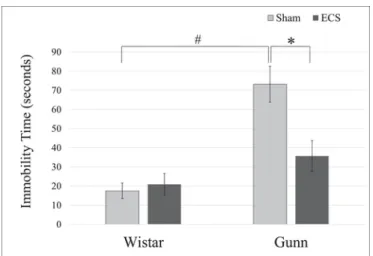
The FST was performed to evaluate the effect of ECS on depres-sive-like behavior of Gunn rats by counting the immobility time. As shown in Fig. 1, the immobility time in the FST was 17.44 ± 4.05s in the WS group, 20.86 ± 5.61s in the WE group, 73.19 ± 9.36s in the GS group, and 35.71 ± 8.08s in the GE group. Immobility time
of the GS group
Fig. 1. Effect of ECS on immobility time of normal rats and Gunn rats in the FST.Immobility time of Gunn rats was significantly longer than that of Wistar rats. The prolonged immobility time of Gunn rats was significantly shortened by ECS. Each value is the mean±S.E.M. (n=6), *p < 0.05, #p < 0.001. ECS, electroconvulsive shock.
UNCORRECTED
PROOF
was significantly longer than that of WS. The ECS administration toGunn rats significantly shortened the duration of immobility compared to that in the GS group. ECS did not affect immobility time significantly on Wistar rats. Both genotype and ECS treatment had significant effects
Fig. 2. Effect of ECS on spontaneous alternation behavior of normal rats and Gunn rats in the Y-maze test.The percentage of spontaneous alternation behavior (SAB) of Gunn rats was significantly decreased compared to Wistar rats. The decreased % SAB of Gunn rats was significantly increased by ECS. Each value is the mean±S.E.M. (n=6), *p < 0.05. SAB, spontaneous alternation behavior; ECS, electroconvulsive shock.
on immobility time in the FST with a significant interactive effect be-tween genotype and treatment.
3.2. Effect of ECS on spontaneous alternation behavior (SAB) in the Y-maze test
Spatial working memory was determined by SAB in the Y-maze test (Fig. 2). The percentage of SAB was 76.62 ± 1.12% in the WS group, 69.51 ± 5.44% in the WE group, 62.44±2.36% in the GS group, and 78.56±3.05% in the GE group. A significant decline in the % SAB was observed in the GS group compared to the WS group. The ECS-admin-istered Gunn rats showed a significant increase of the % SAB compared to the GS group. This finding suggests that ECS improved the spatial memory impairment of Gunn rats. On the other hand, there was no sig-nificant difference between the WS group and the WE group. Neither genotype nor ECS treatment showed a significant effect on SAB in the Y-maze test, even though there was a significant interactive effect be-tween them.
3.3. Effect of ECS on the coverage of brain blood vessels by astrocytic endfeet
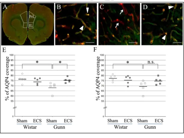
The coverage of brain blood vessels by astrocytic endfeet in both the PrL area and the IL area of the medial prefrontal cortex (mPFC) was investigated. Representative merged fluorescence images show brain blood vessels labelled by lectin (red) and astrocytic endfeet stained with anti-AQP4 antibody (green) in the prelimbic area of the Wistar sham (Fig. 3B), Gunn sham (Fig. 3C) and Gunn ECS groups (Fig. 3D). Overlap
Fig. 3. Effect of ECS on the astrocfigytic-endfoot coverage of blood vessels in the medial prefrontal cortex of normal rats and Gunn rats. The regions of interest consist of the prelimbic (PrL) area and the infralimbic (IL) area (A). Representative merged fluorescence images show brain blood vessels labelled by lectin (red) and astrocytic endfeet stained with anti-AQP4 antibody (green) in the prelimbic area of the normal rat sham (Wistar sham) (B), Gunn sham (C) and Gunn ECS groups (D). The Gunn sham group shows vessel areas uncovered with astrocytic endfeet, that look red (indicated by arrows, C). On the other hand, the Wistar sham and Gunn ECS groups show colocalization of astrocytic endfeet and blood vessels (indicated by arrowheads, B, D), that looks yellowish, suggesting increased vessel areas covered with astrocytic endfeet. Scale bars indicate 50µm. The percentage of the coverage of lectin-positive brain blood vessels by AQP4-positive astrocytic endfeet in the prelimbic area (E) and the infralimbic area (F). Each value is the mean±S.E.M. (n=5), *p≤0.05; n.s., not significant; AQP4, aquaporin 4. (For interpretation of the references to color in this figure legend, the reader is referred to the web version of this article.)
UNCORRECTED
PROOF
area (yellow) of AQP4 immunoreactivity and lectin staining indicatesastrocytic coverage of brain blood vessels. A decrease in the overlap area was discerned in the PrL area of the GS group compared with the other groups. We performed quantitative analysis of astrocytic coverage of brain blood vessels. In the PrL area, the % of AQP4 coverage area was 76.44±1.73% in the WS group, 74.58±1.39% in the WE group, 68.66±2.23% in the GS group, and 75.39±1.13% in the GE group (Fig. 3E). The % of AQP4 coverage of the GS was significantly decreased compared to that of the WS (Fig. 3E). The % of AQP4 coverage in the GE was significantly increased compared to that in the GS (Fig. 3E). In the IL area, the % of AQP4 coverage was 77.72±1.40% in the WS group, 75.66±1.94% in the WE group, 69.43±2.65% in the GS group, and 74.80±1.60% in the GE group (Fig. 3F). The % of AQP4 coverage in the IL area of the GS was significantly less than that of the WS (Fig. 3F). Although the GE group showed a high average of AQP4 coverage com-pared to the GS group, the difference did not reach statistical signifi-cance (p=0.07) (Fig. 3F). Neither genotype nor ECS treatment showed a significant effect on AQP4 coverage in the PrL area, while there was a significant interactive effect between genotype and treatment. There was a significant effect of genotype, but not of ECS treatment, on AQP4 coverage in the IL area, without a significant interactive effect between genotype and ECS treatment. In addition, there was a negative correla-tion between immobility time in the FST and AQP4 coverage of blood vessels in the PrL area (r=−0.476, p=0.034) and IL area (r=−0.508,
p=0.022).
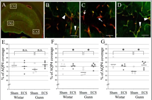
The AQP4 coverage of blood vessels in the hippocampal forma-tion was also evaluated. Representative merged fluorescence images show brain blood vessels labelled by lectin (red) and astrocytic endfeet stained with anti-AQP4 antibody (green) in the CA1 of the Wistar sham (Fig. 4B), Gunn sham (Fig. 4C) and Gunn ECS groups (Fig. 4D). Similar
to the PrL area, a decrease in the overlap area was observed in the CA1 of the GS group compared with the other groups. Quantitative analysis revealed that the % of AQP4 coverage in the DG was 76.68±0.94% in the WS group, 75.40±2.61% in the WE group, 72.29±1.83% in the GS group, 77.28±1.49% in the GE group (Fig. 4E). The % of AQP4 cov-erage in the CA1 was 75.29±2.70% in the WS group, 75.59±0.97% in the WE group, 68.60±1.76% in the GS group, 75.51±1.45% in the GE group (Fig. 4F). In the CA3,% of AQP4 coverage was 77.70±2.17% in the WS group, 75.66±1.10% in the WE group, 69.96±1.37% in the GS group, 76.91±1.88% in the GE group (Fig. 4G). A similar trend was found in the DG (Fig. 4E), the CA1 (Fig. 4F), and the CA3 (Fig. 4G) in comparing the AQP4 coverage among the WS, GS, and GE groups. The scattered plot indicates a significant reduction in the coverage of blood vessels by AQP4-positive endfeet in the CA1 (Fig. 4F) and CA3 (Fig. 4G) of the GS group in comparison with the WS group. However, it did not reach statistical significance in the DG (Fig. 4E). A significant increase in AQP4 coverage was observed in the GE group compared to the GS group in the CA1 (Fig. 4F) and CA3 (Fig. 4G). Although the ECS admin-istration to Gunn rats showed a tendency to increase the AQP4 coverage in the DG, there was no significant difference compared to that of the GS (p=0.07) (Fig. 4E). Like in the medial prefrontal cortex, the % of AQP4 coverage in the WE group did not significantly differ from that in the WS group in all the hippocampal formation regions. Neither geno-type nor ECS treatment showed a significant effect on AQP4 coverage in the hippocampal DG, CA1 and CA3. There was no significant interactive effect between genotype and treatment in the DG and CA1, while that in the CA3 reached significance. In addition, there was a negative corre-lation between the immobility time and the AQP4 coverage in the CA1 (r=−0.488, p=0.029). However, a correlation between the immobil-ity time and the AQP4 coverage was not significant in the DG and CA3.
Fig. 4. Effect of ECS on the astrocytic-endfoot coverage of blood vessels in the hippocampal formation of normal rats and Gunn rats. The regions of interest consist of the dentate gyrus (DG), cornu ammonis (CA)1 and CA3 (A). Representative merged fluorescence images show brain blood vessels labelled by lectin (red) and astrocytic endfeet stained with anti-AQP4 antibody (green) in the CA1 of the normal rat sham (Wistar sham) (B), Gunn sham (C) and Gunn ECS groups (D). The Gunn sham group shows vessel areas uncovered with astrocytic endfeet, that look red (indicated by arrows, C). On the other hand, the Wistar sham and Gunn ECS groups show colocalization of astrocytic endfeet and blood vessels, that looks yellowish, suggesting increased vessel areas covered with astrocytic endfeet, (indicated by arrowheads, B, D). Scale bars indicate 50µm. The percentage of the coverage of lectin-positive brain blood vessels by AQP4-positive astrocytic endfeet in the DG (E), CA1 (F) and CA3 (G). Each value is the mean±S.E.M. (n=5), *p≤0.05; n.s., not significant; AQP4, aquaporin 4. (For interpre-tation of the references to color in this figure legend, the reader is referred to the web version of this article.)
UNCORRECTED
PROOF
3.4. Effect of ECS on the hippocampal expression of AQP4 and claudin-5
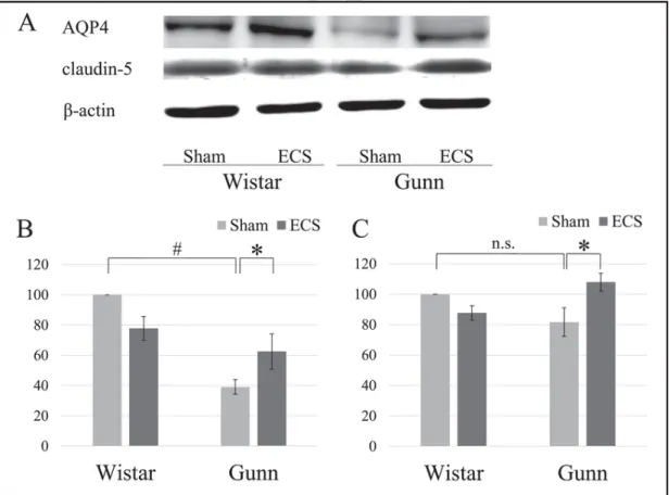
To examine the effect of ECS on the hippocampal expression of AQP4 and the tight junction molecule claudin-5, we also performed quantitative analysis using Western blot. The representative results are shown in Fig. 5A. AQP4 expression levels in the GS group were signifi-cantly lower than those in the WS group (Fig. 5B). However, AQP4 ex-pression in the GE group was significantly increased compared to that in the GS group (Fig. 5B). Claudin-5 expression in the GE group was signif-icantly increased compared to that in the GS group, whereas there was no significant difference in claudin-5 expression between the GS group and the WS group (Fig. 5C).
4. Discussion
There were four major findings in the present study. First, the cov-erage of blood vessels by AQP4-immunoreactive endfeet of astrocytes in the mPFC and the hippocampal formation was significantly reduced in Gunn rats compared to that of Wistar rats, a normal rat strain. Second, ECS significantly reduced immobility time in the FST, suggesting that ECS ameliorated depressive-like behavior of Gunn rats. Third, ECS sig-nificantly improved deficits of working memory of Gunn rats as shown by normalized percentage of SAB in the Y-maze test. The last, ECS sig-nificantly increased the reduced coverage of brain blood vessels by as-trocytic endfeet in Gunn rat. To our knowledge, this is the first study that determines the effect of ECS on the coverage of blood vessels by as-trocytic endfeet in the pathological brain.
Immobility time in the FST can be regarded as behavioral despair which is supposed to reflect depressed moods (Porsolt et al., 1977). The
FST on rodents has been extensively used as a simple animal model for investigating the neurobiology of depression because of its proce-dural simplicity and its high reproducibility (Porsolt et al., 1977). Our finding that repeated ECS administration improved the depressive-like behavior of Gunn rats as shown by shortened immobility time in the FST is consistent with previous studies using the FST, which demon-strated that ECS significantly reduced the duration of immobility in sev-eral genetic rat models of depression, such as Flinders sensitive line rats (Hesselberg et al., 2016), Wistar Kyoto rats (Kyeremanteng et al., 2014) and BDNF knock down rats (Taliaz et al., 2013). Since depressive-like behavior of Gunn rats is ECS-treatable, as in genetic rat models of de-pression, their depressive-like behavior may be associated with common pathophysiological microenvironments which can be modified by ECS. While an antidepressant effect of ECS was observed in diseased Gunn rats, ECS did not affect the normal Wistar rats in the present study. This finding is inconsistent with previous studies which showed that ECS sig-nificantly decreased the immobility time of Wistar rats in the FST (Li et al., 2007; Theilmann et al., 2014). The difference in ECS conditions (e.g., amplitude, voltage, shock duration, the number of shock times and body parts put with electrodes) and anesthetic procedures may attribute to such a discrepancy.
The well-known clinical observation is that major depression signifi-cantly impairs working memory (Pelosi et al., 2000), while ECT has ad-verse effects such as cognitive deficit and amnesia in the acute phase (Semkovska and McLoughlin, 2010). This fact has led to a debate as to whether amnesia associated with ECT is an intrinsic pathological com-ponent of major depression. Under our experimental conditions, ECS ad-ministration on normal Wistar rats showed a trend to decrease the % SAB. To our knowledge, there has been only one study that evaluated the effect of ECS on spatial working memory in rodents under pathologi
Fig. 5. Effect of ECS on the expression of AQP4 and claudin-5 in the hippocampus of normal rats and Gunn rats. The hippocampal homogenates were separated by 10% SDS-PAGE and immunoblotted for AQP4 or claudin-5 (A). Individual expression levels of AQP4 (B) or claudin-5 (C) were normalized to the expression levels of β-actin and presented as percentages of the control group (i.e., Wistar sham) expression, that was defined as 100%. Each value is the mean±S.E.M., (n=5), *p≤0.05; #p≤0.001, n.s., not significant; AQP4, aquaporin 4; ECS, electroconvulsive shock.
UNCORRECTED
PROOF
cal conditions associated with major depression (Henningsen et al.,2013). Our result that repeated ECS treatment significantly amelio-rated the impaired spatial working memory of Gunn rats as shown by increased % SAB in the Y-maze test is congruent with the afore-mentioned study which showed the significant efficacy of ECS in nor-malizing a deficit in % SAB in rats exposed to chronic mild stress (Henningsen et al., 2013).
Our observation that the coverage of brain blood vessels by AQP4-immunoreactive astrocytic endfeet was significantly diminished in Gunn rats presenting depressive-like behavior is consistent with a postmortem study that showed that the coverage of vessels by AQP4 was significantly reduced in the orbitofrontal cortex of patients with ma-jor depression compared to normal controls (Rajkowska et al., 2013). Our result is also in line with an animal study in which rats selectively bred for high anxiety-like behavior, an animal model of depression, showed significantly attenuated coverage of blood vessels in the PFC by AQP4-immunoreactive astrocyte processes compared to non-selected Wistar rats (Di Benedetto et al., 2016). To our knowledge, this is the first study showing that antidepressant treatment, namely ECS adminis-tration in our study, restores the decreased coverage of brain blood ves-sels by astrocytic endfeet in the pathological brain. Based on these find-ings, the astrocytic-endfoot coverage of blood vessels may be negatively correlated with manifesting depressive symptoms. To verify this hypoth-esis, further studies to determine the effects of antidepressant treatment, especially neuropharmacological medication, on the astrocytic-endfoot coverage of brain blood vessels are clearly warranted.
It has been shown that ECT increases serum levels of BDNF and glial cell line-derived neurotrophic factor (GDNF) in patients with major de-pression (Rocha et al., 2016; Zhang et al., 2009). A recent in vitro study demonstrated that GDNF had a protective effect on astrocytes to prevent apoptosis (Wang et al., 2018). Therefore, the ECT-induced production of neurotrophic factors, especially GDNF, may increase the astrocytic-end-foot coverage of blood vessels.
Animal studies using AQP4-knockout mice have demonstrated that AQP4-knockout mice present cognitive deficits similar to those impli-cated in major depression (Skucas et al., 2011) and that AQP4 knockout disrupts fluoxetine treatment-induced improvement of depressive-like behavior caused by chronic mild stress (Kong et al., 2009). Therefore, it could establish a causative role for the decreased endfoot coverage in manifestation of depressive symptoms to examine whether ECS admin-istration to AQP4-knockout mice improves their cognitive deficits. The reduced coverage of brain blood vessels by astrocytic endfeet may be re-lated to reduction in glucose metabolism in the brain, that is mainly me-diated by astrocytes (Kreft et al., 2012). In fact, several neuroimaging studies have demonstrated the decreased glucose metabolism in the pre-frontal cortex of depressed patients (Baxter et al., 1989; Martinot et al., 1990).
Our previous studies have demonstrated that Gunn rats possess astrocytic activation in the hippocampus as shown by increased im-munoreactivity for GFAP and S100B (Arauchi et al., 2018; Limoa et al., 2016). In addition, it has been demonstrated that ECS significantly at-tenuate the increased hippocampal expression of GFAP in Gunn rats, in-dicating ECS has inhibitory effects on activated astrocytes (Limoa et al., 2016). Interestingly, it has been shown that an increase in the num-ber of GFAP-positive astrocytes, that can be referred to as astroglio-sis or astrocytic activation, was accompanied by a decrease in amount of AQP4-immunopositive endfeet colocalized with brain blood vessels in a mouse model of a cerebral small vessel disease with features of gliovascular disruption and cognitive deficits (Holland et al., 2015). Ac-tivated astrocytes may, therefore, contribute toward impaired gliovas-cular units with a decrease in the endfoot coverage of brain blood vessels. Considering our previous observation of the efficacy of ECS in inhibiting activated astrocytes in the hippocampus of Gunn rats,
regenerating the impaired gliovascular
units associated with activated astrocytes may be involved in the thera-peutic mechanisms of ECT. In other words, the therathera-peutic effect of ECS may be exerted, at least in part, by increasing the decreased endfoot coverage of blood vessels, that is presumably related to activated astro-cytes.
In this study, the mPFC was intensively analyzed as the region of interest, since the rat mPFC is supposed to correspond to the hu-man dorsolateral PFC that plays an important role in executive func-tions, including working memory (Barbey et al., 2013). The mPFC sub-regions PrL and IL correspond to human Brodmann areas 32 and 25, respectively (Varea et al., 2005). Dysfunctions of the dorsolateral PFC are involved in various psychiatric disorders, including schizophrenia and major depression (Ferguson and Gao, 2018). We also intensively analyzed the hippocampal formation based on the evidence that hip-pocampal dysfunction along with a small volume is strictly associ-ated with major depression, stress and memory deficits (MacQueen and Frodl, 2011). In addition, alterations in hippocampal anatomy, perfu-sion, and activation have consistently been demonstrated in schizophre-nia (Tamminga et al., 2010).
Although the immobility time in the FST represents desperate behav-ior associated with major depression, it can also be considered as a form of apathy or avolition, which is a negative symptom of schizophrenia (Arauchi et al., 2018). Likewise, impaired spatial working memory, as shown by decreased% SAB in the Y-maze test, can be interpreted as both a cognitive deficit symptom in major depression (Henningsen et al., 2013) and a part of cognitive impairment in schizophrenia (Knox et al., 2017).
Patients with schizophrenia are at an increased risk for the de-velopment of depression (Samsom and Wong, 2015). Overlap in the symptoms and genetic risk factors between schizophrenia and depres-sion implies that a common etiological mechanism may underlie the presentation of comorbid depression in schizophrenia (Samsom and Wong, 2015). In fact, genome-wide genotype data from the Psychi-atric Genomics Consortium show that genetic correlation is high be-tween schizophrenia and bipolar disorder, moderate bebe-tween schizo-phrenia and major depression (Cross-Disorder Group of the Psychiatric Genomics, 2013). Animal studies are also in line with these findings. Dominant-negative Disrupted-In-Schizophrenia-1 transgenic mice, a model for schizophrenia, have been demonstrated to present depres-sive-like behavior as well as schizophrenia-like behavior (Hikida et al., 2007). Our previous studies have revealed that Gunn rats with genetic unconjugated hyperbilirubinemia show impaired sensory-motor gate and recognition memory deficits, which are signs similar to schizophre-nia (Liaury et al., 2014; Limoa et al., 2016), and also present depres-sive-like behavior (Arauchi et al., 2018). Moreover, it has been implied that neonatal hyperbilirubinemia is a vulnerability factor for subsequent development of mental disorders, including schizophrenia, substance abuse, personality disorder and neurosis (Dalman and Cullberg, 1999). Accordingly, Gunn rats seem to be a suitable animal model for studies of a wide range of psychiatric disorders in respect to both pathophysiology and symptomatology.
5. Conclusions
Our findings indicate that repeated ECS administration to Gunn rats ameliorates depressive-like behavior and increases the decreased cover-age of brain blood vessels by astrocytic endfeet in the PrL area of the mPFC, the CA1 and CA3 areas of the hippocampal formation. Therefore, the therapeutic mechanisms of ECT may involve restoration of the im-paired gliovascular units by increasing the decreased astrocytic-endfoot coverage of blood vessels. These findings may provide crucial informa-tion to elucidate roles of impaired gliovascular units with decreased as-trocytic coverage of blood vessels in the pathogenesis of neuropsychi-atric disorders and to develop future therapeutic interventions via mod-ulating astrocytic functions.
UNCORRECTED
PROOF
AcknowledgmentsThis study was supported by JSPS KAKENHI Grant numbers 15550689 (SH and TM) and 19K08018 (SH). We thank Dr. Edith G. McGeer (Kinsmen Laboratory of Neurological Research, The University of British Columbia) for her kind support.
Conflict of interest
All authors declare that they have no conflicts of interest.
Author statement
This animal study was approved by the Shimane University Animal Ethics Committee, under the guidelines of the National Health and Med-ical Research Council of Japan. This manuscript has not been published and is not under consideration for publication elsewhere. All authors have read the manuscript and have approved this submission. There has been no significant financial support for this work that could have influ-enced its outcome.
Contributions
All authors contributed significantly to the design, implementation, analysis, and reporting of this research. All authors have read and ap-proved the final manuscript.
References
Allaman, I., Fiumelli, H., Magistretti, P.J., Martin, J.L., 2011. Fluoxetine regulates the ex-pression of neurotrophic/growth factors and glucose metabolism in astrocytes. Psy-chopharmacology (Berl.) 216, 75–84.
Arauchi, R., Hashioka, S., Tsuchie, K., Miyaoka, T., Tsumori, T., Limoa, E., Azis, I.A., Oh-Nishi, A., Miura, S., Otsuki, K., Kanayama, M., Izuhara, M., Nagahama, M., Kawano, K., Araki, T., Liaury, K., Abdullah, R.A., Wake, R., Hayashida, M., Inoue, K., Horiguchi, J., 2018. Gunn rats with glial activation in the hippocampus show pro-longed immobility time in the forced swimming test and tail suspension test. Brain Behav. e01028.
Barbey, A.K., Koenigs, M., Grafman, J., 2013. Dorsolateral prefrontal contributions to hu-man working memory. Cortex 49, 1195–1205.
Baxter Jr., L.R., Schwartz, J.M., Phelps, M.E., Mazziotta, J.C., Guze, B.H., Selin, C.E., Gerner, R.H., Sumida, R.M., 1989. Reduction of prefrontal cortex glucose metabolism common to three types of depression. Arch. Gen. Psychiatry 46, 243–250. Cabras, S., Saba, F., Reali, C., Scorciapino, M.L., Sirigu, A., Talani, G., Biggio, G., Sogos, V.,
2010. Antidepressant imipramine induces human astrocytes to differentiate into cells with neuronal phenotype. Int. J. Neuropsychopharmacol. 13, 603–615.
Cross-Disorder Group of the Psychiatric Genomics, 2013. Genetic relationship between five psychiatric disorders estimated from genome-wide SNPs. Nat. Genet. 45, 984–994.
Dalman, C., Cullberg, J., 1999. Neonatal hyperbilirubinaemia–a vulnerability factor for mental disorder?. Acta Psychiatr. Scand. 100, 469–471.
Di Benedetto, B., Malik, V.A., Begum, S., Jablonowski, L., Gomez-Gonzalez, G.B., Neu-mann, I.D., Rupprecht, R., 2016. Fluoxetine requires the endfeet protein aquaporin-4 to enhance plasticity of astrocyte processes. Front. Cell. Neurosci. 10, 8.
Ferguson, B.R., Gao, W.J., 2018. PV Interneurons: critical regulators of E/I balance for prefrontal cortex-dependent behavior and psychiatric disorders. Front. Neural Circuits 12, 37.
Gunn, C.K., 1944. Hereditary acholuric jaundice in the rat. CMAJ 50, 230–237. Hamilton, N.G., Frick, R.B., Takahashi, T., Hopping, M.W., 1983. Psychiatric symptoms
and cerebellar pathology. Am. J. Psychiatry 140, 1322–1326.
Hashioka, S., Miyaoka, T., Wake, R., Furuya, M., Horiguchi, J., 2013. Glia: an impor-tant target for anti-inflammatory and antidepressant activity. Curr. Drug Targets 14, 1322–1328.
Henningsen, K., Woldbye, D.P., Wiborg, O., 2013. Electroconvulsive stimulation reverses anhedonia and cognitive impairments in rats exposed to chronic mild stress. Eur. Neu-ropsychopharmacol. 23, 1789–1794.
Hesselberg, M.L., Wegener, G., Buchholtz, P.E., 2016. Antidepressant efficacy of high and low frequency transcranial magnetic stimulation in the FSL/FRL genetic rat model of depression. Behav. Brain Res. 314, 45–51.
Hikida, T., Jaaro-Peled, H., Seshadri, S., Oishi, K., Hookway, C., Kong, S., Wu, D., Xue, R., Andrade, M., Tankou, S., Mori, S., Gallagher, M., Ishizuka, K., Pletnikov, M., Kida, S., Sawa, A., 2007. Dominant-negative DISC1 transgenic mice display schizophrenia-as-sociated phenotypes detected by measures translatable to humans. Proc. Natl. Acad. Sci. U. S. A. 104, 14501–14506.
Holland, P.R., Searcy, J.L., Salvadores, N., Scullion, G., Chen, G., Lawson, G., Scott, F., Bastin, M.E., Ihara, M., Kalaria, R., Wood, E.R., Smith, C., Wardlaw, J.M., Horsburgh, K., 2015. Gliovascular disruption and cognitive deficits in a mouse model with fea-tures of small vessel disease. J. Cereb. Blood Flow Metab. 35, 1005–1014. Jinno, S., Kosaka, T., 2008. Reduction of Iba1-expressing microglial process density in the
hippocampus following electroconvulsive shock. Exp. Neurol. 212, 440–447. Knox, L.T., Jing, Y., Bawazier-Edgecombe, J., Collie, N.D., Zhang, H., Liu, P., 2017. Effects
of withdrawal from repeated phencyclidine administration on behavioural function and brain arginine metabolism in rats. Pharmacol. Biochem. Behav. 153, 45–59. Kong, H., Sha, L.L., Fan, Y., Xiao, M., Ding, J.H., Wu, J., Hu, G., 2009. Requirement of
AQP4 for antidepressive efficiency of fluoxetine: implication in adult hippocampal neurogenesis. Neuropsychopharmacology 34, 1263–1276.
Kreft, M., Bak, L.K., Waagepetersen, H.S., Schousboe, A., 2012. Aspects of astrocyte energy metabolism, amino acid neurotransmitter homoeostasis and metabolic compartmen-tation. ASN Neuro. 4, e00086.
Kyeremanteng, C., MacKay, J.C., James, J.S., Kent, P., Cayer, C., Anisman, H., Merali, Z., 2014. Effects of electroconvulsive seizures on depression-related behavior, memory and neurochemical changes in Wistar and Wistar-Kyoto rats. Prog. Neuropsychophar-macol. Biol. Psychiatry 54, 170–178.
Li, B., Suemaru, K., Cui, R., Araki, H., 2007. Repeated electroconvulsive stimuli have long-lasting effects on hippocampal BDNF and decrease immobility time in the rat forced swim test. Life Sci. 80, 1539–1543.
Liaury, K., Miyaoka, T., Tsumori, T., Furuya, M., Hashioka, S., Wake, R., Tsuchie, K., Fukushima, M., Limoa, E., Tanra, A.J., Horiguchi, J., 2014. Minocycline improves recognition memory and attenuates microglial activation in Gunn rat: a possible hy-perbilirubinemia-induced animal model of schizophrenia. Prog. Neuropsychopharma-col. Biol. Psychiatry 50, 184–190.
Limoa, E., Hashioka, S., Miyaoka, T., Tsuchie, K., Arauchi, R., Azis, I.A., Wake, R., Hayashida, M., Araki, T., Furuya, M., Liaury, K., Tanra, A.J., Horiguchi, J., 2016. Elec-troconvulsive shock attenuated microgliosis and astrogliosis in the hippocampus and ameliorated schizophrenia-like behavior of Gunn rat. J. Neuroinflammation 13, 230. MacQueen, G., Frodl, T., 2011. The hippocampus in major depression: evidence for the
convergence of the bench and bedside in psychiatric research?. Mol. Psychiatry 16, 252–264.
Martinot, J.L., Hardy, P., Feline, A., Huret, J.D., Mazoyer, B., Attar-Levy, D., Pappata, S., Syrota, A., 1990. Left prefrontal glucose hypometabolism in the depressed state: a con-firmation. Am. J. Psychiatry 147, 1313–1317.
Nicchia, G.P., Nico, B., Camassa, L.M., Mola, M.G., Loh, N., Dermietzel, R., Spray, D.C., Svelto, M., Frigeri, A., 2004. The role of aquaporin-4 in the blood-brain barrier de-velopment and integrity: studies in animal and cell culture models. Neuroscience 129, 935–945.
Pelosi, L., Slade, T., Blumhardt, L.D., Sharma, V.K., 2000. Working memory dysfunc-tion in major depression: an event-related potential study. Clin. Neurophysiol. 111, 1531–1543.
Porsolt, R.D., Le Pichon, M., Jalfre, M., 1977. Depression: a new animal model sensitive to antidepressant treatments. Nature 266, 730.
Quesseveur, G., David, D.J., Gaillard, M.C., Pla, P., Wu, M.V., Nguyen, H.T., Nicolas, V., Auregan, G., David, I., Dranovsky, A., Hantraye, P., Hen, R., Gardier, A.M., Déglon, N, Guiard, BP., 2013. BDNF overexpression in mouse hippocampal astrocytes promotes local neurogenesis and elicits anxiolytic-like activities. Transl. Psychiatry 3, e253. Rajkowska, G., Hughes, J., Stockmeier, C.A., Javier Miguel-Hidalgo, J., Maciag, D., 2013.
Coverage of blood vessels by astrocytic endfeet is reduced in major depressive disor-der. Biol. Psychiatry 73, 613–621.
Rocha, R.B., Dondossola, E.R., Grande, A.J., Colonetti, T., Ceretta, L.B., Passos, I.C., Quevedo, J., da Rosa, M.I., 2016. Increased BDNF levels after electroconvulsive ther-apy in patients with major depressive disorder: a meta-analysis study. J. Psychiatr. Res. 83, 47–53.
Samsom, J.N., Wong, A.H., 2015. Schizophrenia and depression co-morbidity: what we have learned from animal models. Front. Psychiatry 6, 13.
Semkovska, M., McLoughlin, D.M., 2010. Objective cognitive performance associated with electroconvulsive therapy for depression: a systematic review and meta-analysis. Biol. Psychiatry 68, 568–577.
Shi, L., Zhang, Z., Li, L., Holscher, C., 2017. A novel dual GLP-1/GIP receptor agonist al-leviates cognitive decline by re-sensitizing insulin signaling in the Alzheimer icv. STZ rat model. Behav. Brain Res. 327, 65–74.
Simard, M., Arcuino, G., Takano, T., Liu, Q.S., Nedergaard, M., 2003. Signaling at the glio-vascular interface. J. Neurosci. 23, 9254–9262.
Skucas, V.A., Mathews, I.B., Yang, J., Cheng, Q., Treister, A., Duffy, A.M., Verkman, A.S., Hempstead, B.L., Wood, M.A., Binder, D.K., Scharfman, H.E., 2011. Impairment of se-lect forms of spatial memory and neurotrophin-dependent synaptic plasticity by dele-tion of glial aquaporin-4. J. Neurosci. 31, 6392–6397.
Taliaz, D., Nagaraj, V., Haramati, S., Chen, A., Zangen, A., 2013. Altered brain-derived neurotrophic factor expression in the ventral tegmental area, but not in the hippocam-pus, is essential for antidepressant-like effects of electroconvulsive therapy. Biol. Psy-chiatry 74, 305–312.
Tamminga, C.A., Stan, A.D., Wagner, A.D., 2010. The hippocampal formation in schizo-phrenia. Am. J. Psychiatry 167, 1178–1193.
Theilmann, W., Loscher, W., Socala, K., Frieling, H., Bleich, S., Brandt, C., 2014. A new method to model electroconvulsive therapy in rats with increased construct validity and enhanced translational value. J. Psychiatr. Res. 53, 94–98.
Varea, E., Nacher, J., Blasco-Ibanez, J.M., Gomez-Climent, M.A., Castillo-Gomez, E., Cre-spo, C., Martinez-Guijarro, F.J., 2005. PSA-NCAM expression in the rat medial pre-frontal cortex. Neuroscience 136, 435–443.
UNCORRECTED
PROOF
Wang, Y., Qin, Y., Guo, T., Tang, C., Liu, L., Gao, D., 2018. High concentration of glial cell line-derived neurotrophic factor protects primary astrocytes from apoptosis. Dev. Neurosci. 40, 134–144.
Weiner, R.D., Reti, I.M., 2017. Key updates in the clinical application of electroconvulsive therapy. Int. Rev. Psychiatry 29, 54–62.
Wolburg, H., Noell, S., Mack, A., Wolburg-Buchholz, K., Fallier-Becker, P., 2009. Brain en-dothelial cells and the glio-vascular complex. Cell Tissue Res. 335, 75–96. Zhang, X., Zhang, Z., Sha, W., Xie, C., Xi, G., Zhou, H., Zhang, Y., 2009. Electroconvulsive
therapy increases glial cell-line derived neurotrophic factor (GDNF) serum levels in patients with drug-resistant depression. Psychiatry Res. 170, 273–275.