50:232 症例報告
播種性骨髄癌症(骨髄癌腫症)にともなう硬膜血管のび漫性腫瘍塞栓
によって硬膜肥厚を生じた 1 例―臨床病理学的検討―
丸田 恭子
1)*脇本 譲二
2)園田 至人
1)内田 裕一
1)梅原 藤雄
3)福永 秀敏
1) 要旨:症例は 64 歳,女性である.頭痛,嘔吐で発症し,1 カ月後昏睡を呈した.一時的に意識は回復したが,ふ たたび傾眠傾向を生じた. 17 年前に胃癌手術の既往がある. 血清 ALP,LDH が高く,血液凝固異常をみとめた. 髄液の蛋白,細胞数,糖は増加していた.頭部 MRI では広範に肥厚した硬膜,脳浮腫,静脈洞の閉塞・狭窄をみと め,胸・腰椎の MRI,67Ga シンチ検査で脊椎への腫瘍転移をうたがった.発症 7 週後に死亡した.病理解剖では肥 厚した硬膜や肺の小血管内に多数の腫瘍塞栓が観察され,多発性骨転移もみとめられたが,原発巣は同定できな かった.硬膜肥厚を呈し,意識障害が急激に増悪したばあい,悪性腫瘍による硬膜血管のび漫性腫瘍塞栓も念頭に 置く必要があると思われる. (臨床神経 2010;50:232-240) Key words:肥厚性硬膜炎,硬膜肥厚,腫瘍塞栓,脳浮腫,播種性骨髄癌症 はじめに 肥厚性硬膜炎は硬膜が慢性炎症により肥厚して,頭痛,多発 性脳神経麻痺や小脳症状などの神経症状をきたす比較的まれ な疾患である.原因として感染症,膠原病,肉芽腫症にともな うものなどが報告されているが,原因不明のものが多く,病態 については不明な点が多い.今回,転移性腫瘍にともない硬膜 肥厚をみとめた 1 症例を経験し,剖検する機会をえたので発 症機序について文献的考察を加えて報告する. 症 例 患者:64 歳,女性 主訴:意識変動 現病歴:2007 年 3 月初めから頭痛,嘔吐を生じ,下旬に某 病院を受診,高 ALP 血症(1,690IU!l)がみとめられたことか ら,精査目的で入院した.入院後,頭部 CT にて側脳室の狭小 がみられたが,その他,胃内視鏡,腹部エコー,胸・腹部 CT では異常はなかった.産婦人科的にも異常はみられなかった. しかし,4 月初旬の某日午後から傾眠状態となり,髄液検査 後,単純ヘルペス脳炎も考慮され,アシクロビルと濃グリセリ ンが開始された.深夜には瞳孔不同(右 3mm,左 5mm)と右 上下肢の筋力低下が出現し,一時昏睡状態になったが,翌日に は瞳孔同大となり,翌々日に意識は正常にもどった.精査のた め本院に救急搬送された. 既往歴:1990 年に胃癌(pT1N1M0.stage IB)のため胃幽門 側切除術を受けた.2007 年 2 月,転倒後に腰・背部痛を生じ, 胸・腰椎圧迫骨折として治療中であった. 入院時所見:身長 145cm,体重 43Kg,血圧 158!76mmHg, 脈拍 72!分整,体温 36.9℃.胸腹部に異常はなく,髄膜刺激症 状はみとめなかった.神経学的には,傾眠状態.瞳孔同大,対 光反射は正常,うっ血乳頭はなく,眼球運動は正常,その他の 脳神経系に異常をみとめなかった.腱反射は上肢が軽度亢進 していたが,運動,感覚,小脳系に異常はなかった. 検査所見:血液検査では赤血球 303 万!µl(正常値 370∼ 490 万),血小板 8.7×10⁴!µl(正常値 15∼35×104)と低値,生化学検査では ALP 2,546IU!l(II 型,III 型が 100%)(正常値 115∼359)と著明な増加,LDH は 320IU!l(LDH5>LDH4) (正常値 119∼229)と増加を示した.凝固検査では,PT(INR) 1.18(正常値 0.9∼1.1),FDP 40µg!ml(正常値<10),さらに 凝 固 系 マ ー カ ー で あ る TAT は 60.0ng!dl 以上(正 常 値≦ 3.0),線溶系マーカーである D-dimer が 23.1µg!ml(正常値< 1.00)といずれもいちじるしく高値で,播種性血管内凝固症候 群(disseminated intravascular coagulation:DIC)をきたし ていた.その他,腫瘍マーカー,抗体検査に異常はなく,ツベ * Corresponding author: 国立病院機構南九州病院神経内科〔〒899―5293 鹿児島県姶良郡加治木町木田 1882〕 1) 国立病院機構南九州病院神経内科 2) 同 研究検査科 3) 鹿児島大学神経内科・老年病学 (受付日:2008 年 8 月 18 日)
Fig. 1 MRIofthe brain.
Gadolinium-enhanced image,demonstrating diffuse enhancementand thickening ofthe dura (TR, 530 msec.TE,12 msec).
ANT POST
Fig. 2 MR venography.
The lefttransverse venoussinusisoccluded,while the straightsinusshowssevere stenosis(arrow) (TR,35 msec.TE,10 msec). ルクリン反応は陰性であった.髄液検査では細胞数 16!mm3 (リンパ球優位,異型細胞なし),蛋白 408mg!dl,糖 82mg!dl (血糖 110mg!dl)でともに増加していた. 画像所見:頭部 CT では著明な脳浮腫をみとめた.頭部 MRI では,脳腫脹に加えて硬膜のび漫性肥厚が観察され,硬 膜は T1強調画像,T2強調画像で低信号を示し,Gd-DTPA 造影によって増強効果を呈したことから,肥厚性硬膜炎と診 断した(Fig. 1).MR venography では,左横静脈洞の閉塞と 直静脈洞の狭窄があり,静脈洞血栓症もともなっていると考
えた(Fig. 2).胸・腰椎 MRI(Fig. 3A)では,T1強調画像に
て低信号を示す椎体をみとめ,さらに,67Ga シンチ(Fig. 3B) で同部位と頭部表面に近い部位に高集積像がみられたことか ら,転移性腫瘍が推察された. 入院後経過:抗浮腫治療をおこない,一時意識障害は改善 した.DIC に対してヘパリンを使用し,肥厚性硬膜炎に対し てステロイドパルス療法を施行したが,意識障害はふたたび 増悪し,4 月某日の傾眠状態出現から 15 日後に呼吸困難を訴 え,呼吸不全にて死亡した.
臨床神経学 50巻4号(2010:4) 50:234
A
T1-weighted image T2-weighted image
B: 67Gallium scintigrams
POST
Fig. 3
A:MRI.Imaging ofthe spine.
The thoracicand lumbarvertebralbodiesshow low intensity (arrows)(T1-weighted image:TR,700
msec.TE,10 msec.T2-weighted image:TR,4,000 msec.TE,120 msec).
B:67Gallium scintigrams.
The dura (smallarrow)and thoracicand lumbarvertebralbodies(large arrows)show multiple fociof increased uptake,indicating the presence ofmetastatictumors.
剖検所見:硬膜は両側び漫性に肥厚しており,組織学的に は外層に膠原線維の増生を,内層に出血と壊死をみとめた (Fig. 4A, B, C, D).硬膜の広範な領域において静脈内に腫瘍 塞栓をみとめたが,硬膜自体への腫瘍細胞浸潤はみられな かった.脳実質の血管内には腫瘍塞栓はみとめられなかった. 静脈洞内には腫瘍塞栓や血栓はなく,血管炎の所見もみられ なかった.腫瘍は,充実性胞巣の中に小腺管の形成をみとめ た.PAS(periodic acid-Schiff reaction)反 応・Alcian blue 染色陽性粘液を有する腺癌であった.上皮性マーカーである epithelial membrane antigen(EMA),carcinoembryonic an-tigen(CEA)は陽性であった.原発巣を推定するために免疫 染色を施行したが,胃癌のマーカーである mucin(MUC)5A! C(Fig. 4E),MUC6,乳癌のマーカーである gross cystic
dis-ease fluid protein ( GCDFP )-15 ,human milk fat globule (HMFG)-1,estrogen receptor(ER),progesterone receptor (PgR),肺癌のマーカーである thyroid transcription factor (TTF)-1,卵巣癌のマーカーである cancer antigen(CA)125 はいずれも陰性であった.17 年前の切除胃癌組織は MUC6 が陰性,MUC5A!C が一部陽性を示した(Table 1,Fig. 4F, G).肺動脈の末梢血管内にも両側び漫性に腫瘍塞栓をみと め,著明な肺内出血がみられたことから直接死因は肺出血で あると考えた(Fig. 4H, I).胸・腰椎,胸骨と縦隔リンパ節に 転移がみられたが,残胃をふくめて,各臓器に原発巣と考えら れる腫瘍性病変はみられなかった.胸・腰椎体および胸骨骨 髄には,腫瘍細胞のび漫性増殖所見をみとめた.
B B C D E F H I G A E S E S B C D E F H I G A E S E S
Fig. 4 Autopsy findings.
A:The dura isdiffusely thickened,with grosshemorrhagesin the areolarlayer.E,epiduralspace;S, subduralspace.
B,C,D:Photomicrographsofthe dura mater(hematoxylin and eosin stain),showing (* )abundantfi -brouscollagenoustissue and (* * )numeroustumoremboliwithin dilated duralveins,and (* * * ) necrosis.
C:Outerdense fibrouslayerofthe dura mater. D:Innerareolarlayerofthe dura mater.
E:Immunohistochemicalstaining showscompletely negative forMUC5A/C.(MUC5A/C stain). F:Photomicrograph showspoorly differentiated adenocarcinoma ofthe stomach resected 17-years earlier.(hematoxylin and eosin stain).
G:Immunohistochemistry ofthe stomach.Cancercellsare weakly positive forMUC5A/C,focally, and the regenerative foveolarepithelium are intensely positive.(MUC5A/C stain).
H:Photomicrograph showstumoremboliin pulmonary arteriole atautopsy.(hematoxylin and eosin stain).
I:Immunohistochemistry ofthe lung showscompletely negative forMUC 5A/C.(MUC5A/C stain).
考 察 本症例は病態として,硬膜の肥厚,静脈洞の閉塞・狭窄,脳 浮腫,脊椎への腫瘍転移および DIC をみとめた.本症例のよ うに,悪性腫瘍にともない硬膜肥厚をみとめた症例はまれで あ り,検 索 し た か ぎ り で は 24 例 が 報 告 さ れ て い る(Ta-ble 2)1)∼20).組織所見については本症例のごとく,腫瘍細胞塊 を硬膜の静脈および毛細血管内にみとめている.MRI 画像は 8 例に記載があり,多くは広範び漫性の硬膜肥厚を呈してい る.本症例は高橋らの報告例と類似している15).高橋らの症例 も DIC や骨転移をともない,硬膜に腫瘍塞栓をみとめてお り,肥厚性硬膜炎の原因は原発巣不明の腺癌であったとし,全 経過 3 カ月で死亡している. 転移性腫瘍にともなう硬膜肥厚の発生機序に関しては明ら かにされていない.組織学的に硬膜は硬膜外層(outer dense
臨床神経学 50巻4号(2010:4) 50:236
Table 1 Histochemicaland immunohistochemicalresults.
Resected gastric adenocarcinoma Autopsy:Dura Metastatic
adenocarcinoma Autopsy:Lung Metastatic
adenocarcinoma Stain + + + PAS/Alcian blue + + + CEA + + + EMA + + (partial) - CA19-9 + (partial) - - MUC5A/C - - - MUC6 Cytokeratin + + + AE1/AE3 + + + CK5/6 + + (partial) - CK7 - - - CK20 + + + CK18 + (partial) + (partial) - CK19
PAS,periodicacid-Schiffreaction;CEA,carcinoembryonicantigen;EMA,epithelialmembrane antigen; CA,carbohydrate antigen;MUC,mucin;AE1/AE3,cytokeratin multi;+ ,positive forstaining;- , nega-tive forstaining. fibrous layer)と,その内側の血管に富んだ硬膜内層(inner areolar layer)から構成されており,硬膜の静脈還流は内層か ら外層に向かう.Russell ら1)は,硬膜転移により硬膜下血腫を きたした症例について,硬膜の静脈に多数腫瘍塞栓をみとめ たことから,腫瘍塞栓が硬膜外層の静脈や毛細血管内に多数 生じ,硬膜内層の血流障害を惹起し,内層の毛細血管の拡張, さらには破綻をひきおこして,硬膜下血腫を生じるとしてい る.この際,内層の浮腫,肥厚をみとめている.一方,Leech ら3)は,内層の肥厚は,腫瘍塞栓に対する angiodesmoplastic response により増生したものと述べている.本症例では,硬 膜静脈内に多数の腫瘍塞栓をみとめ,硬膜は全体に肥厚して いた. さらに硬膜内静脈塞栓型の転移巣形成には,動脈を介した 経路と椎骨静脈叢(Batson s plexus)を介した静脈血行性の経 路がある.椎骨静脈叢は,傍椎体,椎体内および硬膜外の静脈 叢によって構成され,骨盤内や腹腔内腫瘍の硬膜への転移経 路のひとつとなる.椎骨静脈叢へ入った腫瘍細胞は,肝や肺を 経由することなく硬膜へいたる.他臓器への転移がなく,椎体 が初発転移の可能性があるばあい,椎骨静脈叢を介した転移 が示唆されている. 以上から,動脈を介した経路も否定はできないが,本症例で は原発巣の腫瘍細胞が椎骨静脈叢を介して脊椎や硬膜へ転 移,硬膜外層へ腫瘍塞栓を形成し,内層の血流障害を生じたこ とにより硬膜の肥厚をきたしたのではないかと推察した. 本例での静脈洞閉塞の機序は,剖検所見から判断して,単に 硬膜の肥厚による物理的な静脈洞閉塞ではなく,腫瘍の血行 性転移による硬膜静脈内の腫瘍塞栓のほか,DIC などの血液 凝固障害の関与が考えられた.また,MRV でみとめられた横 静脈洞閉塞が剖検時にみられなかったことは,撮像から死亡 までの間にヘパリンを使用したことが影響した可能性があ る. 次に,本症例は DIC を合併していたが,骨転移の特殊型と していちじるしい出血傾向を示す播種性骨髄癌症がある.播 種性骨髄癌症は,貧血,腰背部痛および出血傾向を三主徴と し,検査所見では,“leukoerythroblastic anemia”,DIC または 細血管障害性溶血性貧血(microangiopathic hemolytic ane-mia:MHA)の合併をみとめ,血清 ALP および LDH の著明 な上昇があげられている.予後は 2.3∼4.3 カ月21)22)と急激な経 過をとる.また,本症は広範な骨髄(赤色髄)転移をみとめ, び漫性浸潤傾向が強い腫瘍によると記載されている21)23).本 症例も脊椎,胸骨転移があり,貧血と腰部痛および凝固異常が みられ,発症から死亡まできわめて急激な経過を呈したこと から,播種性骨髄癌症と診断した. 原発巣について,悪性腫瘍にともない硬膜肥厚をみとめた 24 症例では,胃癌 7 例,乳癌 4 例であった(Table 3).River ら24)は MRI で硬膜の造影効果をみとめた 13 例のうち乳癌 7 例,前立腺癌 2 例に悪性腫瘍をみとめたと報告している.これ らでは高率に頭蓋内転移をみとめており,悪性腫瘍の転移,直 接 浸 潤 に よ る 硬 膜 肥 厚 と 考 え て い る.Paakko ら25)も Gd-DTPA にて硬膜の造影をみとめた悪性腫瘍を有する 14 例に ついて検討し,うち前立腺癌 7 例,リンパ腫 5 例と報告してい る. さらに,播種性骨髄癌症をきたす腫瘍は,胃癌が 54.5∼ 92.5%21)∼23)26)とほとんどであると報告されている.辻ら22)は胃 癌術後 6∼15 年に局所再発を欠き本症を呈した症例を,杉本 ら27)は 22 年後に発症した症例を紹介している.本症例は胃癌 術後 17 年が経過している.免疫染色では切除胃癌と完全には 一致しなかったが,17 年を経過して変異を生じた可能性があ る.剖検では乳腺は検索していないが,他臓器に腫瘍性病変は みとめず,原発巣として胃癌の可能性が高いと考えた. 一般に肥厚性硬膜炎の硬膜の肥厚部位は,頭蓋底や小脳テ ント,大脳鎌後部で,通常慢性の経過をたどる.本症例のよう に MRI で広範び漫性に硬膜肥厚を呈し,症状が急激に増悪し たばあい,まれであるが悪性腫瘍の転移も考慮する必要があ
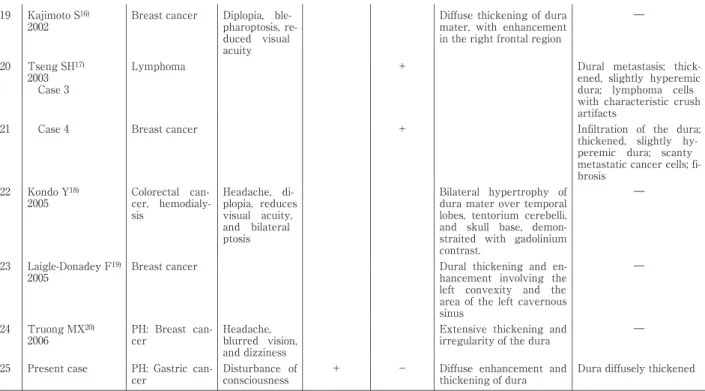
Table 2 Summary of25 caseswith thickened dura associated with metastaticcancer.
Duralpathologicfindings MRIfindings Subdural hematoma Coagulation disorder Clinical symptoms Primary tumor Author Year Case No.
Extensive cartinomatous infiltration. The areolar layerwasthickened. + Diplopia, visual acuity de-creased Carcinoma ofac -cessory nasal si -nuses or prostate RussellDS1) 1934 Case 1 1 Great thickening of the areolar layer also was observed.
+ Delirious,
un-conscious Scirrhous carci
-noma ofstomach Case 2
2
The periostealsurface of the dura was greatly thickened. Delirious Carcinoma of lung Case 3 3 Extensive infiltration of dense and areolarlayers. The areolar layer was greatly thickened by granulation tissue. Nystagmus, leftfacial weak-ness Ewing’ ssarcoma Case 4 4
The dura was diffusely infiltrated. Subdural effusion Headache Hodgkin’ s disease McDonald JV2) 1966 5
The dura was diffusely thickened (2 to 3 mm). Nu-merous tumor nodules filled the vascular c han-nelsofthe innerlayer. +
Thrombocyt -openia Lethargy, ani -socoria,and i n-creased flexor tone in the left arm
Pancreatic c an-cer
Leech RW3)
1974 6
The dura was notably tense and diffusely thickened. Numerous markedly distended ves -sels contained intraluminal tumor nodules. Headache and facialedema Gastriccancer SakakiS4) 1979 7 Adenocarcinoma cells presentin the vessels. + DIC Nausea and vomiting Left hemipar e-sis,coma Gastriccancer
HaraguchiS5)
1981 8
In the inner dural layer, many ofthe innumerable dilated smallvesselsc on-tained tumor.
+ DIC
Left hemipar e-sis, convulsion, coma Adenocarci no-ma of unknown orgin Hirashima Y6) 1983 9
Numeroustumorcell em-boliwere seen in the i n-nervascularlayer.Fibr o-blastic reaction was prominent in the dense outerlayer.
DIC Headache, nau-sea, vomiting, loss of c on-sciousness Gastriccancer Ishikawa I7) 1985 10
Thickening of dura mater. + Headache, stiff neck Adenocarci no-ma ofstomach Tohyama I8) 1986 11
Bone marrow metastasis + Bleeding tendency Right hemi -paresis, coma, anisocoria Adenocarci no-ma ofstomach Kamiya K9) 1987 12 Grossly thickened dural membrane over the left cerebral convexity and base ofskull.
Rim thickestoverthe mi d-temporoparietalarea -
Headache,let h-argy, and inat -tentiveness Pulmonary or gastriointestinal primary tumor TyrrellRL10) 1987 13 On the right, the dura was covered with fresh hemorrhages up to 0.5 cm thick. + Gastric distur -bances and dysphagia Gastriccancer Bergmann M11) 1992 14
Some ofthe largerdural veins contained large clumpsoftumorcells. Diffuse enhancement and
thickening ofdura overl y-ing the right cerebral hemisphere Complex par -tial status epi -lepticus Carcinoma of the bladder Steg RE12) 1993 15
Thickening of parietal dura. + Right hemi -paresis and aphasia Lung cancer KurokiT13) 1994 16 ― Diffuse dural
enhancementand thicking Headache Prostate cancer TerashiH14) 1999 17 Adenocarcinoma,fibrosis, infiltrating lymphocytes, and subdural effusion, correlating with the duralmetastasis Thickened dura and multi
-ple CSF leaks CSF leaks DIC Headache, vomiting Unknown TakahashiS15) 1999 18
臨床神経学 50巻4号(2010:4) 50:238
― Diffuse thickening ofdura
mater, with enhancement in the rightfrontalregion Diplopia, bl e-pharoptosis,r e-duced visual acuity Breastcancer Kajimoto S16) 2002 19 Dural metastasis; thic k-ened, slightly hyperemic dura; lymphoma cells with characteristiccrush artifacts + Lymphoma Tseng SH17) 2003 Case 3 20 Infiltration of the dura; thickened, slightly hy-peremic dura; scanty metastaticcancercells;fi -brosis + Breastcancer Case 4 21 ― Bilateral hypertrophy of
dura materovertemporal lobes, tentorium cerebelli, and skull base, demon-straited with gadolinium contrast. Headache, di -plopia, reduces visual acuity, and bilateral ptosis Colorectal c an-cer, hemodial y-sis Kondo Y18) 2005 22 ― Dural thickening and
en-hancement involving the left convexity and the area ofthe leftcavernous sinus Breastcancer Laigle-Donadey F19) 2005 23 ― Extensive thickening and
irregularity ofthe dura Headache,
blurred vision, and dizziness PH: Breast c an-cer Truong MX20) 2006 24 Dura diffusely thickened Diffuse enhancement and
thickening ofdura -
+ Disturbance of consciousness PH: Gastric c
an-cer Presentcase
25
PH,pasthistory;DIC,disseminated intravascularcoagulation;MRI,magneticresonance imaging;+ ,present;- ,absent.
Table 3 Primary tumorsin caseswith thickened dura associated with metastaticcancer. Paakko (1990)24)** River(1996)23)* Numberofcases Primary tumor 71)4)5)7)~9)11) Gastricca 1 7 416)17)19)20) Breastca 7 2 114) Prostate ca 5 22)17) Lymphoma 21)13) Lung ca 1 118) Colon ca 13) Pancreasca 112) Bladder 11) Ewing’ ssarcoma 1 Nasopharyngealca
1 Malignantmeningioma
1 Medulloblastoma
1 Ovarian ca 41)6)10)15) Unknown 14 13 24 Total ca,carcinoma
*Diffuse duralenhancementand associated cancer **Patientswith duralenhancement
る.
謝辞:本症例の検査についてご教示いただいた国分生協病院内 科,浜田暁子先生に深謝します.
文 献
1)Russell DS, Cairns H. Subdural false membrane of hema-toma (pachymeningitis interna hemorrhagica) in carcino-matosis and sarcocarcino-matosis of the dura mater. Brain 1934; 57:32-48.
2)McDonald JV, Burton R. Subdural effusion in Hodgkin s disease. Arch Neurol 1966;15:649-665.
3)Leech RW, Welch FT, Ojemann GA. Subdural hematoma secondary to metastatic dural carcinomatosis. J Neuro-surg 1974;41:610-613.
4)Sakaki S, Mori Y, Matsuoka K, et al. Metastatic dural car-cinomatosis secondary to gastric cancer. Neurol Med Chir (Tokyo) 1979;19:39-44.
5)原口庄二郎, 坪内隆佳, 蝦名一夫. 胃癌の硬膜転移に続発し た硬膜下出血腫の一例. 上二病医誌 1980;2:177-182. 6)平島 豊, 神山和世, 遠藤俊郎ら. DIC を合併し硬膜下血腫 を呈した硬膜転移癌の 1 例. 脳神経外科 1983;11:651-656. 7)石川育夫, 大川直澄, 山下勢一郎ら. 胃癌の硬膜転移により 硬膜下血腫を呈した 1 症例. 外科 1985;47:1561-1563. 8)遠山育夫, 小川道子, 川又敏男ら. 硬膜下血腫を伴う癌性硬 膜炎例とその文献的考察. 神経内科 1986;24:596-601. 9)上家和子,!川哲二, 長廻 錬. 血腫内容から腫瘍細胞を検 出 し た 胃 癌 転 移 に よ る 慢 性 硬 膜 下 血 腫 の 1 例. Neurol Med Chir (Tokyo) 1987;27:892-898.
10)Tyrrell RL, Bundschuh CV, Modic MT. Dural carcinoma-tosis: MR demonstration. J Comput Assist Tomogr 1987; 11:329-332.
11)Bergmann M, Puskas Z, Kuchelmeister K. Subdural he-matoma due to dural metastasis: case report and review of the literature. Clin Neurol Neurosurg 1992;94:235-240. 12)Steg RE, Frank AR, Lefkowitz DM. Complex partial
status epilepticus in a patient with dural metastases. Neu-rology 1993;43:2389-2392. 13)黒木貴夫, 松元幹郎, 串田 剛ら. 肺癌の硬膜転移による硬 膜下血腫の 1 例―症例報告と文献的考察―. 脳神経外科 1994;22:857-862. 14)赫 洋美, 清水優子, 小宗規子ら. 前立腺癌を伴った慢性肥 厚性硬膜炎の一症例. 頭痛会誌 1999;26:77-82. 15)高橋重人, 阿部光文, 鈴木理恵ら. 肥厚性硬膜炎,多発性髄 液漏を認めた硬膜転移性腫瘍. 神経内科 1999;51:574-577. 16)梶本賀義, 記平為子, 三輪英人ら. 乳癌患者に認められた癌 性肥厚性硬膜炎. 脳神経 2002;54:626-627.
17)Tseng SH, Liao CC, Lin SM, et al. Dural metastasis in pa-tients with malignant neoplasm and chronic subdural
he-matoma. Acta Neurol Scand 2003;108:43-46.
18)近藤裕美, 西村芳子, 田中俊久ら. 大腸癌を伴う慢性肥厚性 硬膜炎を呈した血液透析患者の 1 例. 透析会誌 2005;38:57-60.
19)Laigle-Donadey F, Taillibert S, Mokhtari K, et al. Dural metastases. J Neurooncol 2005;75:57-61.
20)Truong MX, Ting AC, Rossleigh MA, et al. Dural metas-tasis demonstrated on Tc-99m MDP bone scintigraphy in staging workup for a patient with breast cancer. Clin Nu-clear Med 2006;31:223-224.
21)林 英夫, 春山春枝, 江村芳文ら. 播種性骨髄癌症―転移癌 の一病型としての考察ならびに microangiopathic hemo-lytic anemia ま た は disseminated intravascular coagula-tion との関連について―. 癌の臨床 1979;25:329-343. 22)辻 裕二, 早川 滉, 山本匡介ら. 早期胃癌切除後 11 年目 に局所再発なく播種性骨髄癌症をきたした 1 例―播種性 骨髄癌症本邦報告 43 症例(1977∼1980)の検討―. 最新医 学 1982;37:2243-2250. 23)西岡清春, 金武喜子, 分枝孟義ら. 癌のビマン性汎発性骨髄 転移症について. 臨床放射線 1968;13:334-346.
24)River Y, Schwartz A, Gomori JM, et al. Clinical signifi-cance of diffuse dural enhancement detected by mag-netic resonance imaging. J Neurosurg 1996;85:777-783. 25)Paakko E, Patronas NJ, Schellinger D. Meningeal
Gd-DTPA enhancement in patients with malignancies. J Comput Assist Tomogr 1990;14:542-546.
26)Antman KH, Skarin AT, Mayer RJ, et al. Microan-giopathic hemolytic anemia and cancer: a review. A Re-view Medicine 1979;58:377-384.
27)杉本琢哉, 関野考史, 左合 哲ら. 胃癌手術後 22 年目にみ られた播種性骨髄癌症の 1 例. 日臨外会誌 2006;67:64-67.
臨床神経学 50巻4号(2010:4) 50:240
Abstract
An autopsy case of dural thickening caused by widespread dural vein thrombosis associated with disseminated bone marrow carcinomatosis
Kyoko Maruta, M.D.1) , Joeji Wakimoto, M.D.2) , Yoshito Sonoda, M.D.1) , Yuichi Uchida, M.D.1) , Fujio Umehara, M.D.3)
and Hidetoshi Fukunaga, M.D.1) 1)
Department of Neurology, National Hospital Organization Minamikyushu National Hospital
2)
Department of Pathology, National Hospital Organization Minamikyushu National Hospital
3)
Department of Neurology and Geriatrics, Kagoshima University Graduate School of Medical and Dental Sciences
A 64-year-old woman was referred to our hospital because of disturbance of consciousness. She had under-gone distal gastrectomy for gastric carcinoma 17 years previously. General physical examination was unremark-able, neurologic examination disclosed hyperactive deep tendon reflexes in the upper limbs.
Laboratory abnormalities included elevations of alkaline phosphatase ( ALP ) and lactate dehydrogenase (LDH), and findings suggesting disseminated intravascular coagulation (DIC). Lumbar cerebrospinal fluid showed a protein concentration of 408 mg!dl and a glucose concentration of 82 mg!dl (blood: 110 mg!dl), as well as a cell count of 16!mm3
.
Cranial computed tomography indicated brain edema. Magnetic resonance imaging ( MRI ) of the brain showed diffuse thickening of the dura mater, with contrast enhancement upon gadolinium-DTPA administration. These findings suggested hypertrophic pachymeningitis. Magnetic resonance venography (MRV) showed occlu-sion of the left transverse sinus and attenuation of the straight sinus. MRI of the spine as well as gallium scinti-grams demonstrated multiple areas of increased uptake in areas near the skull and spine. We therefore suspected tumor metastasis.
The patient was given heparin as well as pulse therapy with methylprednisolone, but she died 7 weeks after symptom onset. At postmortem examination, the dura was thickened. Histopathologically, numerous tumor cell emboli in the dura were confined to the lumens of veins. The tumor cells were thought to have metastasized to the dura through the vertebral venous plexus (Batson s plexus). Immunostaining demonstrated immunoreactivity of tumor cells to epithelial membrane antigen (EMA) and carcinoembryonic antigen (CEA). The primary origin of the carcinoma was not precisely identified by these findings.
Widespread dural vein tumor emboli should be taken into consideration as a cause in cases that develop rapid deterioration of consciousness associated dura mater thickening.
(Clin Neurol 2010;50:232-240) Key words: hypertrophic pachymeningitis, dural thickening, tumor emboli, brain edema, disseminated bone marrow