内 容 原 著
糞線虫症の治療効果に及ぼすATLウイルス混合感染の影響(英文)
・・佐藤 良也,城間 祥行,喜友名 進,當真 弘,
小林 潤 183−192 航空輸送機関および外国各地公共施設における飲料水の水質(英文)
・・奥村悦之,橋本 恵介,水田 成彦,秦 光孝,
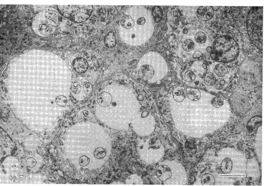
奥村 泰之,岡村 宜典 193−201 実験的リーシュマニア症一エクアドルで分離された,L廊h㎜吻吻翻o朋α感染
ゴールデンハムスターの組織学的検討一(英文)
一A.M.Bhutto,野中 薫雄,E.A.Gomez L.,
橋口 義久,古谷正人 203−215
短 報
無鉤条虫感染症における血清CRP値(英文)
・・大西 健児,村田三沙子 217−218
会報・記録
1992年度第1回幹事会記録 …一……・……・…一………・…………・……… 219−220 1992年度会員名簿……一…………・……一一……・・………・…・……一…………・…… 221−246 第4回熱帯医学と医療を語る会のお知らせ………・一・一・………・……… 247 第17回日本熱帯医学会九州支部大会のお知らせ(第1報) ・…・………・……… 248 投稿規定…………・…・・……・…・………・………一・一………・…………__… 24g
日本熱帯医学会
Jpn J Trop Med Hyg., Vol. 20, No. 3, 1992, pp. 183‑192 183
REDUCED EFFECT OF CHEMOTHERAPY OF
STRONGYLOIDIASIS IN PATIENTS WITHCONCURRENT HTLV‑1 INFECTION IN OKlNAWA, JAPAN
YOSHIYA SATO*'2, *, YOSHIYUKI SHIROMA SUSUMU KIYUNA HIROMU ToMA AND JUN KOBAYASHll
Received March 9 1992/Accepted May 7 1992
Abstract: The effect of concurrent HTLV‑1 infection on the efficacy of anti‑Stron‑
gyloides chemotherapy has been tested in a prognostic study on 96 patients with uncom‑
plicated strongyloidiasis. The efficacy of treatment with pyrvinium pamoate and thiaben‑
dazole was found to be significantly low in the HTLV‑1 seropositive patients, as compared to that in the seronegative group. The cure rates in the HTLV‑1 seropositive patients, when assessed by both faecal examination and serological testing, were only 8.6% for pyrvinium pamoate treatment and 35.0% for thiabendazole treatment. On the other hand, the cure rates were 31.3% for pyrvinium pamoate treatment and 57.1% for thiabendazole treatment in the seronegative group.
Although antibody response against Strongyloides did not decrease among the HTLV‑
I seropositive patients, the total serum lgE Ievels, as well as eosinophil counts in peripheral blood, were relatively low in the seropositive group. Abnormal lymphocytes were demon‑
strated in 38.5% of the patients with concurrent HTLV‑1 infection. The presence of abnormal lymphocytes, however, appeared not to correlate with the therapeutic efficacy.
A possible connection between the poor efiicacy and depres ed immune responses provided by the concurrent HTLV‑1 infection is discussed. The low efficacy in the HTLV‑I seropositive patients also provides a causal explanation for the significant accumulation of patients with concurrent HTLV‑1 infection in Okinawa.
INTRODUCTION
Strongyloidiasis caused by Strongyloides stercoralis infection still remains prevalent in Okinawa Prefecture, Japan, where other parasitic diseases have been almost entirely eradicated in recent years (Sato, 1986). Because of its opportunistic nature, the great majority of patients who are immunocompetent are generally chronic, producing no symptom clearly attributable to the presence of the parasite. On the other hand, under the condition of depressed immune competence, the chronic infection progresses to a massive and often fatal systemic infection as a consequence of increasing autoinfection with larval parasites.
1 Department of Parasitology, and 2 Research Center of Comprehensive Medicine, School of Medicine, University of the Ryukyus, 207 Uehara, Nishihara, Okinawa 903‑01, Japan (*corre‑
spondent author)
3 Izumizaki Hospital, 1‑11‑2 Izumizaki, Naha, Okinawa 900, Japan
Okinawa Prefecture is also known to be an endemic area for human T‑cell leukemia virus (HTLV‑D infection (Clark et al., 1985). The HTLV‑1 is aetiologically associated with adult T‑cell leukemia (ATL) which leads to severe deficiencies in immunological responses.
Recently, it has been demonstrated that the patients with an asymptomatic Strongyloides infection in Okinawa are highly accompanied by the ATL viral infection (Nakada et al., 1984; Fujita et al., 1985; Sato and Shiroma, 1989). Under the condition of concurrent Strongyloides and ATL viral infection, the progression of asymptomatic Strongyloides infec‑
tion to a fatal disseminated state has been often observed among the patients who developed ATL (Takara et al.. 1980; Matsui et al.. 1982; Oura et al.. 1986) . A possible explanation for the high frequency of such a complication, however, has not yet been offered. In this paper we present results showing the depressed efficacy of treatment of strongyloidiasis in patients with concurrent HTLV‑1 infection and thus discuss the possibility that the low therapeutic efiicacy may be attributed to the depressed immune responses provided by the viral infection and also may be responsible for a significant accumulation of strongyloidiasis patients with concurrent HTLV‑1 infection for a long period in Okinawa.
MATERIALS AND METHODS
Patients
A total of 96 patients with strongyloidiasis received medical' treatment for stron‑
gyloidiasis in the lzumizaki Hospital, Okinawa, Japan. They were consisted of 47 males and 49 females. Their age ranged from 30 to 74 years (mean=53.3 years) . They were a ymptomatic or mildly symptomatic cases. Among them, positive rate of antibodies against ATL‑associated antigen (ATLA) was as high as 67.7%.
Treatments
Fifty‑one patients were treated with pyrvinium pamoate suspension (PoquiD , which was administered in a dosage of 5 mg per kg body weight daily for 3 to 5 consecutive days. For the other 27 patients, thiabendazole (25 mg/kg for 4 days) was administered. The above treatment schedules have long been used in Okinawa for treatment of strongyloidiasis. The remaining 18 patients were left without any treatment as a control group. The duration from treatment to follow‑up faecal examination was different for each cases, but it was more than 2 years for all cases.
Faecal examination
The follow‑up faecal examination after the treatment was performed repeatedly for consecutive 3 days by three different method, e.g. direct faecal smear, formol‑ether concen‑
tration method and faecal culture (Harada‑Mori method) . Antibodies to S, stercoralls
In order to assess serologically the efficacy of treatment, the serum antibodies to S.
stercoralis were measured by an enzyme‑linked immunosorbent assay (ELISA). The antigens used were prepared from S. stercoralis filariform larvae collected from faeces of strongyloidiasis patients (Sato et al.. 1983). The conventional technique for the micro‑
ELISA using a microtiter plate was the same as that described in a previous paper (Sato et
185
al., 1985). The sera were tested at a single dilution of 1:50 and the intensity of antibody response was expressed as the absorbancy (OD) at 500 nm. On the basis of the previous criteria, the OD value of over 0.5 was regarded as being antibody positive (Sato et al.. 1990b) .
Detection of antibody to ATL virus
The individuals having anti‑ATLA antibodies have been known to be infected with HTLV‑1 in their peripheral lymphocytes (Gotoh et al.. 1982).
A kit for the particle agglutination test to detect anti‑ATLA antibodies was kindly supplied from Fujirebio Inc., Tokyo. A usual indirect agglutination test using gelatin particles coated with antigens prepared from culture fluid of the virus‑producing cell line was done in U‑bottomed wells of a plastic microplate (Ikeda et al., 1984). The mixtures of the antigen‑coated particles and serially diluted sera in the wells were allowed to stand for 3 hr at room temperature and the resulting patterns formed on the bottom of the wells were read.
The final serum dilution of 1:16 or higher showing agglutination was interpreted as positive.
Serum total lgE
Serum lgE was measured by a standard radioimmunosorbent test using Phadebas lgE test kit (PRIST; Pharmacia Fine Chemicals, Uppsala, Sweden) . Serum lgE Ievels were expressed in ng/ml of serum, assuming that I unit/ml corresponds to approximately 2 ngl ml.
Haematological examinations
Eosinophils from peripheral blood were counted in Wright's‑stained smear and were reported as percentage in 100 Ieukocytes. Morphologically abnormal lymphocytes having lobular or indented nulei were also counted in the sarne srnears.
Statistics
The data were analyzed by X2 test and Student's t test, as appropriate. A P value of more than 0.05 was considered not to be siguificant.
RESULTS
The results of follow‑up examination after treatment are shown in Table 1. The total cure rates, as estimated by both faecal and serological examinations, were 15.7% for pyrvinium pamoate treatment and 40.7% for thiabendazole treatment, respectively. Although three patients in the untreated group were negative in the follow‑up faecal examination, they were serologically equivocal for complete cure and the spontaneous cure determined by exclusion of the equivocal cases was not detected in the untreated group. When the efficacy were compared between the HTLV‑1 seropositive and seronegative patients, the cases with ̲ complete cure were significantly fewer in the HTLV‑1 seropositive group than in the ser‑
onegative group in both treatments.
Table 2 represents total lgE Ievels in sera of 39 patients who were still positive after the treatment. The lgE Ievels were relatively lower in the HTLV‑1 seropositive group than in the seronegative group. Although the difference was not significant statistically, the mean lgE Ievel in the seropositive group was as low as one‑third that of the seronegative group.
Table I Effect of concurrent HTLV‑1 infection on treatment of strongyloidiasis with pyrvinium pamoate and thiabendazole
Faecal examination Positive
Anti‑ATLA No.
Treatment antibody examined (not cured) Negative Significance*
Sero‑positivet sero‑negativet
( cured )
(equivocal) Positive
Pyrvinium Negative
pamoate Total
35 30 (85.7) 16 10 (62.5)
2 (5.7) 1 (6.3)
3 (8.6)
5 (31.3) P<0,05 51 40 (78.4) 3 (5.9) 8 (15.7)
Positive
Thiabenda‑ Negative
zole
20 7
9 (45.0) 2 (28.6)
4 (20.0) 1 (14.3)
7 (35.0)
4 (57.1) N .S.
Total 27 Il (40.7) 5 (18.5) ll (40.7)
Positive
Untreated Negative
10 8
7 (70.0) 8 (lOO)
3 (30,0)
o (o) o (o)
o (o) N.S.
Total 18 15 (83.3) 3 (16.7) o (O)
* Significance between HTLV‑1 seropositive and seronegative groups; t anti‑Strongyloides antibody
Although the difference of cure rate between HTLV‑1 seropositive and seronegative groups was not significant (P<0.1) in thiabendazole treatment, the total cure rate of both treatments was significantly lower (P<0.05) in the HTLV‑1 seropositive group.
Table 2 Comparison of serum total lgE Ievels between HTLV‑I seropositive and seronegative patients
Anti‑ATLA antibody lgE Ievel (ng/ml)
Positive (n=38) Negative (n=12)
< I , OOO 1 , OOO ‑ 3, OOO
3, 100 ‑ 5 ,OOO
> 5 , OOO
34 (89.5) 4 (10.5)
O (O) O (O)
8 (66.7) 2 (16.7) 1 (8.3) l (8.3)
Mean d: SD 415.8:!:523.2 l , 300 d: I , 795 N.S.*
*Significance between HTLV‑1 seropositive and seronegative groups.
Table 3 also shows eosinophil counts in peripheral blood of 25 patients with Strongyloides infection. Eosinophilia of over 6% was detected in 57.8% of patients who were negative for anti‑ATLA aptibody, while the percentage of patients with eosinophilia was 35.0% in the HTLV‑1 seropositive patients. On the other hand, when anti‑Strongyloides antibodies were compared between the HTLV‑1 seropositive and seronegative groups, the antibody levels were relatively higher in the seropositive group, showing no depressed antibody response to Strongyloides among the seropositive group (Fig. 1) .
The presence of abnormal lymphocytes in peripheral blood was determined in 52 patients who were positive for anti‑ATLA antibody and atypical lymphocytes were detected in 20
187
Table 3 Eosinophil counts in peripheral blood of strongyloidiasis with or without concurrent HTLV‑1 infection
patients
Blood
eosinophils (%)
Anti‑ATLA antibody Positive (n=40) Negative (n = 19)
‑6
7 ‑ 10 ll ‑ 15 16 ‑
26 (65.0) 7 (17.5) 6 (15.0) 1 (2.5)
8 7 2 2
(42 .
(36.
(10.
(10.
1) 8) 5) 5)
Eosinophilia ( > 6% ) 14 (35.0) ll (57.8) N.S.*
*Significance between HTLV‑1 seropositive and seronegative groups.
P‑ms (38.5%) patients. The relation between the pres‑
ence of atypical cells and the therapeutic efflcacy is shown in Table 4. The positive rate of atypical cells was relatively higher in the group of unsuc‑
cessful treatment than in the group of complete cure in pyrvinium pamoate treatment, but it were almost similar in thiabendazole treatment.
DISCUSsloN
Strongyloidiasis and ATL are presently high‑
ly prevalent in Okinawa Prefecture, Japan; the prevalence levels appear to be 5 to lO% for Strongyloides (Sato, 1986; Sato et al., 1990b) and about 20% for HTLV‑1 infection (Clark et al..
1985). Recently, it has been pointed out by sev‑
eral researchers that Strongylo,ides carriers in Okinawa are frequently accompanied by HTLV‑I infection (Nakada et al., 1984; Fujita et al.. 1985;
Sato and Shiroma, 1989). In these studies, the positive rates of anti‑ATLA antibody as high as Table 4 Correlation between efficacy of lymphocytes in peripheral blood
inf ection
e
e
,D c::]
O
1
<
u) J
UJ
1 .5
1 .O
0.5
.
dL
$ i
‑
i
‑.
i .
e
A t
a
.
8
Positive Negative Anti‑ATLA antibody
Figure I Comparison of anti‑Strongyloides ELISA values between HTLV‑I seropositive and seronegative patients with strongyloidiasis.
treatment and presence of abnormal of patients with concurrent HTLV‑I
Treatment Efficacy of treatment
Not cured Equivocal Cured Total Pyrvinium pamoate 13/28 (46.4) 0/2
Thiabendazole 3/9 (33.3) l/3
(o) (33 . 3)
1/3 2/7
(33 . (28 .
3) 6)
14/33 6/19
(42.4)
(31 . 6)
Total 16/37 (43.2) 1/5 (20.0) 3110 (33 . 3) 20/52 (38 . 5) No. positive/No. examined (%)
57.8 to 73.6% were detected among the strongyloidiasis patients.
Several explanations have been proposed for the severe complication of these infections.
The participation of antigenic components common to Strongyloides and ATLA which may produce high positive rate of anti‑ATLA antibody among the Strongyloides carriers has been excluded by the authors (Sato and Shiroma, 1989). Possible participation of any epidemiological factors to produce the severe overlap of the two infections has also been excluded in a previous paper (Sato et al.. 1990a).
Alternatively, it can be supposed that the concurrent HTLV‑1 infection may affect on the severity of Strongyloides infection through the depressed immune competence of host during the viral infection. The data presented in this paper demonstrate that the anti‑parasitic effect of the anthelmintics is greatly reduced in strongyloidiasis patients with concurrent HTLV‑I infection. In Okinawa, intractable cases of strongyloidiasis, in which relapse occurs repeated‑
ly after various treatment over a period of many years, have been often observed (Shiroma et al., 1990) . Although the factor responsible for such a resistance to anthelmintic treatment is unclear, it can be postulated that the poor efiicacy of treatment may be attributed to the depressed immune response which provided by the viral infection. As well documented, ATL is characterized by an unique T‑lymphocyte malignancy which leads to severe deficiencies in immune responses, and it is also known that the disorder to a T‑cell mediated immune system has already begun in the stage of virus carriers (Imai and Hinuma, 1983; Yasuda et al.. 1986;
Tanaka et al., 1989; Prince et al.. 1990). Because of the opportunistic nature of the parasitic pathogen, it is reasonable to suppose the immune dependence of anti‑parasitic chemotherapy in strongyloidiasis.
In the past decade, substantial evidence for the immune dependence of chemotherapy has accumulated. It has been known that immunosuppression reduces the efiicacy of chemother‑
apy in several parasitic diseases, such as malaria (Lwin et al.. 1978), trypanosomiasis (Frommel, 1988), onchocerciasis (Bianco et al., 1986) and schistosomiasis (Doenhoff and Bain, 1978). In the case of strongyloidiasis, it is also well documented that the patients with severe infection under the immunocompromised condition often fail to respond to anthel‑
mintic treatment, and that repeated courses of treatment are necessary to obtain a complete cure for such a severe case (Scowden et al.. 1978; Weller et al., 1981; Shelhamer et al.. 1982;
Morgan et al.. 1986). Although the immune factor involved in influencing the drug efficacy is not yet determined, it has been reported that antibodies specific for parasite, when it were administered simultaneously with drug, enhanced the efflcacy of drug against schistosomes and malaria parasite (Doenhoff and Bain, 1978; Targett, 1985; Brindly and Sher, 1987).
When the antibody titers to Strongyloides were compared between HTLV‑1 seropositive and seronegative patients in the present study, however, we could not find any evidence to suppose a depressed antibody response against Strongyloides in the seropositive group. While the drug efiicacy in onchocerciasis reduced markedly in T‑cell deprived mice but not in congenitally B‑cell deficient mice, suggesting that cellular immune effector mechanism other than the serum antibody may contribute to the efficacy of chemotherapy in this parasitic disease (Bianco et al., 1986). With respect to strongyloidiasis, significance of cell‑mediated immu‑
nity in controlling and preventing Strongyloides infection has also been suggested in several clinicopathological studies (Purtilo et al.. 1974; Scowden et al.. 1978; Cohen and Spry, 1979) . As already mentioned above, HTLV‑1 carriers are known to have immunological abnormal‑
ities, such as increased spontaneous lymphoproliferation, decreased mitogenic responses and
189
increased expression of IL‑2 receptor on the surface of T Iyinphocytes. In the present study, morphologically atypical lymphocytes were detected in about 40% of HTLV‑1 seropositive patients examined. In their study on 36 patients, Nakada et al. (1987) has reported that abnormal lymphocytes, as well as monoclonal integration of HTLV‑1 proviral DNA, were detected in many patients and indicated that the presence of abnormal cells and monoclonal integration of proviral DNA correlated with a trend for greater severity of the parasitic infection. However, we could find no correlation between the presence of abnormal cells and the e cacy of treatment in the present study. On the other hand, it was noted that total serum lgE Ievels and eosinophil counts in peripheral blood were relatively low in the HTLV‑I seropositive patients. The lgE and eosinophilic responses are well known to play an impor‑
tant role in protective immunity to parasitic helminths (Capron et al.. 1981; Dessein et al..
1981; Kojima et al.. 1985). Recently, it was also demonstrated in a study with murine infection model that IL‑5‑dependent eosinophilic response was important for protective immunity to Strongyloides infection (Korenaga et al.. 1991). The relative decrease of lgE level and peripheral eosinophils in the HTLV‑1 seropositive patients may participate in the poor efficacy of chemotherapy. Further investigations on a possible connection between the immune responses and drug e :cacy in strongyloidiasis should be intended.
Finally, the reduced efiicacy of drug treatments in HTLV‑1 seropositive patients may provide another explanation for the high concurrency of HTLV‑1. Due to resistance to anthelmintic treatments, which has long been used for strongyloidiasis in Okinawa, the patients with concurrent . HTLV‑1 infection might harbour the infection for many years, resulting a significant accumulation of such patients.
ACKNOWLEDGMENTS
This study was supported in part by grants from the Ohyama Health Foundation and the Chiyoda Mutual Life Foundation. The authors are also grateful to Fujirebio Inc., Tokyo, Japan (Mr. S. Hanzawa) for supplying kits for detection of anti‑ATLA antibody (Serodia‑
ATLA) .
REFERENCES
1 ) Aianco, A.E., Nwachukwu, M.A., Townson, S., Doenhoff, M.J. and Muller, R.L. (1986):
Evaluation of drugs against Onchocerca microfilariae in an inbred mouse model, Trop. Med.
Parasitol., 37, 39‑45
2 ) Brindley, P.J. and her, A. (1987): The chemotherapeutic effect of praziquantel against Schistosoma mansoni is depend on host antibody response. J. Immunol., 139, 215‑220
3 ) Capron, M., Bazin, H., Joseph, M. and Capron, A. (1981): Evidence for lgE‑dependent cytotoxicity by rat eosinophils, J. Immunol., 126, 1764‑1768
4 ) Clark, J.W.. Robert‑Guroff. M., Ikehara, O., Henzan, E. and Blattner, W.A. (1985): Human T‑
cell leukemia‑lymphoma virus type I and adult T‑cell leukemia‑lymphoma in Okinawa, Cancer Res., 45, 2849‑2852
5 ) Cohen, J. and Spry, C.J.F. (1979): Strongyloides stercoralis infection and small intestinal lymphoma, Parasite Immunol., 1, 167‑178
6 ) Dessein. A.J., Parker, W.L., James, S.L. and David, J.R. (1981): IgE antibody and resistance to infection.
I. Selective suppression of the lgE antibody response in rats diminishes the resistance and the eosinophil response to Trichinella spiralis infection, J. Exp. Med., 153, 423‑436
7 ) Doenhoff, M.J. and Bain, J. (1978): The immune‑dependence of schistosomicidal chemother‑
apy: relative lack of efflcacy of an antimonial in Schistosoma mansoni‑infected mice deprived of their T‑cells and the demonstration of drug‑antiserum synergy, Clin. exp. Immunol., 33, 232‑
238
8 ) Fujita. K., Tajima, K., Tominaga, S., Tsukidate, S., Nakada, K.. Imai, J. and Hinuma, Y.
(1985): Seroepidemiological studies of Strongyloides infection in adult T‑cell leukemia virus carriers in Okinawa Island, Trop. Med., 27, 203‑209
9 ) Frommel. T.O. (1988) : Tr)panosoma brucei rhodesiense: Effect of immunosuppression on the efficacy of melarsoprol treatment of infected mice. Exp. Parasitol., 67, 364‑366
10) Gotoh, Y., Sugamura, K. and Hinuma, Y. (1982): Healthy carriers of a human retrovirus, adult T‑cell leukemia virus (ATLV): Demonstration by clonal culture of ATLV‑carrying T cells from peripheral blood, Proc. Natl. Acad. Sci. USA, 79, 4780‑4782
lD Ikeda, M., Fujino, R., Matsui, T.. Yoshida, T., Komoda, H. and Imai, J. (1984): A new agglutination test for serum antibodies to detect T‑cell leukemia virus. Gann, 75, 845‑849 12) Imai, J. and Hinuma, Y. (1983): Epstein‑Barr virus specific antibodies in patients with adult
T‑cell leukemia and healthy ATLV‑carriers, Int. J. Cancer, 31, 197‑200
13) Kojima, S., Yamamoto, N.. Kanazawa, T. and Ovary, Z. (1985): Monoclonal lgE‑dependent eosinophil cytotoxicity to haptenated schistosomula of Schistosoma japonicum: enhancement of the cytotoxicity and expression of Fc receptors for lgE by Nippostrongylus brasiliensis infection, J. Immunol., 134, 2719‑2722
14) Korenaga, M., Hitoshi, Y., Yamaguchi, N.. Sato, Y., Takatsu, K. and Tada, I. (1991): The role of interleukin‑5 in protective immunity to Strongyloides venezuelensis infection in mice, Im‑
munology, 72, 502‑507
15) Lwin, M., Targett, G.A.T. and Doenhoff, M.J. (1987): Reduced efiicacy of chemotherapy of Plasmodium chabaudi in T cell deprived mice, Trans. Roy. Soc. Trop. Med. Hyg., 81, 899‑902 16) Matsui, K.. Sakihara, H.. Toyama, K. and Ito. E. (1982): Clinicopathological studies of strongyloidiasis in the Okinawa Prefecture. Ryukyu Univ. J. Health Sci. Med., 5, 19‑32 (in Japanese)
17) Morgan, J.S., Schaffner, W. and Stone, W.J. (1986): Opportunistic strongyloidiasis in renal transplant recipients, Transplantation, 42, 518‑524
18) Nakada, K.. Kohakura. M.. Komoda, H. and Hinuma, Y. (1984): High incidence of HTLV‑
antibody in carriers of Strongyloides stercoralis. Lancet, 1, 633
19) Nakada, K., Yamaguchi, K., Furugen, S., Nakasone, T., Nakasone, K., Oshiro, Y.. Kohakura, M., Hinuma, Y., Seiki, M., Yoshida, M.. Matutes, E.. Catovsky, D., Ishii, T. and Takatsuki, K.
(1987): Monoclonal integration of HTLV‑1 proviral DNA in patients with strongyloidiasis, Int. J. Cancer, 40, 145‑148
20) Oura, T., Kadena, M., Irei, M., Higa. S., Mimura, G. and Matsui. K. (1986) : Fatal stron‑
gyloidiasis, ‑TWO autopsy cases‑, Internal Med., 58, 1243‑1246 (in Japanese)
21) Prince, H., Kleinman, S., Doyle, M., Lee, H. and Swanson, P. (1990): Spontaneous lymphocyte proliferation in vitro characterizes both HTLV‑1 and HTLV‑II infection, JAIDS, 3, 1199‑1200 22) Purtilo, D.T., Meyers, W.M. and Conner, D.H. (1974): Fatal strongyloidiasis in immunosup‑
pressed patients, Am. J. Med., 56, 488‑493
23) Sato. Y. (1986): Epidemiology of strongyloidiasis in Okinawa, Collected Papers on the Control of Soil‑transmitted Helminthiasis, 3, 20‑31
24) Sato, Y. and Shiroma, Y. (1989) : Concurrent infections with Strongyloides and T‑cell leukemia virus and their possible effect on immune responses of host, Clin. Immunol. Immunopathol., 52, 214‑224
191
25) Sato, T., Takai, A., Maeshiro, J., Otsuru, M. and Shiroma, Y. (1983): Studies on the prepara‑
tion of antigen and application of enzyme‑linked immunosorbent assay (ELISA) to im‑
munodiagnosis of strongyloidiasis, Ryukyu Med. J., 6, 35‑49 (in Japanese)
26) Sato, Y., Takara, M. and Otsuru, M. (1985): Detection of antibodies in strongyloidiasis by enzyme‑linked immunosorbent assay (ELISA), Trans. Roy. Soc. Trop. Med. Hyg., 79, 51‑55 27) Sato, Y., Toma, H., Takara, M., Kiyuna, S. and Shiroma, Y. (1990a): Seroepidemiological studies on the concomitance of strongyloidiasis with T‑cell leukemia viral infection in Okinawa Japan, Jpn. J. Parasitol., 39, 376‑383
28) Sato, Y., Toma, H., Takara, M. and Shiroma, Y. (1990b): Application of enzyme‑linked immunosorbent assay for mass examination of strongyloidiasis in Okinawa, Japan, Int. J.
Parasitol., 20, 1025‑l029
29) Scowden, E.B., Sbhaffner, W. and Stone, W.J. (1978): Overwhelming strongyloidiasis; an unappreciated opportunistic infection, Medicine (Baltimore) , 57, 527‑544
30) Shelhamer, J.H., Neva, F.A. and Finn. D.R. (1982): Persistent strongyloidiasis in an immunodeficient patients, Am. J. Trop. Med. Hyg., 31, 746‑751
31) Shiroma, Y., Kiyuna, S. and Sato, Y. (1990): Clinical studies on human strongyloidiasis in Okinawa, Japan, Jpn. J. Parasitol., 39, 277‑283
32) Takara, M., Hirata, R., Maeshiro, H., Nakamura, M., Shiroma, Y., Akaboshi, N., Nakaji, S.
and Murata, S. (1980) : Five cases of malignant lymphoma be6ame overt during the course of strongyloidiasis, Okinawa Med. J., 18, 129‑131 (in Japanese)
33) Tanaka, Y., Oda, S., Nagata, K., Mori, N., Sakamoto, H., Eto, S. and Yamashita, U. (1989):
Immunological functions and phenotypes of peripheral blood lymphocytes from human T‑cell leukemia virus‑1 carriers, J. Clin. Immunol., 9, 477‑484
34) Targett, G.A.T. (1985): Chemotherapy and the immune response in parasitic infections, Parasitology, 90, 661‑673
35) Weller, I.V., Copland, P. and Gabriel, R. (1981): Strongyloides stercoralis in renal transplant recipients, Brit. Med. J., 1, 524
36) Yasuda, K., Sei, Y., Yokoyama, M.M., Tanaka, K. and Hara, A. (1986): Healthy HTLV‑I carriers in Japan: the haematological and immunological characteristics, Brit. J. Haematol., 64, 195‑203
糞線虫症の治療効果に及ぼすATLウイルス混合感染の影響
佐藤 良也 ・2・*・城間 祥行3・喜友名 進3・當真 弘1・小林 潤1
沖縄では糞線虫保有者の多くに,ATL病原ウイルスの混合感染が見られることが知られてい る。ATLウイルスの混合感染が,糞線虫に対する駆虫効果にどのような影響を与えているかにつ いて,96名の糞線虫保有者を対象に検討した。治療はピルビニーム・パモエート(PP)とサイア ベンダゾール(TB)を用いて行い,いずれの場合でもATLウイルスの感染が陽性である糞線虫 保有者において,治療後の治癒率が著明に低いことを確認した。すなわち,ATLウイルス陽性者 での治癒率は,PP治療でわずか8.6%,TB治療の場合で35.0%であったのに対し,ATLウイル ス陰性の糞線虫保有者では,おのおの3L3%,57.1%であった。
これらの糞線虫保有者について,抗一糞線虫抗体値を比較したところ,ATLウイルス陽性群に おいて抗体値が低いことを確認することはできなかったが,末梢血好酸球と血清総IgEレベル は,ATLウイルス陽性群において低い傾向が認められた。また,ATLウイルス陽性の対象者に おいて,その約40%に異型リンパ球の出現を認めたが,異型リンパ球の存在と駆虫効果の間には 特に関連を認め得なかった。かかる治癒率の低下は,ATLウイルスの感染による何らかの免疫低 下状態によってもたらされた可能性と,これが長年にわたりATLウイルス陽性の糞線虫保有者
を選択的に蓄積させ,今日の高い混合感染状態をもたらした,主要な原因である可能性が考えら
れた。
琉球大学医学部寄生虫学教室(*別冊請求先:〒903−01沖縄県中頭郡西原町上原207)
琉球大学医学部付属地域医療研究センター 泉崎病院
Jpn. J. Trop. Med. Hyg., Vol. 20, No. 3, 1992, pp. 193‑201 193
ANALYSIS OF DRlNKING WATER IN AIRPORTS, AIRPLANES AND PUBLIC WATER FACILITIES
ETSUSHI OKUMURA , *, KEISUKE HASHIMOT02, NARUHIKQ MizUTA' MITSUTAKA HATA', YASUSHI OKUMURA' AND YOSHISUKE OKAMURA
Received April 8 1992/Accepted June 4 1992
Abstract: The results indicated that drinking water samples from 4 (44.4%) of 9 airports and from 2 (22.2%) of 9 airplanes gave a positive qualitative test for at least any one of ammoniacal nitrogen, nitrite nitrogen and nitrate nitrogen. Samples from 8 (57.1%) of 14 airports, from 8 (66.7%) of 12 airplanes and from 10 (62.5%) of 16 public facilities showed a value for the consumption of potassium permanganate of 20 mg/1 or above. Those from 5 (31.3%) of 16 airports, from 3 (18.8%) of 16 airplanes and from 3 (15.0% ) of 20 public facilities had a hardness of 200 mg/1 or above. A total bacterial count of 1.0 x 10²/ml or above was obtained for samples from 6 (66.7%) of 9 airports, from 7 (63.6%) of 11 airplanes and from ll (61.1 ) of 18 public facilities; a positive coliform group culture was noted for samples from 5 (62.5%) of 8 airports, from 6 (60.0%) of 10 airplanes and from 4 (36.4%) of ll public facilities. Residual chloride was not detected in any measurable amount in samples from 9 (56.3% ) of 16 airports, from 2 (40.0% ) of 5 airplanes and from 4 (57.1%) of 7 public facilities.
Bacteriological examination of 29 samples positive for total bacteria and coliform bacilli revealed that Escherichia coli was detected most frequently, followed by A cinetobacter calcoaceticus. Aeromonas hydrophilia. Pseudomonas spp., Klebsiella pneumoniae.
Proteus mirabilis. Enterobacter cloacae and Citrobacter freundii in that descending order.
Striking correlation were noted to exist between negativity for residual chloride and positivities for total bacteria or coliform group; and then positivities for ammoniacal nitrogen, nitrite nitrogen, nitrate nitrogen, a heightend value of the consumption of potassium permanganate and positivities for total bacteria or coliform group.
INTRODUCTION
The number of Japanese traveling abroad has reportedly been increasing steadily to exceed 10 million per year in these latest years. As a natural consequence, persons who are affected with a diarrheal or feverish disease while staying abroad or after returning home are also on the increase and this growing population with so‑called imported infections poses a
1
2 3
5
Department of Health Science, Kochi Gakuen Junior College, 292‑26 Jingamori, Asahi‑Tenjin‑
machi, Kochi 780, Japan (*correspondent author)
Association of Tropical Medicine, Osaka Medical College
Department of Clinical Technology, Minami Osaka General Hospital Okumura Medical Clinic
Department of Animal Health Service Center, Kochi
serious socio‑medical ploblem (Okumura and Nunode, 1980; Kimura et al., 1987; Weinke et al., 1988). Of these infectious diseases, the main source of those involving the digestive organs, needless to say, is insanitary foods and drinks, plays a major etiologic role (Sunaga and Sawada, 1986; Okumura et al.. 1990).
From this viewpoint, we have carried out analysis of drinking water offered to Japanese residents as well as natives in under‑developed countries and reported the results thus obtained during the past decade (Okumura and Watanabe, 1985; Okumura, 1986) . In the present study, we undertook analyses, with bacteriological examination, of those types of drinking water to which travelers in general can easily gain access, i.e. offered in airplanes, in airports and via public water facilities in noted places for sight‑seeing in an effort to contribute to the prevention of water‑borne infections occurring during overseas traveling.
The purpose of this communication is to present the finding thus yield.
MATERIALS AND METHODS
Chosen by us for examination were samples of drinking water serviced in airplanes, from water coolers at the airports and from water faucets at railway stations, parks, noted places and historic sites. These samples were analyzed or determined for ammoniacal nitrogen, nitrite nitrogen, nitrate nitrogen, chloric ion, residual chloride, consumption of potassium permanganate, fluorine and total hardness in accordance with "Method of Water Analysis" ‑ Explanation of Hygienic Test Methods ed. the Pharmaceutical Society of Japan.
For quantification of total bacteria and coliform group, water specimens were filtered through membrane filters (Millipore, Water‑tester MT 25 & MC 25, Millipore Corp.), washing of the filters were incubated in media for 24‑48 hr at 37'C and colonies formed were counted. Specimens were also incubated in blood agar, BTB and Butzler's medium for the formation of colonies, which submitted themselves t() identification procedures utilizing media for identification (TSI‑agar, etc.) and simple identification kits (Thermo Co., Ltd., BL‑E21 Enterogram, BL‑Nl8 Nonfagram, etc.) (Pharmaceutical Society of Japan, 1983) .
RESULTS
Drinking water samples from water coolers installed in 4 (44.4% ) of 9 airports (including their boarding lounges) , from 2 (22.2%) of 9 airplanes and from 10 (56.2%) of 16 public water facilities (railway station and noted place) studied were found positive, both qualitatively and quantitatively, for all or any one or two of ammoniacal nitrogen, nitrite nitrogen and nitrate nitrogen.
High values for the consumption of potassium permanganate in excess of 20 mgll were obtained for drinking water samples from 8 (57.1%) of 14 airports, from 8 (66.7%) of 12 airplanes and from 10 (62.5%) of 16 public water facilities studied.
A total hardness of 200 mgll or greater was noted for drinking water samples from 5 (31.3%) of 16 airports, from 3 (18.8%) of 16 airplanes and from 3 (15.0%) of 20 public water facilities.
Drinking water samples from 6 (66.7%) of 9 airports, from 7 (63.6%) of ll airplanes and 11 (61.1%) of 18 public water facilities were shown to contain 1.0 X 10'/ml of total bacteria.
Drinking water samples from 5 (62.5% ) of 8 airports, from 6 (60.0% ) of 10 airplanes and from
195
Table l Ratio of abnormality on chemical and biological test in drinking water Airport
(%)
Airplane (%)
Water facilrtles Total
(%) (%)
NH3‑N, N02‑N, N03: positive (44 . 4) 4/9 2/9
(22 . 2)
9/16
(56 . 2)
15/34
(44 , l)
KMnO*: over 20 mg/l 8114
(57 . l)
8/12
(66 . 7)
10/16
(62 . 5)
26/42
(61 . 9)
Hardness: over 200 mg/l 5/16
(31 . 3)
3/16 (18.8)
3/20 (15.0)
ll/52 (21.2)
Total bacteria: positive 6/9
(66 . 7)
7/11
(63 . 6)
11/18
(61 . 1)
24/38
(63 . 2)
Coliform group: positive 5/8
(62 . 5)
6/10
(60 . O)
4111 (36.4)
15/29
(51 . 7)
Residual Cl: negative 9/16
(56 . 3)
2/5
(40 . O)
417
(57 . 1)
15/28
(53 . 6)
4 (36.4%) of ll public water facilities proved positive for coliform group.
Also samples from 9 (56.3%) of 16 airports, from 2 (40.0%) of 5 airplanes and 4 (57.1%) of 7 public water facilities were noted to be negative for residual chloride on qualitative testing (Table l).
Bacteriological examination of colonies of total bacteria and coliform group with culture media for their identification and simple identification kits revealed that E. coli was detected in drinking water samples from 11 sources (5 airports, 4 airplanes and 2 public water facilities) , A. calcoaceticus from 6 sources, A. hydrophila from 4 sources, Pseudomonas spp.
from 4 sources, K. pneumoniae from 3 sources, P. mirabilis from 2 sources, E. cloacae from 2 sources, C. freundii from 2 sources, and non‑haemolytic streptococci, Bacillus spp., Sta‑
phylococcus epidermidis, NF‑GNR (dextrose non fermentant Gram‑negative rods) , Campylobacter jejurd. Chromobacterium spp., Klebsiella oxytoca. Hafnia alvei and Acinetobacter lwoffii from I source each (Tables 2, 3, 4 and 5).
Table 2 Bacterial dose and isolated stains from drinking water in airports Airport Dose (CFU/ml) Isolated strains
A 3 . O X 106 Acinetobacter calcoaceticus, Escherichia coli
B 1 . 6 x 10⁶ A, calcoaceticus, non‑haemolytic streptococcii
C 2 . 7 x 10⁶ Aeromonas hydrophila, E. co li, Bacillus sp.
D 2 . O X 105 E, coli
E 8 . 2 x 10⁵ Pseudomonas sp., Proteus mirabil is
F 7 . 4 x 10³ Pseud omonas ce pacia
G 1 . O x 104 E. coli, Pseudomonas sp.
H 2 . 8 x 10⁴ E. coli
I 3 . O x 103 Enterobacter cloacae
(media MacConkey‑agar, YCA, DHL, BTB)
Table 3 Bacterial dose and isolated strains from drinking water in airplanes Airpl ane Dose (CFU/ml) Isolated strains
A 7 . O X 102 E. coli. Staphylococcus e pid ermid is. Citrobacter freundii
B 1 . O X 102 E. coli
C 5 . O X 106 A. calcoaceticus. A. h yd ro phila
D 5 . O X 107 Pseudomonas sp., Klebsiella pneumoniae
E 3 , O X 102 .E. coli, E. cloacae
F 7 . O X 102 E, coli
G 4 . 3 x 10⁵ Campylobacter jejuni, Chromobacterium sp.
H 7 . 8 x 10⁵ P, mirabllrs NF‑GNR*
(media : MacConkey‑agar, YCA, DHL, BTB) ': dextrose non fermentant Gram‑negative rods
Table 4 Bacterial dose and isolated strains from drinking water in public water facilities
Place Dose (CFU/ml) Isolated strains
Bus stop in Thailand 3 . 2 x 10³ K. pneumoniae
National park in Moritania 2 . O X 103 A cinetobactor lwoffii, Hafnia alvei
Oasis in Argeria 2 . O X 104 E. coli
National park in Ethiopia l . 5 x 10³ A. hydrophila
Water job in Nigeria 2 . O X 106 A. calcoaceticus. C, freundii Central park in Tanzania 2 . O x 103 Klebsiella oxytoca
Central park in lraq 6 . 4 x 10⁴ A calcoaceticus, E. coli
Market in Saudi Arabia 2 . O X 103 A. calcoaceticus Central park in Israel 5 . O X 106 A. hydrophila Park in Pakistan 1 . 4 x 10³ K. pneumoniae (media MacConkey‑agar, YCA, DHL, BTB)
The relationships between positivities for total bacteria and coliform group vs.
nitrogenous compounds, consumption of potassium permanganate and residual chloride are shown in Table 6, indicating that the ratio of drinking water positive to negative for ammoniacal nitrogen, nitrite nitrogen and nitrate nitrogen was 9:5 for samples positive for total bacteria and 6:2 for those positive for coliform group. Similarly, the ratio of drinking water samples showing a consumption of potassium permanganate of 20 mg/1 to those with a corresponding value of > 20 mgll was 10:6 for samples positive for total bacteria and 7:3 for those positive for coliform group. The positive‑to‑negative ratio of samples for residual chloride was 9:1 and 8:O, respectively, for those positive for total bacteria and coliform group
197
Table 5 Total of isolated strains in each place
Airport Air plane Public water facilities Total E. coli
A . calcoaceticus A. hydrophila Pseudomonas sp.
K. pneumaniae P. mirabilis E. cloacae C, freundii
non‑haemolytic streptococcii Bacillus sp.
S. epidermidls
NF‑GNR
C. jejuni
Chromobacterium sp.
K. oxytoca H, alvei A. Iwoffii
5 2
3 o
o
o o o o o o o
4 1 1 1 1 1 l l O O 1 1 1 1 O O O
2 3 2 o 2 o o
o o o O o o
ll 6 4 4 3 2 2 2 1 1
1
l 1 1 1
Table 6 Correlation between chemical test and isolated bacteria
Airport Airplane Public
water facilities Total
Total Bacteria
Colif orm group
Total Bacteria
Colif orm group
Total Bacteria
Colif orm grQu p
Total Bacteria
Colif orm grou p
NH3‑N, N02‑N,
N03‑N '
positive negative
3 1
2 l
l 3
2 l
5 1
2 o
9 5
6 2
KMn0+ 20 mg/1: over under
3 3
3 2
2 1
3 l
5 2
1 O
lO 6
7 3
Residual Cl: negative positive
6 o
5 o
1 O
2 o
2 l
l O
9 1
8 o
DlscussroN
While the main source of intestinal infection in Japanese travelers and residents in foreign countries is contaminated foods and drinks in many instance, it is an undeniable fact that contaminated drinking water also plays a causative role not infrequently. From this viewpoint we have carried out analyses of drinking water mainly in underdeveloped countries during the past 10 years and, based on the results thus obtained, have referred to possible relations between regional peculiarities and the quality of drinking water in climatic zones of vegetation (Okumura, 1986) .
In the present study which was conducted over a 10 year period from 1981 to 1991,
samples of drinking water readily accessible to Japanese tourists while traveling abroad, e.g., offered in airplanes, airports and sight‑seeing place of various countries, were collected, analyzed and examined bacteriologically in an attempt to contribute to the prevention of drinking water‑bone bacterial infections occurring in Japanese travelers in foreign countries.
Testing for the quality of drinking water is done in two ways, i.e. by an environmental research to investigate water fiow, geographic and geologic features and the way of using and treating water and by physicochemical tests of water, and on the basis of the results of these studies combined the potability of water and the most appropriate methods of treating and using it are determined. Therefore, with the intention of supplying water of good quality, Article 4 of Japanese Water Works Law (Health & Welfare Ministry's Ordinance No. 56, August 1978) stipulates quality standards of drinking water. While offlcial quality standards of drinking water very somewhat from one country to another, the Japan's law requires that the content of nitrite nitrogen and of nitrate nitrogen be below 10 mg/1, although there is no regulation content of ammoniacal nitrogen. The consumption of potassium permanganate must be below 10 mg/1, free residual chloride must be 0.1 mg/1 or above and the hardness be less than 300 mg/1. The absence of coliform bacteria and less than 100 of total bacteria per mililiter are mandatory.
However, ammoniacal nitrogen which results from contamination with excreta, factory drainage, etc. is a valuable index for estimation of water contamination. In addition, nitrite nitrogen which is generated mainly by oxidation of ammoniacal nitrogen produced by contamination with excreta and sewage and nitrate nitrogen which is an end‑product of oxydation of various nitrogenous compounds are also hallmarks of hygienic importance. The fact that drinking water samples from 15 (44.1% ) of 34 facilities studied were positive for all three or any one or two of these forms of nitrogen (even though a substantial number of samples gave less than the regulation value) may be considered as suggesting the possibility that samples were contaminated to some extent.
The Germany's standards require that the consumption of potassium permanganate be below 20 mg/1. Drinking water samples from 26 (61.9%) of 42 facilities showed higher values even compared to this rather conservative standard level. The consumption of potassium permanganate usually is increased by contamination with sewage, factory drainage or urine and feces, though occasionally by reasons of geological factors.
The presence of residual chloride in demonstrable quantities is an indication of the efnciency with which drinking water has been sterilized. The fact that samples from 15 (53.6% ) of 28 facilities were negative for residual chloride can be constructed as evidencing that chlorination of drinking water as an administrative procedure was not practiced at all or only incompletely.
Moreover, a more than 1.0 X 10² cell count was noted of total bacteria for drinking water samples from 24 (63.2%) of 38 facilities and coliform group for those from 15 (51.7%) of 29 facilities. This fact implies that tap water offered at these facilities was inadequately sterilized or was contaminated with excreta of man and/or animal or some other agents or organisms in the water distribution system. A compar,ative study of positivities for total bacteria and coliform group vs. nitrogenous compounds, consumption of potassium perman‑
ganate and residual chloride showed that drinking water samples that were found positive for nitrogenous compounds as well as those showing higher values for the consumption of potassium permanganate gave higher positive rates for total bacteria and coliform group