原 著
Surface Lectin Binding Characteristics of Developing Stages ofβn g勿
in且卿卿召SS伽16α嬬:IB耀飾釦伽9げ
一Zahedi,M.,Denham,D.A.and Ham,P.J.
Surface Lectin Binding Characteristics of Developing Stages ofβ㎎宛 in/17 Z磐6名6S S麗ゐα1わ Z云%S:IIβ7%9凌Z㎜んZ鉾
一Zahedi,M.,Denham,D.A.and Ham,P.」.
271−283
285−293
タイ産ヒロクチ肺吸虫動矧goπ加薦h6伽o舵吻粥のラットおよびマウスヘの感染試験(英文)
…杉山 広,柴原 壽行,片平じゅん,堀内 貞治,
冨村 保,吾妻 健,波部 重久,川島健治郎,
Ketudat,PりThaithong,S. 295−300 フィラリア感染好適宿主としてのMongolian g6rbil
一被毛色突然変異体の成長と繁殖に関する比較および被毛色の遺伝様式について(英文)
・・清水 眞澄,七戸 和博,月舘 説子,藤田紘一郎 大平肺吸虫外皮抗原を認識するモノクローナル抗体の他種肺吸虫への反応性(英文)
一池田 照明,及川陽三郎 熱帯熱マラリア原虫47kD抗原の疫学的意義(英文)
一狩野 繁之,El Gadda1,A.Aゆ鈴木 守
301−310
311−316
317−324
丁肋躍oso㎜6耀2」から得た2種のAmastigoteを抗原として用いたELISA法の試み(英文)
一Maldonado,Mり一瀬休生,Samudio,M.,de Arias,A.,
坂本 信,Azorero,R.M。神原 廣二 32卜332 症例報告
静岡県藤枝地域に発生したランブル鞭毛虫症
・・市澤 知子 加藤 裕美,持塚いずみ,栗田
瀬野尾一孝,鈴木 了司
雅史,
333−339
(裏面に続 く)
日本熱帯医学会
SURFACE LECTIN BlNDING CHARACTERISTICS OF DEVELOPING STAGES OF BRUGIA IN ARMIGERES SUBALBA TUS= I BR UGIA PAHANGI
M. ZAHEDll'3, D.A. DENHAMI AND P.J. HAM2 Received March 23 1990/Accepted September 7 1990
Abstract: Surface characteristics of microfilariae and developing stages of Brugia
pahangi in its natural vector, Armigeres subalbatus were assayed using fiuorescein isoth‑
iocynate conjugated lectins. The following lectins were used: wheat germ agglutinins, lentil agglutinins, Helix aspersa agglutinins, Concanavalin A agglutinins, kidney bean agglutinins, asparagus pea agglutinins and pea nut agglutinins. It was observed that developing stages of B. pahangi the mosquito showed a dynamic surface carbohydrate
characteristics. The larvae change their surface coat configuration frequently during the
10 day observation period. However, blood dwelling microfilariae and fully maturedinfective larvae obtained from the mosquito's head showed little or no binding afiinity for the lectins tested. It's postulated that the rapid tumover of the surface carbohydrates,
while development of the larvae is taking place, is the worm's response to a 'hostile' mosquito environment.INTRODUCTION
Studies on the structural as well as histochemical nature of filarial worm surface (Mclaren, 1972; Lumsden, 1975; Martinez‑Palomo, 1978; Bird, 1980; Cherian et al., 1980;
Sayers et al., 1984) suggest that the cuticle is a fiuid structure in which surface components are continuosly being shed and replaced (Philipp et al., 1980; Maizels et al., 1984) . However, the evidence for turnover of surface components of filarial nematode is scarce (Marshall and Howells, 1986; Howells and Blainley, 1983).
Recently, a number of lectin binding studies on surface characteristics of filarial worms have been reported (Furman and Ash, 1983a, b; Forsyth et al., 1984; Kaushal et al., 1984;
Devaney, 1985; Taylor et al., 1986; Nwachukwu et al., 1987; Rao et al., 1987a, b, 1988;
Schraemeyer et al., 1987). Histochemical studies have revealed carbohydrate residues on their cuticular surface (Simpson and Laurence, 1972; Laurence and Simpson, 1974; Cherian et al., 1980; Sayers et al., 1984) . Though these reports are numerous most were done on either the microfilariae (mf) , infective stages (L3) or the adults. Little is known about the composition and properties of the surface coats of developing stages of filarial worms in their
1 2 3
London School of Hygiene and Tropical .Medicine, Keppel Street, London WGIE 7HT, U.K.
Liverpool School of Tropical Medicine, Pembroke Place. Liverpool L3 50QA, U.K.
Current address and correspondence to: Dr. M. ZAHEDI. Department of Parasitology and Medical Entomology, Medical Faculty, U.K.M., Jalan Raja Muda Abdul Aziz, 50300 Kuala Lumpur. Malaysia
insect hosts. Ham et al. (1988) had observed dynamic changes in surface carbohydrate moieties of larval stages of Onchocerca lienalis in Simulium ornatum.
The aim of the present study was to determine whether developmental stages of Brugia pahangi in its natural vector, Armigeres subalbatus, possess different carbohydrate groups on their surface coats. The significance of these changes in relation to the evasion of the mosquitoes immune responses by the parasite is discussed.
MATERIALS AND METHODS
Ar. subalbatus at ages 7‑10 days were fed directly onto an anaesthetized B. pahangi infected‑cats. At the same time, 0.5 ml of the cat blood was withdrawn through jugular puncture, mixed with a drop of EDTA (1 mg per drop) and kept for isolation of mf. The peripheral mf concentration of the cats ranged from 120‑200 mf/20 pl.
Isolation of mf'
A half ml blood from a cat infected with B. pahangi was thoroughly mixed with 9.5 ml of distilled water. The mixture was then passed through a 5 pm Nuclepore membrane, followed by two saline washes. The Nuclepore membrane was then removed from the filter‑
chamber and placed in 2 ml PBS and kept at 37'C u ntil used.
Both sheathed and exsheathed mf were used in the lectin‑binding studies. Exsheathed mf were obtained from two sources i.e. (i) from the haemocoel of the mosquitoes and (ii) chemically exsheathed mf. Exsheathment of mf was done by exposing the mf to high concentration of calcium ions in medium 199. In the original protocol of Devaney and Howells (1979) phosphate‑free HBSS with 10 mM Ca++ and above gave an in vitro exsheath‑
ment rate of more than 80% within an hour. In the present study, the Ca++ concentration in medium 199 was increased to 20 mM (normal medium 199 contained apprQximately 1.8 mM Ca++). Magnesium sulphate was also added to the mediuin at 0.2 mg/ml. Microfilariae begun to exsheath within 60 min at 37'C; most of the worms were half‑body length through the sheath. More than 72% of the mf exsheathed within 90 min (Table 1). Many mf were covered with calcium phosphate crystals at the end of the incubation period; two or more washings in normal medium 199 or PBS removed these crystals.
Developing larval stages:
Mosquitoes were dissected every 24 hours post‑infection (p.i.) over a period of 10 days.
Table I Exsheathment of microfilariae in Medium‑199 containing 20 mM calcium
chloride
Incubation
period
(min)
Percentage exsheathment (No. exsheathed/Total mf)
1
Re plicate
2 3
x +̲ s.e.
30 60 90 120 180 240
2.9 (3/102) 35.7 (15142) 61.2 (60/98) 94.4 (51154)
90.2 (165/183) 92.0 (138/150)42.4 (75/177) 42.9 (48/112) 84.6 (33/39) 88.5 (115/130) 80.7 (50/62) 94.2 (97/l03)
O (0/76)
5.9 (3/51) 70.2 (33147) 77.4 (48/62)
94.7 (72/76) 100 (43/43)15 . I 13 . 7 28 . 2 11 . 3 72 . O 6 . 8
86.8i4.9
88 . 5: : 4 . 1
95.4 2.4
Larvae were removed with the aid of flame‑drawn capil'lary tubes and placed into individual cavity slides ・containing 25 pl of PBS (pH 7.2) .
Though L3 was found in the thorax as early as day 7 p,i., Iarvae were not assayed until day 10 when the infective stages were found in all parts of the mosquitoes. Thus L3 tested on day 10 were in different stages of maturation.
Lectins:
Fluorescein isothiocynate (FITO ‑conjugated lectins (Sigma Co., U.K.) and their com‑
plementary sugars used were as listed in Table 2. All lectins were made up to a concentration of 50 pg/ml in PBS (pH 7.2) and the inhibitory sugars to a 0.1 M concentration.
Lectin‑binding experiments:
The binding and inhibition assays were carried out in glass cavity slides. At least.5 parasites were placed per cavity. The PBS‑suspending medium was removed prior to the addition of 50 pl of either lectins or lectin‑sugar mixture. The cavity was then topped with a glass coverslip, sealed with vaseline and incubated in a humid chamber at 25'C for 30 min
(Rao et al., 1987) . PBS and sugar controls were included in each assay.
After incubation, the parasites were washed twice in PBS before being examined.
Washed parasites were resuspended in PBS in cavity slides and examined as a wet mount under an incident UV Iight microscope (Reichert‑Jung‑Polyvar; Filter system Bl) .
Fluorescent staining was gtaded as O = Negative, no staining; + = Positive, weak stain‑
ing; + + = Bright fiuorescence; + + + = Intense fluorescence.
Observations were made in duplicate. Whenever possible, parasites were obtained from the same mosquitoes for each series of lectins.
Table 2 Lectins and their complementary sugar inhibitors
Lectin (Abbrevation)
Sugar specificity
Wheat germ agglutinins, Triticum vulgaris (WGA)
Lentil, Lens culinaris (LCH)
Helix aspersa (HAA)
Concanavalin A, Canavalia enstformis (Con A)
Kidney bean, Phoseolus vulgaris (KBA)
Asparagus pea, Tetragonolobus purpureus (APA)
Pea nut, Arachis hapogaea (PNA)
N ‑acetyl‑D‑glucosamine N‑acetyl‑D‑glucosamine,
a‑D ( + ) *glucose, D‑mannose,
a ‑methyl‑D‑mannoside N‑acetyl‑D‑glucosamine, N ‑acetyl‑D‑galactosamine N‑acetyl*D‑glucosamine,D ( + ) ‑glucose, D ( + ) ‑mannose,
Methyl‑a‑D‑manno pyranoside N ‑acetyl‑D‑glucosamine N ‑acetyl‑D‑glucosamine,
L ( ‑ ) ‑fucose, D ( + ) ‑mannose,
Methyl ‑ a ‑D‑manno pyranosideD ( + ) ‑galactose
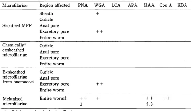
Table 3 FITC‑conjugated lectin binding on Brugia pahangi
microfilariae
Microfilariae
Region affectedPNA WGA LCA
APA HAA Con A KBASheathed MFF
Sheath
Cuticle
Anal pore Excretory pore Entire worm+
++
Chemicallyt exsheathed
microfilariae
Cuticle
Anal pore Excretory pore Entire worm Exsheathedmicrofilariae
from haemocoelCuticle
Anal pore Excretory poreEntire worm ++
Melanized
microfilariae
Entire wormt
++
1
+ ++
2, 3 ++
t: Calcium exsheathed microfilariae.
*: Binding was onto the melanized capsular material.
Inhibitors: I = D ( + ) ‑galactose, 2 = N ‑acetyl‑D‑galactosamine, 3 = N ‑acetyl ‑D‑glucosamine
Fluorescence mtensrty + + + mtense, + + moderate, + poor
RESULTS
None of the lectins tested except wheat germ agglutinin (WGA) bound onto the sheathed and exsheated mf (Table 3). A + and + + fluorescence with WGA was observed on the sheath and excretory pore respectively. Haemocoel‑exsheathed and Ca++ exsheathed mf did not show any significant differences in their cuticular surface lectin binding characteristics;
none bound any of the lectins tested. The excretory pore of haemocoel‑exsheated mf however bound WGA whereas Ca++ exsheathed mf did not. N‑acetyl‑D‑glucosamine
(GlcNAC) inhibited neither WGA binding to the sheath nor the excretory pore.
All lectins except lentil lectin (LCA) , asparagus pea agglutinin (APA) and kidney bean agglutinin (KBA) bound onto the surface of melanized mf. A + + fluorescent staining of melanized granular deposits on the melanized mf was observed. The staining by Helin aspersa agglutinin (HAA) was blocked by GlcNAC and N‑acetyl‑D‑Galactosamine (GalNAc) , and pea nut agglutinin (PNA) by D ( + ) ‑galactose (GaD. WGA and Concanavalin A (Con A) binding were not inhibited by any of their complementary sugars.
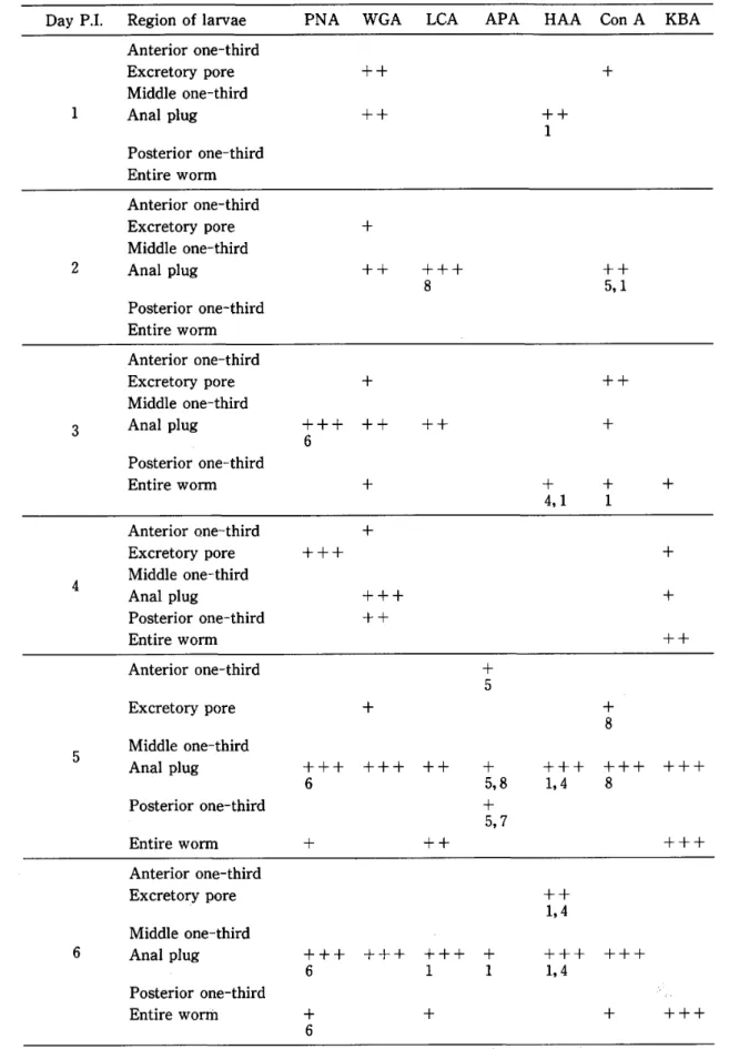
Fluorescent staining of developmental stages of B, pahangi in Ar. subalbatus is shown in Table 4. The degree of staining and types of lectins involved varied as differentiation and development of these stages occurred. Con A, HAA, KBA and WGA bound weakly to the worm on day 3 p.i. A strong positive fiuorescent staining was not detected on the cuticular surface of larval developing stages till day 4 p.i. when KBA bound onto the entire surface of the worm. Also WGA binding was seen at day 4 p.i. at both the anterior and posterior end̲*
Table 4 FITC‑conjugated lectin binding onto developing stages of Brugia pahangi
Day P.1.
Region of larvae PNAWGA LCA
APAHAA
Con A KBAl
Anterior one‑third Excretory pore Middle one‑third Anal plug
Posterior one‑third
Entire worm++
++ ++ l
+
2
Anterior one‑third Excretory pore Middle one‑third Anal plug
Posterior one‑third
Entire worm+
++ +++ 8
++5, l
3
Anterior one‑third Excretory pore Middle one‑third Anal plug
Posterior one‑third
Entire worm+++ 6
+
++
+
++
+ 4, 1
++
+
+ l +
4
Anterior one‑third Excretory pore Middle one‑third Anal plug
Posterior one‑third
Entire worm+++ + +++ ++
+ +
++
5
Anterior one‑third Excretory pore Middle one‑third Anal plug
Posterior one‑third
Entire worm+++ 6
+
+
+++
++++
+ 5
+ 5, 8
+ 5, 7
+++ 1, 4
+ 8
+++ 8 +++
+++
6
Anterior one‑third Excretory pore Middle one‑third Anal plug
Posterior one‑third Entire worlil
+++ 6
+ 6
+++ +++ l
+
+ l
++ 1, 4
+++ 1, 4 +++
+ +++
Table 4
(Cont.)
Day P.1.
Region of larvae PNAWGA LCA
APAHAA
Con A KBA7
Anterior one‑third Excretory pore Middle one‑third Anal plug
Posterior one‑third
Entire worm++
6
+ 6
+++ 6 +++
++ 9 +++
+ ++ l, 4
+++ 1 ++ 1
+++
8
Anterior one‑third Excretory pore Middle one‑third Anal plug
Posterior one‑third
Entire worm+++ 6
++6
+++ +++ 1, 4 ++
+ 5
++
+
9
Anterior one‑third Excretory pore Middle one‑third
Anal plug
+++ 6
++1
++1, 9 +++ 1, 4 ++ 1 + 1
Pesterior one‑third
Entire wormInhibitors: I = N ‑acetyl‑D‑glucosamine, 4 = N ‑acetyl‑D‑galactosamine,
5 = Methyl‑a‑D‑mannopyranoside, 6 = D ( + ) ‑galactose, 7 = L ( ‑ ) ‑fucose, 8 = D ( + ) ‑mannose, 9 = a‑D ( + ) ‑glucose
Fluorescence intensity: + + + intence, + + moderate, + poorof the larvae. Both GalNAc and GlcNAC inhibited HAA binding to the cuticular surface at day 3 p.i. whereas Con A was inhibited only by GlcNAC (T ble 4). Neither WGA nor KBA were inhibited by any of the sugars tested at day 4 p.i. At day 4, the larvae were in their late
first‑stage (L1) .
During the first four days of development of L1, all lectins tested except APA showed positive binding to either the excretory pore or the anal pore. GlcNAc did not interfere with WGA binding, neither to excretory pore nor to the anal pore. D‑mannose (Man) and methyl‑
a‑D‑mannopyranoside (ManMe) reduced the intensity of LCA and Con A binding to the anal pore respectively at day 2 p.i. GlcNAC did not interfere with Con A binding to the excretory pore at day I p.i. HAA and Con A binding to the anal pore at day I p.i. and day.2 p.i.
resp ctively. were both inhibited by GlcNAc. A different set of lectin receptors for WGA, LCA and Con A appeared at day 3; none of the sugar inhibitors interfered with their binding tQ either the excretory or anal pore. PNA binding to the anal pore was blocked by Gal.
Con A, HAA and KBA all gave a weak fluorescent staining along the entire length of the worm at day 3 p.i. Both GalNAc and GlcNAC inhibited HAA‑binding to the cuticular surface
though Con A was blocked only by GlcNAc.
Only WGA and KBA bound onto larvae isolated on day 4 p.i. Intense binding of WGA to the excretory pore and anus was observed in contrast to a weak binding seen on larvae on d'ay 3. KBA bound weakly along the entire length of the larvae surface. Neither WGA nor KBA binding were inhibited by their complementary sugars.
At day 5 p.i., PNA, LCA and KBA bound along the entire length of the worm whereas APA bound only to the anterior and posterior ends. The anal pore bound all lectins tested.
Though the intensity of PNA binding was + on the cuticular surface and + + + on the anal plug, both were equally blocked by the sugar. WGA, LCA and KBA showed on change in binding when incubated with their complementary inhibitory sugars. ManMe blocked APA binding to the anterior and posterior cuticular surfaces and the anus, whereas Man and L ( ‑ ) ‑Fucose (Fuc) inhibited binding to only the anus and the posterior cuticular surface respectively. Both GlcNAC and GalNAc interfered with HAA binding to the anus. Man inhibited Con A binding to both the excretory pore and the anal plug.
At day 6, Con A (in addition to PNA. LCA and KBA) bound to the entire worm. At the same time the anterior and posterior ends lost their receptors for APA. Gal blocked binding of PNA onto the worm; none of the sugar inhibitors of Con A and KBA interfered with lectin binding. The anal plug bound all lectins except KBA at day 6 p.i. HAA, APA and LCA were all inhibited by GlcNAc; GalNAc blocked HAA binding to the anal plug.
At day 7 p.i., further changes to the surface carbohydrates occurred. Whereas APA and HAA Iost their receptors on the anal plug, KBA receptors reappeared. Only LCA and Con A, in addition to HAA, bound the entire cuticular surface. LCA binding to the middle‑third of the cuticular surface of the worm was blocked by Glc whereas the rest of its surface did not. Though the intensity of Con A binding to anal plug and cuticular surface differed, both were equally blocked by GlcNAc. Man inhibited Con A binding to the middle‑ and posterior
‑third of the cuticular surface but not to the anal plug; the anal plug was blocked instead by GlcNAc. Both GalNAc and GlcNAC blocked HAA binding to the worm. PNA binding to the middle‑third of the cuticular surface of the worm, the anal plug and excretory pore were inhibited by Gal.
The worms lost their LCA and HAA receptors on day 8 p.i. At the same time, cuticular receptors for KBA and PNA reappeared. PNA binding was inhibited by Gal; GlcNAC did not interfere with KBA binding onto the worm. Whereas ManMe blocked Con A binding onto the worm, Man blocked only Con A binding to the middle‑third of the cuticular surface. A11 lectins except LCA and APA bound onto the anal plug. Both GalNAc and GlcNAC inhibited HAA binding and none of the sugar inhibitors of Con A and WGA blocked their binding onto the anal plug.
A dramatic change in lectin binding was observed at day 9 p.i. The entire cuticular surface seemed to have lost its carbohydrate moieties. All lectins tested except APA bound only to the anal plug. Con A, HAA, KBA, LCA and WGA binding to the anal plug were inhibited by GlcNAc and PNA by Gal. Also, LCA and HAA were inhibited by Glc and GalNAC, respectively. All larvae used in these experiments were in their late second‑stage
(L2). Some very advanced L2 were seen though none had moulted to L3 at day 9 p.i.
At day 10 p.i., L3 were seen in the head, thorax and abdomen. Early L3 from the thorax gave fluorescent staining of the anus and preanal cuticular surface with WGA (Table 5) . None of the mature L3 in the thorax bound any lectins tested. Stage‑3 Iarvae (L3) isolated
Table 5 FITC‑conjugated lectin
cuticular surface
binding onto Brugia pahangi
inf ective st age
larvae(L3)
Source Region of larvae PNAWGA LCA
APA H AA Con AKBA
Head
Anterior one‑third Middle one‑third Anus and anal cavity
Posterior one‑third Entire one‑third
Entire worm++
++
Thorax
Anterior one‑third Middle one‑third Anus and anal cavity
Posterior one‑third
Entire wormAbdomen
Anterior one‑third Middle one‑third Anus and anal cavity
Posterior one‑third
Entire worm+++
++
+
Fluorescence intensity: +++ intence,
++ moderate, + poorfrom the abdomen gave positive fiuorescent staining of the anus with WGA, buccal cavity with KBA and APA and the cuticular surface of the anterior end with LCA. An L3 obtained from the abdomen bound weakly with lentil along its entire cuticular surface. Most of the L3 obtained from the head did not bind any of the lectins tested. One L3 however bound LCA onto its anterior end and another bound Con A onto its anus. The worm which bound Con A to its anus was an early L3 stage; probably it had accidentally migrated from the thorax into the head before attaining full maturity. Buccal cavity contents of L3 from the head bound all except PNA and HAA.
None of the sugar inhibitors blocked lectin binding to the buccal cavity (and its contents) or anterior end of L3 isolated from the head of the mosquitoes. However, binding of APA to the buccal cavity of L3 from the abdomen was blocked by Man and GlcNAc; KBA binding to the buccal cavity was not inhibited by GlcNAc. None of the complementary sugar inhibitors blocked WGA and LCA binding to the anus or the anterior‑third of the cuticular surface of the worm.
Lectins also bound weakly to the buccal cavity (and its contents) of various larval stages (Table 6). HAA bound to the pharyngeal thread of one‑ and 3‑day olds and to the buccal cavity of late L2. The carbohydrate moieties responsible for HAA binding differed with the stage of developing larvae. PNA bound onto the buccal cavity of 3‑day olds and the binding was inhibited by Gal. None of the larvae dissected‑out on day 4 and day 5 p.i. bound any of the lectins tested. Six‑day olds larvae bound PNA and WGA and 7‑day olds with KBA, LCA and WGA; none of these bindings were inhibited by their respective complementary sugars.
Eight‑day olds bound HAA, KBA and WGA; both KBA and WGA bindings were not inhibited by their specific sugars. Late L2 on day 9 bound all lectins except APA. GlcNAC interfered
with LCA, HAA and Con A binding. PNA binding to the buccal cavity was blocked by Gal;
and LCH and Con A were also blocked by Man and a‑D ( + ) ‑glucose (Glc) respectively.
None of the L3 collected from the thorax of the mosquitoes bound any lectins onto their buccal cavity. Those from the abdomen bound only KBA. L3 obtained from the head bound all except HAA and PNA; APA binding to the buccal cavity was inhibited by Man and GlcNAc.
DrscussloN
Surface lectin binding characteristics of mf and developing stages of B. pahangi in Ar.
subalbatus were assayed using FITC‑labeled lectins. Blood derived B. pahangi mf bound only WGA onto their sheaths and excretory pore; similar observations were reported by others (Furman and Ash, 1983a,b; Devaney, 1985; Rao et al., 1988). Also Furman and Ash (1983b) and Sayers et al. (1984) observed Con A binding onto the sheath. In addition, Sayers et al.
(1984) noted Con A binding onto the epicuticle of blood‑derived B. pahangi mf while Furman and Ash (1983a) did not observe binding of any of the lectins tested (Limulus pol ,hemus, pea nut, Ricinus communis and soybean lectins) to either the epicuticle of mature or in utero derived mf. The sheath of in utero‑derived mf, however, bound Limulus p01 iphemus, pea nut, Ricinus communis and soybean lectins (Furman and Ash, 1983) .
Exsheathment of mf, either naturally in the mosquito's haemocoel or chemically‑induced, Table 6 FITC‑conjugated lectin binding to
developing stages of Brugia pahangi
the buccal cavity (and
in Armigeres subalbatus
its
contents) ofDay P.1. PNA WGA LCA APA
H AA Con A KBAl
+ +a2
3 +++ 6
++l, 4
4 5
6 + +
7 + + +
8 + +
4 +
9 +
6 + +
8, 1
+'
1 +
9, 1 +
10
+b +b +b8, 1
+b +c
a: Pharyngeal thread.
b: Infective stage larvae from head.
c: Infective stage larvae from head and abdomen.
Inhibitors:
1 = N ‑acetyl‑D‑glucosamine, 4 = N ‑acetyl‑D‑galactosamine,
6 = D ( + ) ‑galactose, 8 = D‑mannose, 9 = a‑D ( + ) ‑glucose
Fluorescence intencity: + + + intense, + + moderate, + poordid not cause any significant changes in the surface characteristics. None of the lectins tested bound onto the cuticle of exsheathed mf. Similar results have been reported in B. pahangi by others (Furman and Ash, 1983a,b; Devaney, 1985; Rao et al., 1988) and in Wuchereria bancrofti and Litomosoides carinii (Rao et al., 1987a) . However, melanized mf of B. pahangi
bound PNA, WGA, HAA and Con A; the fluorescent staining was most intense on the
capsular material rather than on the surface of the worm itself. Encapsulation and melaniza‑
tion of mf involve both the humoral and cellular components of the mosquito defence system (Chen and Laurence, 1985; Forton et al., 1985; Christensen and Forton, 1986). Recently, Nappi and Christensen (1986) had demonstrated transformation of mosquito blood cells or haemocytes in response to a parasitic invasion. They observed an increase in the number of haemocytes exhibiting WGA binding in mosquitoes eliciting a melanization response against inoculated mf. Surface carbohydrates of melanized mf identified by Con A, HAA, PNA and WGA in the present study may well represent aggregations of specific haemocyte population involved in encapsulation and melanization or a byproduct of such a response. Futher, Chen and Laurence (1987) had shown that the material around an encapsulated mf is a protein‑
carbohydrate complex; our sugar inhibition studies had indicated that several carbohydrate molecules or haemocyte types were present on the surface of an encapsulated and melanized mf. The reasons why most of the mf of B. pahangi are not encapsulated are largely unknown.
It has been suggested that mf acquire midgut antigens on their surface during migration through the midgut wall (Sutherland et al., 1984; LaFond et al., 1985) . Christensen et al.
(1987), however, failed to detect midgut antigens on the surface of midgut‑penetrated mf though a significant loss of microfilarial surface electronegativity was observed.
Table 4 shows that surface carbohydrate moieties associated with Con A, HAA, PNA and WGA dissappeared and reappeared at different times during the developmental period.
Certain parts of the larvae e.g. the anal pore or plug did not change lectin binding specificities
throughout the period; Con A, PNA and WGA associated moieties were the most frequently encountered. Similar results were reported with developmental stages of O. Iienalis in Simulium (Ham et al., 1988) .Whereas the carbohydrate moieties associated with most of the lectins tested could be identified, those for WGA were beyond the range of sugars used in the present study. WGA specifically recognizes GlcNAC and binds to a sequence of three P‑ (1‑4) ‑linked residues of the sugar (Allen et al., 1973; Goldstein et al., 1975) . However, GlcNAc itself has a lower afiinity for WGA than oligomers of the sugar, thus WGA will not be completely inhibited by GlcNAC (Peters and Latka, 1986) . Kaushal et al. (1984) showed that GlcNAC could reduced WGA‑binding by only 50‑70% though Devaney (1985) reported complete inhibition by GlcNAc. The surface molecules of developing stages of B. pahangi may bear several sugar residues or these molecules may exist as oligomers of lectins with differing carbohydrate binding specificities. Also, WGA is known also to interact with sialic acid residues (Bhavanandan and Katlic, 1979) . However, Furman and Ash (1983a) , Kaushal et al. (1984) and Rao et al. (1988) failed to detect sialic acid residues on blood‑derived mf. Smail (1987) reported inhibition of WGA binding onto L1 of O. Iienalis by neuraminic acid though removal of sialic acid residues using neuraminidase failed to produce such inhibition. The results implied that WGA bound onto oligomers of GlcNAC or onto a receptor site resembling neuraminic acid; it explains also the failure o.f some sugars to inhibit their complementary
lectins.
The fluidity of the surface carbohydrates may be a consequence of the mosquito's haemocyte responses to the developing larvae. Interaction between mosquito haemocytes and the worm's surface carbohydrates may determine its successful development. The fact that Con A, HAA, PNA and WGA‑associated moieties were the most commonly encountered on both melanized mf and developing larval stages suggest that the larvae may be evading host responses through molecular mimicry. However, there is a possibility that the observed differences in surface lectin binding characteristics were due to larval age differences. In the present study, the larvae were assayed every 24 hours and due care was taken such that dissection for the larvae was performed at a precise hour everyday; Iarvae from a single mosquito were used for each series of lectin binding assay. Nevertheless the possibility of some larvae being more mature than others in the series could not be ruled out.
It is interesting also to note that lectin binding was observed on the pharyngeal thread of early developmental stages of B. pahengi. A wider range of lectins bound onto the buccal cavity and the pharynx as the larvae approach maturity. Lectin bindings were most likely to the contents of the tract. The pharyngeal thread consists largely of a protein‑mucosub‑
stance complex (Simpson and Laurence, 1972) and these were probably stored waste prod‑
ucts. Waste materials are not being expelled until the rectal plug is excreted during the last moult.
In conclusion, developing stages of B, pahangi in Ar. subalbatus showed very dynamic surface carbohydrate characteristics. The larvae change their surface coat configuration frequently over the 10‑day observation period. On the other hand, blood‑dwelling mf and fully matured infective larvae showed little or no binding afflnity for the lectins tested;
apparently the worms response differently in their vertebrate host. Infective stages of O.
lienalis obtained from the head also bound less lectins than those from the thorax (Ham et al., 1988). Rao et al. (1987c), however, reported that the cuticular surface of L3 of W.
bancrofti bound WGA.
These observations suggest that the rapid turnover of the surface carbohydrates, while development of the larvae is taking place, is the worms' response to a 'hostile' host environ‑
ment. Encapsulation and melanization of invading mf always occur in the haemocoel and have never been reported in the thorax of a vector mosquito. Rapidly changing surface carbohydrates may assist the larvae in evading the mosquito's immune system. On the other hand an active mosquito lectin system (e.g. in the form of opsonins or agglutinins) acting in concert with haemocytes may result in the successful elimination or isolation of the parasites.
ACKNOWLEDGEMEN T
M.Z. was supported by a studentship from the Government of Malaysia, D.A.D. is an Medical Research Council external staff at London School of Hygiene and Tropical Medicine and P.J.H. is a research fellow of the Wellcome Trust. Financial support from the respective bodies is gratefully acknowledged.
REFEREN CES
1 ) Allen, K., Neuberger, A. and Sharon, N. (1973): The purification, composition and specificity
of wheat germ agglutinin, Biochem. J., 131, 155‑162
2 ) Bhavanandam, V.P. and Katlic, A.W. (1979): The interaction of wheat germ agglutinins with
sialoglycoproteins, J. Biol. Chemist., 254, 4000‑4008
3 ) Bird, A.F. (1980): The nematode cuticle and its surface. In: Nematodes as Biological Models,
B.M. Zuckerman (ed), Academic Press, New York, 2, 213‑2364 ) Chen, C.C. and Laurence, B.R. (1985): An ultrastructural study on the encapsulation of microfilariae of Brugia pahangi in the haemocoel of Anopheles quadrinaculatus. Inter. J.
Parasitol., 15, 421‑428
5 ) Chen, C.C. and Laurence, B.R. (1987): In vitro study on humoral encapsulation of
microfilariae: Establishment of technique and description of reaction, Inter. J. Parasitol., 17, 781 ‑787
6 ) Cherian, P.V., Stromberg, B.E., Weiner, O.J. and Soulsby, E.J.L. (1980): Fine structure and
cytochemical evidence for the presence of polysaccharides surface coat of Dirofilaria immitis microfilariae, Inter. J. Parasitol., 10, 227‑233
7 ) Christensen, B.M. and Forton, K.F. (1986) : Haemocyte‑mediated melanization of microfilariae in Aedes aeg pti. J. Parasitol., 72, 222‑225
8 ) Christensen, B.M., Forton, K.F., LaFond, M.M. and Grieve, R.B. (1987): Surface changes on Brugia pahangi microfilariae and their association with immune evasion in Aedes aeg)pti. J.
Inverteb. Pathol., 72, 216‑219
9 ) Devaney, E. (1985): Lectin binding characteristics of Brugia pahangi microfilariae, Trop. Med.
Parasitol., 36, 25‑28
lO) Forsyth, K.P. Copeman, D.B. and Mitchell, G.F. (1984) : Differences in the surface radioiodinat‑
ed proteins of skin and uterine microfilariae of Onchocerca gibsoni. Mol. Biochem. Parasitol., 10, 217‑229
11) Forton, K.F., Christensen, B.M. and Sutherland, D.R. (1985): Ultrastructure of the melaniza‑
tion response of Aedes trivittatus against inoculated Dirofilaria immitis microfilariae, J.
Parasitol., 71, 331‑341
12) Furman, A. and Ash, L.R. (1983a): Analysis of Brugia pahangi microfilariae surface carbo‑
hydrates: Comparison of the binding of a panel of fluoresceinated lectins to mature in vivo‑
derived and immature in utero‑derived microfilariae, Acta Tropica, 40, 45‑51
13) Furman, A. and Ash, L.R. (1983b) : Characterization of the exposed carbohydrates on sheath surface of in vitro‑derived Brugia pahangi microfilariae by analysis of lectin binding, J.
Parasitol., 69, 1043‑l047
14) Goldstein, I.J., Hammarstrom, S. and Sunblad, G. (1975): Precipitation and carbohydrate‑
binding specificity studies on wheat germ agglutinin, Biochi. Biophy. Acta, 405, 53‑61
15) Ham, P.J., Smail, A.J. and Greoger, B.K. (1988): Surface carbohydrate changes on Onchocercalienalis larvae as they develop from microfilariae to the infective third‑stage in Simulium
ornatum. J. Herminth., 62, 195‑205
16) Howells, R.E. and Blainley, L.J. (1983): The moulting process and the phenomenon of
intermoult growth in the filarial nematode, Brngia pahangi. Parasitol., 87, 493‑505
17) Kaushal, N.A., Simpson, A.J.G., Hussain, R. and Ottesen, E.A. (1984): Brugia malayi: Stage‑
specific expression of carbohydrates containing N‑acetyl‑D‑glucosamine on the sheath surface of microfilariae, Exp. Parasitol., 58, 182‑187
18) LaFond, M.M., Christensen, B.M. and Lasee, B.A. (1985): Defence reactions of mosquitoes to filarial worms: Potential mechanisms for avoidance of the response by Brugia pahangi
microfilariae, J. Invert. Pathol., 46, 26‑30
19) Laurence, B.R. and Simpson, M.G. (1974): The ultrastructure of the microfilariae of Brugia,
Nematoda: Filarioidea, Inter. J. Parasitol., 4, 523‑536
20) Lumsden, R.D. (1975): Surface ultrastructure and cytochemistry of parasitic helminths, Exp.
Parasitol., 37, 267‑339
2D Marshall, E. and Howells, R.E. (1986) : Turnover of the surface proteins of adult and third and forth stage larvae of Brugira pahangi. Mol. Biol. Parasitol., 18, 17‑24
22) Martinez‑Palomo, A. (1978): Ultrastructural characterization of the cuticle of Onchocerca
volvulus microfilariae, J. Parasitol., 64, 127‑136
23) Maizels, R.M., DeSavigny, D. and Ogilvie, B.M. (1984): Characterization of surface and
excretory‑secretory antigens of Toxocara canis infective larvae, Parasite Immunol., 6, 23‑37
24) McLaren, D. (1972): Ultrastructural studies on microfilariae (Nematoda: Filarioidea),Parasitol., 65, 317‑332
25) Nappi, A.J. and Christensen, B.M. (1986): Haemocyte cell surface changes in Aedes aeg pti in response to microfilariae of Dirofilaria immitis. J. Parasitol., 72, 875‑879
26) Nwachukwu, M.A., Mackenzie, C.D., Litchfield, T.M. and Howard, G. (1978): Lectin binding
to the developing forms of Onchocerca gutturosa microfilariae, Trop. Med. Parasitol., 38, 131‑
134
27) Peters, W. and Latka, I. (1986): Electron microscopic localization of chitin using colloidal gold
labelled with WGA, Histochem., 84, 155‑16028) Philipp, M., Parkhouse, R.M.E. and Ogilvie, B.M. (1980): Changing proteins on the surface of parasitic nematodes, Nature, 287, 538‑540
29) Rao, U.R., Chandrashekar, R. and Subrahmanyam, D. (1987a): Litomosoides carinii: charac‑
terization of surface carbohydrates of microfilariae and infective larvae, Trop. Med. Parasitol., 38, 15‑18
30) Rao, U.R. Chandrashekar, R., Parab, P.B., Rajasekariah, G.R. and Subramanyam, D. (1987b):
Lectin binding characteristics of Wuchereria bancrofti microfilariae, Acta Tropica, 44, 35‑42
31) Rao, U.R., Chandrashekar, R., Rajasekariah, G.R. Subrahmanyam, D. (1987c): Wheat germagglutinin specifically binds to the surface of infective larvae of Wuchereria bancrofti. J.
Parasitol., 73, 1256‑1257
32) Rao, U.R., Chandrashekar, R. and Subrahmanyam, D. (1988): Developmental changes in the
surface carbohydrates of filariae, Ind. J. Med., 87, 9‑14
33) Sayers, G., Mackenzie, C.D. and Denham, D.A. (1984): Biochemical surface components of
Brugia pahangi microfilariae, Parasitology, 89, 425‑434
34) Schraermeyer, U., Peters, W. and Zehner, H. (1987): Lectin binding studies on adult filariae,
intrauterine developing stages and microfilariae of Brugia malayi and Litomosoides carinii.Parasitol. Res., 73, 550‑556
35) Simpson, M.G. and Laurence, B.R. (1972): Histochemical studies on microfilariae, Par‑
asitology, 64, 61‑88
36) Smail; A.J. (1987): Studies on lectin binding characteristics of developing stages of Onchocerca
lienalis in Simulium species and their possible interaction with haemolymph lectins, Unpub‑lished, MSc, thesis, University of London, pp. 84
37) Sutherland, D.R., Christensen, B.M. and Forton, K.F. (1984): Defence reactions of mosquitoes to filarial worms: Role of the microfilarial sheath in the response of mosquitoes to inoculated Brugia pahangi inicrofilariae, J. Invest. Pathol., 44, 275‑281
38) Taylor, D.W., Goddard, J.M. and McMahon, J.E. (1986) : Surface components of O. volvulus,
Mol. Biol. Parasitol., 18, 283‑300
SURFACE LECTlN BlNDING CHARACTERISTICS OF DEVELOPlNG STAGES OF BRUGIA IN ARMIGERES SUBALBATUS= 11 BRUGIA MALA YI
M. ZAHED11'3, D.A. DENHAMI AND P.J. HAM2 Received March 23 1990/Accepted September 7 1990
Abstract: Surface lectin binding characteristics of microfilariae and developing stages of Brugia malayi in Armigeres subalbatus are presented. Ar. subalbatus is not a vector of B. malayi. The following lectins were used: wheat germ agglutinins, Ientil agglutinins, Helix aspersa agglutinins, Concanavalin A agglutinins, kidney bean agg‑
lutinins, asparagus pea agglutinins and pea nut agglutinins. Frequent change in surface carbohydrate moieties on developing stages of B. malayi was observed. However, blood dwelling microfilariae and mature infective larvae showed little binding a nity for the
lectins tested. It is postulated that B. malayi larvae evaded the mosquito immune system
by continuously changing their surface carbohydrate moieties.INTRODUCTION
In a previous communication, we have reported lectin binding characteristics of the cuticular surfaces of Brugia pahangi in Armigeres subalbatus. Larval stages of B. pahangi showed dynamic surface characteristics; frequent change in cuticular carbohydrate configuration was seen during their development in the mosquito. Similar observations were made with larval stages of Onchocerca lienalis in Simulium ornatum (Ham et al., 1988) .
The present paper describes the surface characteristics of developing stages of B. malayi in B. malayi‑susceptible Ar. subalbatus. The aim of this study was to elucidate and to compare these characteristics with the larval stages of B. pahangi in Ar, subalbatus (previous communication) ; Ar. subalbatus is not a natural vector of B. malayi.
MATERIALS AND METHODS
B. malayi‑susceptible Ar. subalbatus at ages 9 to 10 days were fed directly onto an anaesthetized B. malayi‑infected cats. The peripheral microfilariae (mf) concentration of the cats ranged from 80‑120 mf/20 pl blood.
The methods of isolation of B. malayi larvae, Iectin binding experiments and types of lectins used were as described previously for studies with B. pahangi (Zahedi et al. 1990) .
2 3
London School of Hygiene and Tropical Medicine. Keppel Street, London WGIE 7Hlt, U.K.
Liverpool School of Tropical Medicine. Pembroke Place, Liverpool L3 50QA, U.K.
Current address and correspondence to: Dr. M. ZAHEDI, Department of Parasitology and Medical Entomology, Medical Faculty, U.K.M., Jalan Raja Muda Abdul Aziz, 50300 Kuala Lumpur. Malaysia
Observations were made in duplicate and whenever from the same mosquitoes for each series of lectins.
possible parasites were obtained
RESULTS
Sheathed and Ca++ exsheathed B. malayi mf bound KBA, LCA and WGA (Table 1).
Faint fluorescent staining of cast sheaths by these lectins were also detected. Haemocoel‑
derived exsheathed mf bound KBA and LCA, while WGA bound only onto the excretory pores. Melanized mf bound Con A, HAA and PNA. It was noted that lectin‑binding was onto the capsular envelope rather than the exsheathed mf itself. A weak binding of WGA onto the melanized capsule was also observed. The lectin‑bindings could not be blocked by any of their complementary sugars.
Table 2 shows lectin binding characteristics of developing larval stages of B. malayi in Ar. subalbatus. All lectins tested bound weakly onto the cuticular surface of 24‑hr old larvae.
In addition to the worm's surface, Con A and PNA also bound onto both anal and excretory pores of the larvae. HAA, KBA and PNA binding onto the worm were completely blocked by their complementary sugars (in this issue, p. 273) . Both Methyl‑a‑D‑mannopyranoside (ManMe) and a‑D(+)‑glucose (Glc) inhibited Con A binding onto the cuticle, anal and excretory pores of the worm. Also Con A binding to the cuticle and excretory pores was blocked by N‑acetyl‑D‑glucosamine (GlcNAC) and D‑mannose (Man) , respectively. None of the complementary sugars of APA, LCA and WGA interfered lectin binding onto the worm's cuticular surface.
The larvae lost all except APA and LCA binding onto their cuticular surface on day 2 Table l FITC‑conjugated lectin binding on Brugia malayi
microfilariae
Microfilariae
Region affected PNAW GA LCA
APAHAA
Con A KBASheathed MFF
Sheath
Cuticle
Anal pore Excretory poreEntire worm
+ + +
Chemicallyt exsheathed
microfilariae
Cuticle
Anal pore Excretory poreEntire worm
+ + +
Exsheathed
microfilariae
from haemocoelCuticle
Anal pore Excretory poreEntire worm ++
++
+ +
Melanized
microfilariae
Entire worm$ +++
++++
t: Calcium exsheathed microfilariae.
$: Binding was onto the melanized capsular material.
Fluorescence intensity: + + + intense, + + moderate, +̲ poor
Table2 FITC−conjugated lectin binding onto developing stages ofβ耀g勉㎜五の學
Day P.1. Region of larvae
PNAWGA LCA APA HAA
Con A KBA1
Anterior one−third Excretory pore Middle one−third Anal plug
Posterior one−third
Entire wom1十十 6
十十十 6
十 6 十 十 十
十4 4 十1 十1 9
9
8 十9
5
十5 十5 十1
十十 1
2
Anterior one−third Excretory pore Middle one−third Anal plug
Posterior one third
Entire worm十6十6 十 十
十十
十9 十 十8 十8
十 1,8,9
十 1,5,8
8 十1
十十十 1,4
十十十
1,5,8,9
十十十 十十
1 十
1,5,8,9
3
Anterior one−third Excfet・rゾP・re
Middle6ne−thirdAnal plug
Posterior one−third
Entire椥orm十 十6 十6 十
十十
十十
十
十
、十
十7 十7 十 十1 十1 十1
十4 十4 4十 十十
5,8,9
十十十
十 5,8,9
十十
十十
4
Anterior one−third Excret6ry pore Middle one−third Anal plug
Posterior one−third
Entire wom十 十6 十6 十
十
十十
十1
8,9
十十十
十9 5 十1
十十十 4,1
十 1,4
十 1,5,8,9
十十
十5 十十
5
Anterior one−third Excretory pore Middle one−third Anal plug
Posterior one−third
Entire worm十十十 6
十
十十 十十十
十8
十 1,7,8
十 1,7,き
十1 十1 十4 十4 十
十一 5,8
十十十
十十
十十
十十
十十
6
Anterior one−third
Excreto可pore
Middle one−third Anal plug
Posterior one−third Entire form
十十十 6
十
十十 十十十
十8
十 1,7,ε
十 1,7,8
十十十
1,4
十十 1,4
十 5,8
十十十
十十
十十
十十
十十
7
Anterior one−third Excretory pore Middle one−third Anal plug
Posterior one−third
Entire worm十十十 6
十6
十
十
十十
十十
十1十1 十4十4 十
十十十 1
十十
1,5 十十十
Table 2
(Cont.)
Day P.1.
Region of larvae PNAWGA LCA
APAHAA
Con AKBA
8
Anterior one‑third Excretory pore Middle one‑third Anal plug
Posterior one‑third
Entire worm+++ 6
++ +++ l, 9
+++ l. 4
+++ l, 8, 9
9
Anterior one‑third Excretory pore Middle one‑third Anal plug
Pesterior one‑third
Entire worm+ 6
+++ 6
++ +++ l, 8, 9
+++ + l
+
Inhibitors: I = N‑acetyl‑D‑glucosamine, 4 = N‑acetyl D galactosamme
5 = Methyl‑a‑D‑mannopyranoside, 6 = D ( + ) ‑galactose, 7 = L ( ‑ ) ‑fucose, 8 = D ( + ) ‑mannose, 9 = a ‑D ( + ) ‑glucose
Fluorescence intensity: + + + intence, + + moderate, + poorp.i. The binding could be blocked by both Man and GlcNAc; in addition Glc too blocked LCA binding onto the cuticle. A very weak fiuorescent staining with Con A and PNA was also detected at the posterior third of the cuticular surface of the larvae. PNA binding to the surface could be inhibited by D ( + ) ‑galactose (GaD and Con A binding by ManMe, Man, Glc and GlcNAc. All lectins tested bound onto the anal pore and only Con A, LCA and WGA onto the excretory pore. LCA and PNA binding was inhibited by Gal and Man respectively. APA binding was inhibited by ManMe, Man and GlcNAC and HAA binding was inhibited by N‑
acetyl‑D‑galactosamine (GalNAC) and GlcNAc. Further, GlcNAC caused a reduction in the intensity of KBA fluorescent staining. Con A binding to the excretory pore could be blocked by all its complementary sugars and LCA binding only by Man and Glc.
Larvae obtained on day 3 p.i. bound all lectins tested onto their cuticular sprfaces. Gal
and L‑(‑)‑Fucose (Fuc) inhibited APA and PNA binding, respectively, and HAA was blocked by both GalNAc and GlcNAc. APA binding onto the anterior‑half of the larvae could be blocked by GlcNAc. Similarly, GlcNAC also blocked KBA binding to the middle‑
third of the worm. Glc, Man and ManMe inhibited Con A binding onto the worm's cuticle.
None of the complementary sugars of LCA and WGA interfered with the binding. As was the case with larvae dissected on day 2, 72‑hr old larvae bound all the lectins tested onto their anal pore. PNA binding could be blocked by Gal; APA by Fuc and HAA by both GalNAc and GlcNAc. None of the other lectin bindings could be inhibited by any of the sugars tested.
Fluorescent staining of the excretory pore was observed with Con A, HAA and WGA. HAA binding was inhibited by GalNAc and GlcNAC and Con A by Glc, Man and ManMe.
Late first‑stage larvae (Ll) dissected on day 4 p.i. bound all lectins except WGA onto its cuticular surface. Con A, LCA and PNA binding were inhibited by Gal, Glc and ManMe respectively. Also Con A binding to the anterior half of the larvae could be inhibited by GlcNAc; the posterior‑half was inhibited by Glc. APA binding was inhibited by ManMe and