JAPANESE JOURNAL OF
TROPICAL MEDICINE AND HYGIENE
JAPANESE SOCIETY OF TROPICAL MEDICINE
日 本 熱 帯 医 学 会
第3 0巻 第4号 平成1 4年1 2月
内 容
原 著
末梢血単核球と多核白血球によるインターロイキン8の産生と熱帯熱マラリア原虫の発育阻害作用 大屋ゆりジュリエッタ,松岡 裕之,向田 直史,笠原 忠,石井 明………351 日本人輸入サイクロスポーラ症の1例
大西 健児,加藤 康幸,井関 基弘………357
Plasmodium yoelii 17XL感染ICRマウスでのジョウザンアジサイ葉水煮沸抽出液の抗マラリア活性の
季節的相違,特にフェブリフジン・イソフェブリフジン含有量に関して
石井 明,藤井 香,酒井美香子,飯干 昌美,宮瀬 敏男,寺田 護………361
短 報
プエルトリコ産及びブラジル産Biomphalaria貝体内に於ける広東住血線虫幼虫の発育の比較
岩永 襄………365
会報・記録
2002年度(平成14年)日本熱帯医学会役員名簿(2002年12月1日現在)………371 投稿規定………375 著作権複写に関する注意………380
30巻総目次
日本熱帯医学会表紙 ! − " MK 中島 芦塚
日熱医会誌
VOL. 30 No. 4 DECEMBER 2002
CONTENTS
Original article
Ohya, J-Y., Matsuoka, H., Mukaida, N., Kasahara, T., and Ishii, A.
Growth inhibition of Plasmodium falciparum and interleukin-8 production by peripheral blood
mononuclear and polymorphonuclear cells in vitro ………351
Ohnishi, K., Kato, Y., and Iseki, M.
An imported Japanese case of cyclosporiasis ………357
Ishih, A., Fujii, K., Sakai, M., Iiboshi M., Miyase, T., and Terada, M.
Seasonal differences in antimalarial activity of hot-water extract of Dichroa febrifuga leaves against Plasmodium yoelii 17XL in ICR mice, with reference to febrifugine and isofebrifugine content………361
Short Communication Iwanaga, Y.
Comparative studies on the development of larval Angiostrongylus cantonensis in Puerto Rican and
Brazilian Biomphalarla snails. ………365
背は2mm (決定)
Jpn. J. Trop.
Med. Hyg.
JapaneseJournalofTropicalMedicineandHygieneVol.30No.4DECEMBER2002
GROWTH INHIBITION OF PLASMODIUM FALCIPARUM AND INTERLEUKIN-8 PRODUCTION BY PERIPHERAL BLOOD MONONUCLEAR AND POLYMORPHONUCLEAR CELLS IN VITRO
JULIETAYURIOHYA1, HIROYUKIMATSUOKA1, NAOFUMIMUKAIDA2, TADASIKASAHARA3and AKIRAISHII1
Accepted November, 19, 2002
Abstract: Peripheral blood mononuclear and polymorphonuclear cells have been demonstrated to kill and inhibit malaria parasite proliferation in vitro, but most of the reports required activation of the cells by cytokines or pres- ence of immune sera or opsonins. In our study, peripheral blood leukocytes from non-immune donor efficiently in- hibited Plasmodium falciparum growth, depending on the effecter/target ratio. Moreover, these cells produced a large amount of interlenkin-8 (IL-8) under stimulation with infected erythrocytes or supernatant of the Plasmodium falciparum culture. IL-8 secretion in the culture supernatant of polymorphonuclear cells was noted from 6-9 hr of stimulation with the parasites, with a substantial increase over 24 hours of culture. Attempt to elucidate whether IL- 8 was involved in malaria suppression was done, and the result suggested that IL-8 was not directly involved in the anti-malarial activity of the mononuclear and polymorphonuclear cells. The other mechanism besides IL-8 may work for inhibiting parasite growth in the culture system of malaria parasites associated with leukocytes.
Key words: cytokine, interleukin-8, malaria, mononuclear cell, Plasmodium falciparum, polymorphonuclear cell
INTRODUCTION
Anti-malarial activity of peripheral blood mononuclear cells (MNC) and polymorphonuclear cells (PMN) have been demonstrated (Vernes, 1980; Celada et al., 1983;
Nnalue and Friedman 1988; Bouharoun-Tayoun et al., 1990; Kumaratilake et al., 1997), but in most of the reports, presence of opsonising agents (Nnalue and Friedman 1988), antibodies (Celada et al., 1983; Bouharoun-Tayoun et al., 1990) or complements (Kumaratilake et al., 1997) were re- quired. There are considerable evidences showing involve- ment of cytokines such as tumor necrosis factorα(TNFα), interferon-gamma (IFNγ), interleukin lα(IL-Iα), and others in the immunity and pathophysiology of malaria (Ocken- house et al., 1984; Kossodo and Grau, 1993; Kwiatkowski, 1995). The involvement of interleukin-8 (IL-8)and its ligand in Plasmodium vivax infection has been suggested (Horuk, 1994), and this chemokine has been detected in the serum of patients infected with Plasmodium falciparum (P.
f.) (Friedland et al., 1993; Looareesuwan et al., 1999), but the roles of IL-8 in falciparum malaria are not clear.
We performed in vitro experiments to study the anti- malarial activity of the non-immune peripheral blood leuko- cytes. Moreover, we studied the IL-8 production of periph-
eral blood MNC and PMN induced by P.f., and tried to elu- cidate the role of this cytokine in immunity to malaria.
MATERIALSANDMETHODS Malaria parasites
Plasmodiun falciparum FCR-3 strain has been main- tained in our laboratory in a continuous culture according to previous described methods (Trager and Jensen, 1976), ex- cept for the serum supplemented to the medium. The para- sites were sowed in red blood cells (RBC) at an initial he- matocrit of 5% in RPMI 1640 medium (Sigma Chemical Co, St. Louis, MO, USA) supplemented with l0% heat- inactivated horse serum (Cosmo Bio Co., Tokyo, Japan).
Substitution of the human sera with horse sera greatly fa- cilitate the malaria culture, because this permitted the use of all types of human erythrocytes, without checking for com- patibility.
Separation of MNC and purification of monocytes
50 ml of peripheral venous blood was collected from healthy non-immune donors in a heparinized syringe. MNC were separated by Ficoll/Urografin density gradient cen- trifugation, and suspended in a plastic dish with RPMI 1640
Jpn. J. Trop. Med. Hyg., Vol.30, No.4,2002, pp.351‐355
1Department of Medical Zoology, Jichi Medical School, Kawachi-gun, Minamikawachi-machi, Tochigi 329-0498, Japan
2Cancer Research Institute, Kanazawa University, Takara-machi, Kanazawa 920-0934, Japan
3Kyoritsu College of Pharmacy, Minato-ku, Tokyo l05-8512, Japan
351
supplemented with 5% fetal calf serum. Two hours after in- cubation at 37 °C, non-adherent cells were removed from the plate by washing with phosphate buffered saline and ad- herent cells (mainly monocytes) were detached from the plate with a rubber policeman and resuspended in the same medium of MNC.
Separation of PMN
Once the MNC removed, PMN (about 95% neutro- phils) were separated from RBC by sedimentation in 1.25%
gelatin. Mingled erythrocytes were hemolysed in lysing re- agent (Ortho Diagnostic Systems, New Jersey, NJ, USA) and PMN were resuspended in the same medium of MNC.
P.f. growth inhibition assay
Growth inhibition assay was done by a radiometric method, described by Kumaratilake et al. (1997). Briefly, the experiments were conducted in 96 well flat-bottomed microplates (Becton Dickinson Co., Franklin Lakes, NJ, USA). The cells (MNC or PMN) were subsequently diluted and placed to the wells in 100µl aliquots, where the para- sites (1x10⁵parasites in 5x10⁷RBC in 100µl of medium) were added 2 hours later. Following 48 hours incubation at 37°C in a CO2 incubator containing a gas mixture of 5%
CO2in air, each well was pulsed with lµCi of [3H] hypox- anthine and harvested 24 hours later on a glass fiber sheet.
The disks on the sheet were air dried, placed in a 96 well polyethylene plate with 100 µl of toluene scintillant and counted by a microplate scintillation counter (TopCount, Packard Instrument Co., Meriden, CT, USA). Each dilution was made in triplicate and the average was calculated.
Growth inhibition rate was calculated using the formula as follows:
scintillation count of experimental well
Growth inhibition(%)=100− x100
scintillation count of P.f. alone well
Microscopic studies
Interaction of the phagocytic cells with parasites was observed by preparing cytocentrifuged smears of the culture.
Parasites were cultured together with PMN or MNC and smears were made 48 hours later. MNC were detached from the culture plate with a rubber policeman before cytocen- trifugation. The smears were air dried and stained with Gi- emsa for microscopic study at 1000x magnification.
Stimulation of the PMN and MNC with parasites and parasite-derived products
P. faciparum induced IL-8 secretion by the PMN and MNC was investigated by measuring IL-8 in the superna- tant of the culture. PMN or MNC (1x10⁵cells in 100µl of medium) were cultured with viable parasitized erythrocytes
(1x10⁵parasites in 5x10⁷RBC in 100 µl of medium), and the supernatant was collected by 24 hr. For positive control, PMN (1x10⁵ cells in 100 µl of medium) were stimulated with human recombinant TNFα (20 ng in 100 µl of me- dium), which was kindly provided by Dainihon Pharmaceu- tical Co. (Osaka, Japan).
To investigate the fraction responsible for IL-8 induction, PMN (1x10⁵ cells in 100 µl of medium) were stimulated respectively with intact infected erythrocytes (1x 10⁵parasites in 5x10⁷RBC in 100µl of medium), 100µl of supernatant of the P.f. culture, 100µl of hemolysate of the infected erythrocytes (originally 1x10⁵ parasites in 5x10⁷ RBC), and partially purified 1x10⁵ parasites obtained by hemolysing and washing out the RBC ghost. As negative control, uninfected 5x10⁷RBC in 100µl of medium were added to the cell culture.
IL-8 assay
IL-8 in the supernatant was measured by an enzyme- linked immunosorbent assay (ELISA) method as described elsewhere (Ko et al., 1992).
Neutralization of IL-8 activity
To study the role of IL-8 on parasite suppression by PMN, the cells were pretreated, 2 hours before addition of the parasites, with a mouse anti-human IL-8 monoclonal antibody WS-4 (Ko et al., 1992) at a concentration of 50µg /ml. For the control groups, we used monoclonal antibodies for IL-1α (Kasahara et al., 1987) and an unspecific IgG.
After 3 days culture, proliferation of the parasite was as- sessed by incorporation of [3H] hypoxanthine.
RESULTS P. f. growth inhibition
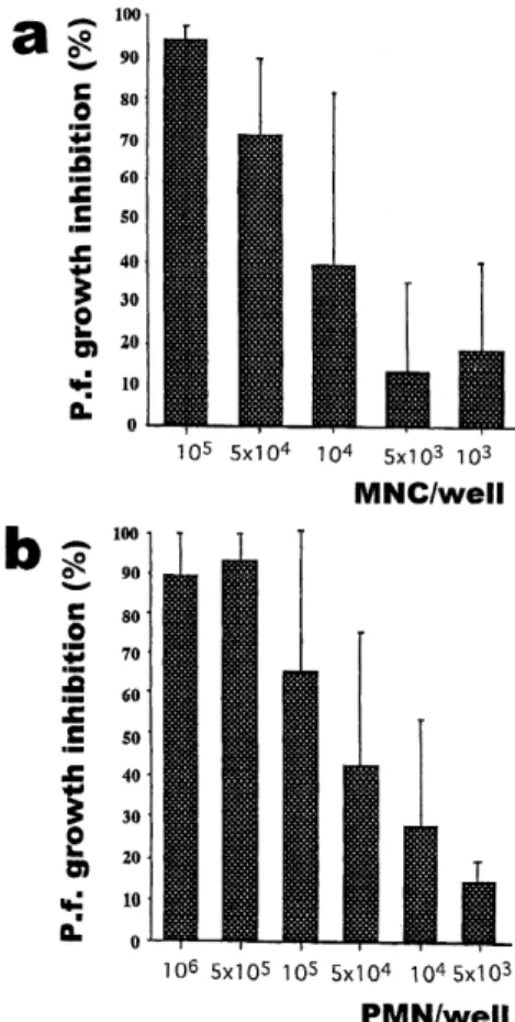
Inhibitory effect of the MNC or PMN on P.f. prolifera- tion depended upon the effecter to target ratio (E/T) (Fig. 1 a). 1x10⁵ MNC almost inhibited the parasite proliferation (parasite number at the start was 1x10⁵/well), and to get 50% growth inhibition of the parasites, we needed 0.2x10⁵ cells, meaning the E/T for IC50was about 0.2. PMN also ef- ficiently inhibited parasite growth with the E/T for IC50of 0.7 (Fig. 1b). Monocytes (adherent cells) were then sepa- rated from non-adherent cells (mainly lymphocytes) and re- spectively cultured with the parasites. The E/T of the mono- cytes for IC50 was 0.9 (Fig. 2). Non-adherent cells alone showed no or minimal inhibitory effect on parasite growth.
This suggests that both monocytes and lymphocytes are re- quired to inhibit P.f. growth.
352
Morphological studies
Morphologically, 2 days after P.f. stimulation, the monocytes greatly increased in size, and most of them con- tained a large number of engulfed parasites. PMN also ac- tively phagocytosed parasites, but the number of engulfed parasites per cell was not noticeable as did the monocytes.
IL-8 production of the PMN and MNC induced by P.f.
A remarkable amount of IL-8 was detected in the su- pernatant of P.f. stimulated MNC. After 24 hours culture, IL -8 level in the supernatant of P.f. stimulated culture was 9 times higher than that of unstimulated one (Fig. 3). Al- though less prominent than the MNC, P.f. stimulated PMN also secreted a considerable amount of IL-8 (Fig. 4). The IL -8 was detectable in the supernatant after 6-9 hours of stimulation, increasing progressively with time. 24 hours later, IL-8 concentration in the supernatant of P.f. stimulated PMN was 80 times higher than that of negative control.
TNFα at a concentration of l µg/ml also induced IL-8
production in PMN but no stronger than P.f. parasites did.
Experiments were also designed to determine the frac- tion responsible for IL-8 induction. Among the fractions tested, P.f. infected erythrocytes and supernatant of the P.f.
culture were the most potent stimulants for IL-8 production in the PMN cells. Hemolysate of the infected erythrocytes, partially purified parasites, and uninfected RBC had little or no stimulative effect on IL-8 production (Table 1).
Figure l In vitro inhibitory effect of normal donors (a) MNC and (b) PMN on P.f. growth. The starting parasite number was 1x10⁵/well. Results are cal- culated as percent inhibition in relation to the [3H]
hypoxanthine uptake of the P.f. alone, and pre- sented as the mean ±standard error from 5 iso- lated experiments.
Figure 2 Effect of the monocytes (black bars) and non- adherent mononuclear cells (dotted bars) for P.f.
growth inhibition.
Figure 3 IL-8 detection in the supernatant of the P.f. stimu- lated MNC (circles). Squares represent the value of IL-8 in the supernatant of MNC cultured with uninfected RBC.
353
P.f. proliferation after neutralization of the IL-8
Treatment of the PMN or MNC with anti-IL-8 or anti- IL-1α monoclonal antibodies did not influence the inhibi- tory effect of these cells on parasite growth (data not shown), suggesting that IL-8 and IL-1αare not directly in- volved in suppression of the parasite by these cells.
DISCUSSION
Some authors have demonstrated that peripheral blood monocytes can kill malaria parasites in vivo and in vitro (Vernes, 1980; Bouharoun-Tayoun et al., 1990), but mono- cytes required presence of immune sera (Druilhe and Khus- mith, 1987; Lunel and Druilhe, 1989) or addition of cytoki-
nes (Ockenhouse et al., 1984) for activation of the cells. Pe- ripheral blood neutrophils are also reported to be effective against P.f. in the presence of immune sera (Celada et al., 1983) or opsonins, and this activity was enhanced by TNF- α, lymphotoxin and IFNγ(Kumaratilake et al., 1997).
We demonstrated that mononuclear cells and neutro- phils from non-immune individuals efficiently inhibited P.f.
growth in vitro in absence of anti-malarial antibodies or complements. Growth inhibition was dependent on the ef- fecter/target ratio. The mechanism how the leukocytes re- strain the parasite growth is not yet clear. For monocytes and macrophages, phagocytosis may be the major mecha- nism, as observed by morphology. The non-adherent mono- nuclear cells, mostly lymphocytes, did not have anti- parasitic activity, but when they were incubated with mono- cytes, they strongly enhanced the ability of the monocytes to inhibit parasite growth. This suggests the interaction be- tween monocytes and lymphocytes by direct contact or via cytokines. For neutrophils, the number of engulfed parasites per cell was not conspicuous. This suggests that, besides the phagocytic activity, neutrophils may produce soluble sub- stances that may contribute to suppress parasite growth.
Cytokines have been demonstrated to be important in immunity and pathogenesis of malaria. IL-8 is a CXC type chemokine responsible for activation of a variety of cells in- cluding neutrophils (Yoshimura et al., 1987), and the recep- tor of IL-8 has been shown to be related to Plasmodium vi- vax infection (Horuk, 1994). Duffy antigen on erythrocytes has long been reported as a target molecule for vivax ma- laria infection (Miller et al., 1975). Interestingly, IL-8 receptor was shown to be homologous to the Duffy antigen, and the involvement of IL-8 and its receptor in infection with vivax malaria was suspected (Horuk, 1994). Some authors have detected IL-8 in the sera of P.f. infected pa- tients (Friedland et al., 1993; Looareesuwan et al., 1999), and Wahlgren et al., (1995) have detected various cytokines, including IL-8, in the culture supernatant of P.f. stimulated peripheral blood MNC and PMN.
Our experiments demonstrated IL-8 in the supernatant of PMN after P.f. stimulation. Since collected fraction of PMN was neutrophils by 95% and contamination with some mononuclear cells might occur, we histochemically con- firmed IL-8 expression in PMN. Intracellular IL-8 was de- tected from 4 hours after P.f. stimulation (data not shown).
Whether this IL-8 production resulted from a direct stimula- tion by parasites or an intermediated stimulation by a secon- dary cytokine has to be investigated.
In our study, we did not investigate the time required for IL-8gene expression in the P.f. stimulated PMN, but the elapsing time for IL-8 secretion may suggest presence of in- termediating cytokines rather than a direct stimulation by Figure 4 Time course of IL-8 secretion in1x105 PMN
stimulated with 1x10⁵ parasites (circles). Squares represent IL-8 detected in the PMN cultured with uninfected RBC, and triangles represent IL-8 se- cretion after TNF-α(100 ng/ml) stimulation.
Table 1 Concentration of IL-8 (ng/ml) in the culture superna- tant of PMN stimulated with infected RBC or P.f.
products
Period of calture
18 hr 24 hr 32 hr 42 hr
PMN + medium 5.1 6.6 8.3 12.1
PMN +
uninfected RBC 5.4 6.0 5.1 6.7
PMN +
infected RBC 140 610 1100 4500
PMN + supernatant
of P.f. culture 140 370 1700 2200
PMN + hemolysate
of infected RBC 16 68 130 250
PMN + partially
purified parasites 34 100 160 490
354
the parasite. Cytokines such as TNF-α, IL-1 and IFNγare demonstrated to be produced in P.f. infections (Brown et al., 1990; Wahlgren et al., 1995), and we have to consider the possibility of an autocrine stimulation by these cytokines.
TNF-α also induced IL-8 production in the PMN (Fig.4), but PMN responded earlier and stronger to P.f. than did to TNF-α. Thus, TNF-αshould not be responsible for the in- duction of IL-8.
To elucidate the involvement of IL-8 in the immunity to falciparum malaria, we neutralized IL-8 with anti-IL-8 monoclonal antibody in the culture mixture of P.f. and PMN, resulting no abrogation of inhibitory ability to PMN. There- fore, we conclude that IL-8 has no direct influence on P.f.
inhibitory activity of the PMN. The other mechanism be- sides IL-8 may work for inhibiting parasite growth in the culture system of malaria parasites associated with leuko- cytes.
ACKNOWLEDGEMENTS
We are grateful to Ms. C. Seki and Ms. M. Okada for their technical assistance.
REFERENCES
1)Bouharoun-Tayoun, H., Attanath, P., Sabchareon, A., Chongsuphajaisiddhi, T. and Druilhe, P. (1990): Antibod- ies that protect humans against Plasmodium falciparum blood stages do not on their own inhibit parasite growth and invasion in vitro, but act in cooperation with mono- cytes. J. Exp. Med., 172, 1633-1641
2)Brown, A.E., Webster, H.K., Teja-Isavadharm, P. and Keeratithakul D. (1990): Macrophage activation in falci- parum malaria as measured by neopterin and interferon- gamma. Clin. Exp. Immunol., 82, 97-101
3)Celada, A., Cruchaud, A. and Penin, L.H. (1983): Phago- cytosis of Plasmodium falciparum-parasitized erythro- cytes by human polymorphonuclear leukocytes. J. Parasi- tol., 69, 49-53
4)Druilhe, P. and Khusmith, S. (1987): Epidemiological cor- relation between levels of antibodies promoting mero- zoite phagocytosis of Plasmodiam falciparum and malaria-immune status. Infect. Immun., 55, 888-891 5)Friedland, J.S., Ho, M., Remick, D.G., Bunnag, D., White,
N.J. and Griffin, G.E. (1993): Interleukin-8and Plasmo- dium falciparum malaria in Thailand. Trans. R. Soc. Trop.
Med. Hyg., 87, 54-55
6)Horuk, R. (1994): The interleukin-8-receptor family: from chemokines to malaria. Immunol. Today, 15, 169-174 7)Kasahara, T., Mukaida, N., Shinomiya, H., Imai, M., Mat-
sushima, K., Wakasugi, H. and Nakano, K. (1987):
Preparation and characterization of polyclonal and mono- clonal antibodies against human interleukin lα(IL-lα). J.
Immunol., 138, 1804-1812
8)Ko, Y.C., Mukaida, N., Panyutich, A., Voitenok, N.N., Matsushima, K., Kawai, T. and Kasahara, T. (1992): A sensitive enzyme-linked immunosorbent assay for human interleukin-8. J. Immunol. Methods., l49, 227-235 9)Kossodo, S. and Grau, G.E. (1993): Profiles of cytokne
production in relation with susceptibility to cerebral ma- laria. J. Immunol., 151, 4811-4820
10)Kumaratilake, L.M., Ferrante, A., Jaege, r T. and Morris- Jones, S.D. (1997): The role of complement, antibody, and tumor necrosis factor alpha in the killing of Plasmo- dium falciparum by the monocytic cell line THP-1. Infect.
Immun., 65, 5342-5345
11)Kwiatkowski, D. (1995): Malarial toxins and the regula- tion of parasite density. Parasitol. Today, l1, 206-212 12)Looareesuwan, S., Sjostrom, L., Krudsood, S.,
Wilairatana, P., Porter, R.S., Hills, F. and Warrell, D.A.
(1999): Polyclonal anti-tumor necrosis factor-alpha Fab used as an ancillary treatment for severe malaria. Am. J.
Tro.p Med. Hyg. 61, 26-33
13)Lunel, F. and Druilhe, P. (1989): Effector cells involved in nonspecific and antibody-dependent mechanisms di- rected against Plasmodium falciparum blood stages in vi- tro. Infect. Immun., 57, 2043-2049
14)Miller, L.H., Mason, S.J., Dvorak, J.A., McGinniss, M.H.
and Rothman, I.K. (1975): Erythrocyte receptors for (Plasmodium knowlesi) malaria: Duffy blood group de- terminants. Science, l89, 561-563
15)Nnalue, N.A. and Friedman, M.J. (1988): Evidence for a neutrophil-mediated protective response in malaria. Para- site Inmunol., 10, 47-58
16)Ockenhouse, C.F., Schulman, S. and Shear, H.L. (1984):
Induction of crisis form in the malaria parasite Plasmo- dium falciparum by γ-interferon-activated, monocyte- derived macrophages. J. Immunol., 133, 1601-1608 17)Trager, W. and Jensen, J.B. (1976): Human malaria in
continuous culture. Science, 193, 673-675
18)Vernes, A. (1980): Phagocytosis of P. falciparum parasit- ised erythrocytes by peripheral monocytes. Lancet, 8207, 1297-1298
19)Wahlgren, M., Abrams, J.S., Fernandez, V. Bejarano, M.
T., Azuma, M., Torii, M., Aikawa, M. and Howard, R.J.
(1995): Adhesion of Plasmodium falciparum-infected erythrocytes to human cells and secretion of cytolkines (IL-1β, IL-1RA, IL-6, IL-8, IL-10, TGFβ, TNFα, G-CSF, GM-CSF). Scand. J. Immunol. 42, 626-636
20)Yoshimura, T., Matsushima, K., Tanaka, S., Robinson, E.
A., Appella, E., Oppenheim, J.J. and Leonard, E.J.
(1987): Purification of a human monocyte-derived neu- trophil chemotactic factor that has peptide sequence simi- larity with other host defense cytokines. Proc. Natl. Acad.
Sci. USA 84, 9233-9237
355
AN IMPORTED JAPANESE CASE OF CYCLOSPORIASIS
KENJIOHNISHI1), YASUYUKIKATO1)and MOTOHIROISEKI2)
Accepted December, 5, 2002
Abstract: A stool examination revealed oocysts of Cyclospora cayetanensis in a 54-year-old Japanese man who had recently returned from the Phillipines. He suffered from watery diarrhea for about two weeks prior to visiting our hospital. An oral dose of 1,600 mg sulfamethoxazole and 320 mg trimethoprim was administered daily for nine days, and his diarrhea had disappeared by the fifth day of medication. Most Japanese physicians and laboratory technicians are unfamiliar with this protozoan disease, and need to be reminded of its existence when they encoun- ter patients with diarrhea who have recently returned from a cyclosporiasis endemic area.
Key words: Cyclospora cayetanensis, diarrhea, Japanese
INTRODUCTION
The protozoan Cyclospora cayetanensis is recognized as causing diarrhea (Herwaldt, 2000), and has mainly been identified in residents and travelers of North, Central, and South America, the Caribbean Islands, Eastern Europe, Pakistan, India, Nepal, Southeast Asia, the Solomon Islands, Papua New Guinea, Morocco, and South Africa (Wurtz, 1994; Soave, 1996). To our knowledge, the first Japanese case of cyclosporiasis upon returning from Thailand and Cambodia was reported in 1996 (Iseki, 1996), since then, few Japanese patients with cyclosporiasis have been re- ported (Iseki et al., 1998; Oki et al., 2001; Masuda et al., 2002; Ohnishi et al., 2002; Shiota et al., 2002; Yamaura et al., 2002). This, perhaps, is due to the fact that cyclosporia- sis is unfamiliar to Japanese physicians. Here we report the case of a Japanese patient with cyclosporiasis who had re- cently returned from the Philippines. The purpose of this re- port is to emphasize the importance of taking into account cyclosporiasis in the differential diagnosis of diarrhea, espe- cially when related to countries in tropical and subtropical areas.
CASEREPORT
A 54-year-old Japanese man stayed in the Philippines from April 11 to June 30, 2002. He was admitted to a hospi- tal in Mandaluyong City, Philippines on June 21, 2002, be- cause of watery diarrhea and fever lasting for 2 days. He
was treated with metronidazole and ciprofloxacin, but the watery diarrhea did not improve. He was discharged on June 25, 2002, and visited our hospital on July 2 because of the diarrhea. His medical history was unremarkable except for a fatty liver (bright liver had been identified by ultra- sonography in another hospital in Japan), and he had been well before visiting the Philippines. When we saw him on July 2, he was afebrile, alert, and without palpable neck lymph nodes. Auscultation of the heart and lungs was also unremarkable, and bowel sounds were mildly hyperactive.
Laboratory data on July 2, 2002, showed normal hemogram and blood chemistry, except for slightly increased white blood cell counts of 9,200/mm3, AST 47 IU/l, ALT 64 IU/l, ALP 432 IU/l, and C-reactive protein 0.5 mg/dl. Serum HIV antibody was not checked. No pathogenic bacteria were cultured from the stool. Microscopic stool examina- tion, using a formalin-ether concentration technique for helminth ova and protozoan, revealed a lot of spherical or- ganisms that were 8-10 µm in diameter and contained nu- merous globules enclosed within a wall (Fig.1). The organ- isms appeared as bright-blue circles under ultraviolet epifluorescence microscopy (Fig.2). When unpreserved or- ganisms isolated from the stool specimen were incubated in 2.5% potassium dichromate for 7 days at 27°C, a cluster of globules inside the organism was transformed into two el- lipsoidal structures (Fig.3). On the basis of these character- istics, the organisms were identified as oocysts of C. cay- etanensis. Our patient was treated with a daily oral dose of 1,600 mg sulfamethoxazole and 320 mg trimethoprim for
Jpn. J. Trop. Med. Hyg., Vol.30, No.4,2002, pp.357‐359
1)Department of Infectious Diseases, Tokyo Metropolitan Bokutoh General Hospital
2)Department of Parasitology, Graduate School of Medical Science, Kanazawa University
Correspondence to: Kenji Ohnishi, M.D., Department of Infectious Diseases, Tokyo Metropolitan Bokutoh General Hospital, 4-23-15 Kohtohbashi, Sumida City, Tokyo 130-8575, Japan
357
nine days, and his diarrhea disappeared by the fifth day af- ter starting the medication.
DISCUSSION
Infections with C. cayetanensis can be detected in 0.07 -2.8% of European diarrheal patients returning from devel- oping countries (Gascon, et al., 1995; Clarke and McIntyre, 1996; Jelinek, et al., 1997). Many Japanese people have traveled to areas where cyclosporiasis is endemic, but to our knowledge, only nine other cases of Japanese travelers in- fected with C. cayetanensis upon returning from these countries have been reported (Table 1)(Iseki, 1996; Iseki et al., 1998; Oki et al., 2001; Masuda et al., 2002; Ohnishi et al., 2002; Shiota et al., 2002; Yamaura et al., 2002). These facts indicate that some Japanese patients with diarrhea upon returning from developing countries may, infact, be infected with C. cayetanensis, and some of them with cy- closporiasis may be overlooked. This oversight may be due to the fact that most Japanese physicians and laboratory technicians are unfamiliar with this protozoan disease. Our case indicates that C. cayetanensis is one of the causative organisms associated with diarrhea, especially when no im- provement is observed, in spite of the administration of an- tibacterial agents and metoronidazole, as is similar to our case. Most cases with cyclosporiasis resolve themselves spontaneously in immunocompetent hosts, but cyclosporia- sis is one of the diarrheal diseases which physicians should be aware of, since cyclosporiasis is fatal in immunocom- promised patients. C. cayetanensis is probably indigenous to Japan, since a Japanese patient with cyclosporiasis who had no history of traveling to other countries was found in 2001 (Shiota et al., 2002). Therefore, we have to be aware of the fact that people have a chance of contracting cy- closporiasis even in Japan.
REFERENCES
1)Clarke, S. C. and McIntyre, M. (1996): The incidence of Fig. 1: Immature oocyst of Cyclospora cayetanensis
isolated from the patient’s stool specimen; bar
=8µm
Fig. 2: Autofluorescence of the oocyst wall; bar=8µm.
(Ultraviolet epifluorescence microscopy using a 365-nm excitation filter)
Fig. 3: Mature oocyst with two sporocysts; bar=5µm.
(Nomarski interference-contrast microscopy)
Table 1: Reported Japanese cases of imported cyclosporiasis Case Age(years) Sex Visited countries References
1 22 M Thailand and Cambodia 4, 7
2 50 M Myanmer, Singapore,
Thailand, and Cambodia 7
3 26 M Thailand 5
4 70 M Southeast Asia 9
5 46 M Indonesia 7
6 36 M Thailand and Cambodia 7
7 35 F Hong Kong and Indonesia 10
8 36 M Philippines 8
9 53 M Mexico 13
Present case 54 M Philippines 358
Cyclospora cayetanensis in stool samples submitted to a district general hospital. Epidemiol. Infect., 117, 189-193 2)Gascon, J., Corachan, M., Bombi, J. A., Valls, M. E. and Bordes J. M. (1995): Cyclospora in patients with travel- ler’s diarrhea. Scand. J. Infect. Dis., 27, 511-514
3)Herwaldt, B. L. (2000): Cyclospora cayetanensis: a review, focusing on the outbreaks of cyclosporiasis in the 1990s.
Clin. Infect. Dis., 31, 1040-1057
4)Iseki, M. (1996): The first human case of Cyclospora in- fection in Japan. I. A. S. R.,17, 241-242 (in Japanese) 5)Iseki, M., Kimata, I., Nakamura, S., Matsumoto, Y.,
Kuroki, T. and Koezuka T. (1998): A case of traveler’s di- arrhea due to Cyclospora cayetanensis. J. J. A. Inf. D., 72, 1138 (in Japanese)
6)Jelinek, T., Lotze, M., Eichenlaub, S., Loescher, T. and Nothdurft, H. D. (1997): Prevalence of infection with Cryptosporidium parvum and Cyclospora cayetanensis among international travelers. Gut, 41, 801-804
7)Masuda, G., Ajisawa, A., Imamura, A., Negishi, M. and Iseki, M. (2002): Cyclosporiasis: four case reports with a review of the literature. J. J. A. Inf. D.,76, 416-424 (in Japanese with English Abstract)
8)Ohnishi, K., Kato, Y., and Iseki, M. (2002): An imported case of Cyclospora cayetanensis in Japan. J. J. A. Inf. D., 76, 118-120
9)Oki, K., Fujiue, Y., Watanabe, Y., Shimizu, S. and Moro, K. (2001): A case of traveler’s diarrhea with Cyclospora cayetanensis. J. Jpn. Soc. Clin. Microbiol., 11, 130 (in Japanese)
10)Shiota, T., Yamada, M., Wakabayashi, N. and Funakoshi, H. (2002): Cyclospora cayetanensis: first domestic infec- tion and an imported infection in Japan. Program and Ab- stracts of the 71stAnnual Meeting of the Japanese Society of Parasitology, 158 (in Japanese)
11)Soave, R. (1996): Cyclospora: an overview. Clin. Infect.
Dis., 23, 429-437
12)Wurtz, R. (1994): Cyclospora: a newly identified intesti- nal pathogen of humans. Clin. Infect. Dis., 18, 620-623 13)Yamaura, H., Kikuchi, K., Itoda, I., Yasunami, T., Totsuka,
K., Kobayashi, M., Takasaki, K., Isoda, N., Shimada, K., Miyazawa, M. and Kobayakawa, T. (2002): A case of cy- closporiasis detected after returning from Mexico. Pro- gram and Abstract of the 13th Congress of the Japan Clinical Parasitology Meeting, 51 (in Japanese)
359
Seasonal differences in antimalarial activity of hot-water extract of Dichroa febrifuga leaves against Plasmodium yoelii 17XL in ICR mice, with reference to
febrifugine and isofebrifugine content
Akira Ishih1*, Kaori Fujii1, Mikako Sakai1, Masami Iiboshi2, Toshio Miyase2, and Mamoru Terada1
Accepted November, 15, 2002
Abstract: The antimalarial activity of the hot-water extract of leaves and roots of Dichroa febrifuga was evaluated against Plasmodium yoelii 17XL in ICR mice. Untreated control mice died with a gradual body weight loss and in- crease of parasitemia by day 9 after infection. The hot-water extract of leaves collected in June showed an antima- larial activity, and furthermore the possible adverse side effect was also observed. All mice given orally the extract of leaves (0.1 g/ml) collected in June died by day 9 without parasite multiplication. The mice given the lower con- centration (0.025 g/ml) of the same leaf extract showed low parasitemia levels during administration. Following a transient increase of malaria parasites in the bloodstream, no parasites could be detected by a microscopic examina- tion, and all mice survived during the experiment. On the other hand, the extract of leaves (0.1 g/ml) collected in December showed no activity. The extract of roots (0.1 g/ml) of D. febrifuga collected in December, however, had an antimalarial activity, and three mice out of four survived during the experiment. The leaves collected in June contained about 30 times as much febrifugine and isofebrifugine mixture as those in December.
Key words: Plasmodium yoelii 17XL,Dichroa febrifuga, febrifugine, antimalarial activity
INTRODUCTION
Malaria is one of the most important tropical diseases in the world. World-wide emergence of chloroquine- resistant Plasmodium falciparum has stimulated the devel- opment of new effective treatments against malaria (Payne, 1987; Winstanley, 2000). Of several strategies on malaria control it is a new interest to investigate the folk medicine exhibiting a potent antimalarial activity. Isolation of artem- isinin from leafy portions of Artemisia annua L. in 1972 has led to further trial for identifying other medicinal com- pounds of potential as antimalarial agents from natural products (Iwu et al., 1986; Ajaiyeoba et al., 1999; Isaka et al., 1999; Rahman et al., 1999; Boonlaksiri et al., 2000).
In our continuation of investigation of plants having antimalarial activity, the hot-water extract of leaves of Hy- drangea macrophylla Seringe var. Otaksa Makino showed a high in vivo antimalarial activity against the rodent ma- laria parasite P. yoelii 17XL though the extract of roots had no activity (Sakai et al., 2000; Ishih et al., 2001). Further- more the fraction including the febrifugine and isofebrifug- ine mixture having an antimalarial activity was isolated from the leaves of this plant. These compounds had already
been isolated from the roots of Dichroa febrifuga Lour.
(Koepfli et al., 1947) and from the leaves of Hydrangea umbellata Rehd. Et Wils. (Ablondi et al., 1952). It is there- fore of interest to clarify the distribution and/or content of these principles in the different parts of the plant. This paper deals with a preliminary in vivo antimalarial evaluation of the hot-water extract of leaves of D. febrifuga collecting in June or in December using ICR mice infected with P. yoelii 17XL.
MATERIALS ANDMETHODS Animals and parasites
All animal experiments were performed according to the Guidelines for Animal Experimentation, Hamamatsu University School of Medicine. Outbred male ICR mice,8 weeks old, purchased from SLC Inc. (Hamamatsu, Japan), were used. For experiments, the blood stage parasites of Plasmodium yoelii 17XL from frozen stock were injected into two mice. The mouse showing 10-15% of parasitemia was bled under ether anaesthesia to collect P. yoelii 17XL- parasitized blood. Experimental mice were infected intrape- ritoneally with1x105parasitized erythrocytes. Infected mice
Jpn. J. Trop. Med. Hyg., Vol.30, No.4,2002, pp.361‐364
1Department of Parasitology, Hamamatsu University School of Medicine, Hamamatsu 431-3192, Japan
2Department of Pharmacognosy, School of Pharmaceutical Sciences, University of Shizuoka, Shizuoka 422-8526, Japan
361
treated with extracts were monitored for % parasitemia and days of survival relative to control mice up to 60 days post- infection.
Preparation of the hot-water extract of leaves and roots of D. febrifuga
Leaves (before fallen, dark green and hard) and roots of D. febrifuga were collected from the Botanical Garden, University of Shizuoka in December, 1999, and leaves (in full bloom, green and soft) of the same plant were also col- lected in June, 2000. Five g of air-dried leaves or roots of the plant put in a herbal bag (Tokiwa Industry, Ehime, Ja- pan) was added with 500 ml of distilled water and boiled.
When the fluid volume came to about 250 ml, leaves or roots were taken out and the extract was finally concen- trated to 50 ml. Each hot-water extract (equivalent to the 0.1 g of dried material /ml extract) was stored at 4°C until use.
Quantitative analysis of febrifugine and isofebrifugine mix- ture
Dried leaves or roots (1 g) of D. febrifuga collected in December, 1999 and leaves in June, 2000 were extracted with 50 ml of water under boiling for1hr and the extract was filtered and the residue was washed with 20 ml of boil- ing water. The combined filtrate was acidified with hydro- chloric acid (1 ml) and extracted with chloroform (10 ml) three times. To the water layer, 40% of potassium hydroxide aq. was added until the solution became to alkali. The alka- line solution was extracted with chloroform (10 ml) three times. The chloroform layer was concentrated to dryness af- ter washing with water. The residual chloroform extract was analysed by liquid chromatography (HPLC) comparing the peak area. HPLC condition: Column, Develosil ODS - 74.6 mm x 25 cm (Nomura Chemical, Aichi, Japan); Solvent, 20 mM NaH2PO4- 20 mM H3PO4 (pH 2.1) / CH3CN (9:1);
Flow rate 1.0 ml/min; UV, 276 nm; Retention times; 7.4 min, 9.2min.
In vivo antimalarial activity of the hot-water extract of Fig.1. Body weight profile of each mouse in response to treat-
ment with the hot-water extract of roots or leaves of D.
febrifuga. P. yoelii 17XL-infected mice were orally given each extract at 75µl/10 g body weight, twice a day, for 5 consecutive days from day 3 to day 7 after in- oculation intraperitoneally with 105parasitized erythro- cytes. Control group (A, n=5) of mice received an equivalent volume of distilled water. Each symbol rep- resents an individual mouse. Extract of leaves collected in December (B, n=5;0.1 g/ml), extract of roots col- lected in December (C, n=4;0.1 g/ml) and extract of leaves collected in June (D, n=5;0.1 g/ml and E, n=5;
0.025 g/ml).
Fig.2. Parasitemia profile of each mouse in response to treat- ment with the hot-water extracts of roots or leaves of D.
febrifuga. P. yoelii 17XL-infected mice were orally given each extract at 75µl /10 g body weight, twice a day, for 5 consecutive days from day 3 to day 7 after in- oculation intraperitoneally with 105parasitized erythro- cytes. Control group (A, n=5) of mice received an equivalent volume of distilled water. Each symbol rep- resents an individual mouse. Extract of leaves collected in December (B, n=5;0.1 g/ml), extract of roots col- lected in December (C, n=4;0.1 g/ml) and extract of leaves collected in June (D, n=5;0.1 g/ml and E, n=5;
0.025 g/ml).
362
leaves collected in June and of leaves or roots collected in December
To evaluate antimalarial activity of hot-water extract, infected ICR mice were divided into four to five per group for activity assay of each extract and untreated control.
From3days after injection of parasitized erythrocytes, mice were orally given each hot-water extract at 75µl/10 g body weight in the treated group and distilled water in the control one, respectively, twice a day for5consecutive days.
RESULTS
Antimalarial activity of the hot-water extract of leaves or roots of D. febrifuga
As shown in Fig.1, mice in the untreated control and groups treated with the extract of leaves (0.1 g/ml) collected either in June or in December died from day 7 to day 9 with a gradual body weight loss. However, in the group given the root extract (0.1 g/ml), the mouse body weight transiently decreased, but turned to increase in several days, and three mice out of four survived during the experiment. All mice given the extract of leaves (0.025 g/ml) collected in June showed a transient body weight loss but survived during the experiment. Malaria parasites appeared from day 3 of infec- tion in the bloodstream of the control and treated groups (Fig.2). Parasitemia of each mouse in the control and the group given the extract of leaves collected in December gradually increased, and all mice died by day 9. On the other hand, mice in the group given the extract of leaves (0.1 g/ml) collected in June showed low parasitemia levels during medication but all mice died by day 9. Autopsy of dead mice showed that the livers became whitish wholly, in- dicating the hepatotoxicity by higher concentration of the leaf extract. Mice treated with the lower concentration (0.025 g/ml) of the extract of leaves collected in June showed low parasitemia levels during medication. After a transient increase of parasitemia, malaria parasites in the bloodstream of mice could not be detected by a microscopic examination from day 16 to day 22, and all mice survived during the experiment. Mice treated with the root extract also showed low parasitemia levels during medication. On day 10, malaria parasites increased in the bloodstream of the treated mice and one mouse died on day 17. Then the parasitemia level of three surviving mice decreased and the parasites in the bloodstream could not be detected by a mi- croscopic examination from day 16 to day 30.
Preparation of alkaloid fraction from leaves and roots One g of dried leaves of D. febrifuga collected in De- cember, 1999 and in June, 2000 afforded 0.024 and 0.718 mg of febrifugine and isofebrifugine mixture, respectively.
One g of dried roots collected in December, 1999 afforded
0.14 mg of alkaloids.
DISCUSSION
The roots of D. febrifuga have been used as a treat- ment for malaria in Chinese traditional remedies, and the mixture of febrifugine and isofebrifugine having an antima- larial activity was first isolated from the roots of D. febri- fuga (Koepfli et al., 1947). In the present study, the hot- water extract of the roots of D. febrifuga collected in De- cember had a direct antimalarial effect on P. yoelii 17XL in- fection in ICR mice; namely, suppression of multiplication of parasites during medication. Besides the extract of the roots, this effect was shown in the hot-water extract of the leaves of D. febrifuga collected in June, but not in Decem- ber. The leaves collected in June contained the febrifugine and isofebrifugine mixture about 5 times as much as the roots collected in December did. Furthermore, the former contained the alkaloid mixture about 30 times as much as the leaves in December did. Kuehl et al . (1948) reported that the total alkaloidal contents having the antimalarial ac- tivity were 0.1 to 0.15% of the dried roots of D. febrifuga but the yield of alkaloids from the stem and leaf material was invariably much lower. There is no description avail- able about a season of harvesting leaves, and if the leaves were collected after flowering, the present results coincide with their report. Previously, a solid extract of the leaves of D. febrifuga (Shuu Chi) were found equally effective against P. gallinaceum infection in chicks in doses of only about one-fifth of the root extract (Ch’ang Shan) (Jang et al., 1946). Our results also agree with their results. It was also reported that the root samples from India contained only about one-tenth of the alkaloidal fraction present in the Chi- nese samples. The present results hence suggest that the seasonal changes in activity of hot-water extract of leaves of D. febrifuga might be due to the movement of active com- pounds from leaves to other parts or the biological and/or chemical inactivity of compounds.
Acute toxicity tests of Ch’ang Shan, the roots of D.
febrifuga, made on dogs showed a 50% lethal dose value of 20 g/kg (Jang et al., 1946). Fatal dose produced in dogs in- tense congestion with numerous hemorrhagic patches throughout the whole gastrointestinal tract. Aside from some congestion, no specific lesions were found histologi- cally in the liver, spleen and kidneys. Toxic deaths in mice were observed at 10 mg/kg for febrifugine (Chien and Cheng, 1970). In the present experiment, the hepatotoxicity of alkaloids is suggested by autopsy of mice given the higher dose of the hot-water extract (0.1 g of dried material /ml extract) of leaves collected in June. Further experiments are at present being undertaken to examine histologically 363
and pharmacologically the toxicity of febrifugine and isofebrifugine mixture in detail.
Recently, the evaluation of antimalarial activity of me- dicinal plants against malaria was extensively studied, and various active natural plant products were discovered (Iwu et al., 1986; Ajaiyeoba et al., 1999; Rahman et al., 1999;
Boonlaksiri et al., 2000). The present study showed that the marked difference in the antimalarial activity of the hot- water extract of D. febrifuga leaves collected in between June and December, and thus it should be very important to consider the material conditions such as parts of plants, the geographic conditions, and the season of harvesting, in an ongoing effort to detect the antimalarial activity of medici- nal plants and to identify novel antimalarial agents from them.
REFERENCES
1)Ablondi, F., Gordon, S., Morton, J. II and Williams, J.H.
(1952): An antimalarial alkaloid from hydrangea. II. Iso- lation. J. Org. Chem., 17, 14-18
2)Ajaiyeoba, E.O., Abalogu, U.I., Krebs, H,C. and Oduola, A.M.J. (1999): In vivo antimalarial activities of Quassia amara and Quassia undulata plant extracts in mice. J.
Ethnopharmacol., 67, 321-325
3)Boonlaksiri, C., Oonanant, W., Kongsaeree, P., Kittakoop, P., Tanticharoen, M. and Thebtaranonth, Y. (2000): An antimalarial stilbene from Artocarpus integer. Phyto- chemistry, 54, 415-417
4)Chien, P-L. and Cheng, C.C. (1970): Structural modifica- tion of febrifugine. Some methylenedioxy analogs. J.
Med. Chem., 13, 867-870
5)Isaka, M., Punya, J., Lertwerawat, Y., Tanticharoen, M.
and Thebtaranonth, Y. (1999): Antimalarial activity of macrocyclic trichothecenes isolated from the fungus My- rothecium verrucaria. J. Nat. Prod., 62, 329-331
6)Ishih, A., Ikeya, C., Yanoh, M., Takezoe, H., Miyase, T.
and Terada, M. (2001): A potent antimalarial activity of Hydrangea macrophylla var. Otaksa leaf extract against Plasmodium yoelii 17XL in mice. Parasitol. Int., 50, 33- 39
7)Iwu, M.M., Obidoa, O. and Anazodo, M. (1986): Bio- chemical mechanism of the antimalarial activity of Azadi- rachta indica leaf extract. Pharmacol. Res. Commun., 18, 81-91
8)Jang, C.S., Fu, F.Y., Wang, C.Y., Huang, K.C., Lu, G. and Chou, T.C. (1946): Ch’ang Shan, a Chinese antimalarial herb. Science, 103, 59
9)Koepfli, J.B., Mead, J.F. and Brockman, J.A. Jr. (1947):
An alkaloid with high antimalarial activity from Dichroa febrifuga. J. Am. Chem. Soc., 69, 1837
10)Kuehl, F.A.Jr., Spencer, C.F. and Folkers, K. (1948): Al- kaloids of Dichroa febrifuga Lour. J. Am. Chem. Soc., 70, 2091-2093
11)Payne, D. (1987): Spread of chloroquine resistance in Plasmodium falciparum. Parasitol. Today, 3, 241-246 12)Rahman, N.N.N.A., Furuta, T., Kojima, S., Takane, K.
and Mohd, M.A. (1999): Antimalarial activity of extracts of Malaysian medicinal plants. J. Ethnopharmacol., 64, 249-254
13)Sakai, M., Ishih, A., Takezoe, H., Fujii, K., Sano, M., Asanuma, N., Miyase, T. and Terada, M. (2000): A potent antimalarial activity of hot-water extracts of plants be- longing to the family Saxifragaceae against Plasmodium yoelii 17XL in ICR mice [Abstract no A-36]. Jpn. J. Trop.
Med. Hyg., 29, 43-44
14)Winstanley, P.A. (2000): Chemotherapy for falciparum malaria: The armoury, the problems and the prospects.
Parasitol. Today, 16, 146-153 364
Short communication
COMPARATIVE STUDIES ON THE DEVELOPMENT OF LARVAL ANGIOSTRONGYLUS CANTONENSIS IN PUERTO
RICAN AND BRAZILIAN BIOMPHALARIA SNAILS.
YUZURUIWANAGA Accepted November, 28, 2002
Since the first report of angiostrongylosis cantonensis by Nomura and Lin (1945), Angiostrongylus cantonensis has been recognized as a causative agent of eosinophilic meningoencephalitis in humans in South Asian and Pacific areas (Roux et al., 1987; Purohit et al., 1991; Alibhoy et al., 1999; Cooke-Yarborough et al., 1999; Re and Gluckman, 2001). The diagnosis of the disease is usually based on clinical manifestations and immuno-serological test.
Immuno-serological tests require a large amount of antigens.
Therefore, it is a paramount importance to establish the life cycle of A. cantonensis in the laboratory. For this purpose, it is necessary to maintain the intermediate hosts in the laboratory. It is well known that various molluscan species can serve as intermediate hosts for A. cantonensis (Lim and Hyneman, 1965; Liat et al., 1965; Hori et al., 1976;
Iwanaga et al., 1983). In general, snail species used to es- tablish the life cycle of the parasite in the laboratory is Biomphalaria sp. that is easy to breed them. Although the larval development of A. cantonensis in Biomphalaria glabrata (Yousif and Lammler, 1977), Achatina fulica (Hori and Yamaguchi, 1982) and Ampullarium sp (Uchikawa et al., 1986) has been described, few detailed observations on the location of larvae and susceptibility of the pigmented and albino types of Biomphalaria snails are available.
The present paper examines this relationship using laboratory colonies of the pigmented and albino types of Puerto Rican and Brazilian B. glabrata and pigmented B.
straminea collected in Brazil. Only adult snails were used.
The pigmented and albino types of Puerto Rican strains of B. glabrata were obtained from Puerto Rico via NIH in U.S.
A. The Brazilian strains of snails were from the following areas in Brazil: Pigmented B. glabrata from Jaboatao, Per- nambuco, albino B. glabrata from Belo Horizonte and pig- mented B. straminea from Sao Lourenco da Mata. The snails have been reared in our laboratory by the modified
method of Iwanaga and Tsuji (1972). Albino rats, Rattus norvegicus experimentally infected with A. cantonensis served as a source of the first stage larvae (L1) for snail in- fection. A. cantonensis L1 were isolated from feces of in- fected rats by means of a Baeruman apparatus. About 200 snails each were individually exposed to 50 L1 in 10 ml beaker for 20 hrs at 24 - 26°C. Snails were then washed with dechlorinated tapwater and kept in 24 - 26°C in a soil fil- trated aquarium. To delineate the larval infection sites in the snail, the snail was divided into four regions: the foot (re- gion 1), seminal receptacle sac and vas deferens (region 2), prostate gland and oviduct (region 3), and ovotestis and di- gestive gland (region 4). 10 snails each were examined 10, 15, 20, 25 and 30 days after larval exposure. Some snails were examined with a dissecting microscope, while others were fixed in Bouin’s solution, serially sectioned at 8 - 10 µm, and stained with hematoxylin-eosin for histological ob- servations. Furthermore, some of the third stage larvae (L3) obtained from each snail were tested for infectivity by feed- ing rats on them using a stomach tube. The second stage larvae (L2) and L3 were distinguished by morphological examinations after fixation in 10% formalin; The intestine of L2 is composed of large cells filled with refractile food granules, whereas L3 have two well-developed chitinous rods with expanded knob-like tips near the anterior end and their intestinal cells are clear without food granules.
The number of L1 penetrated into pigmented B.
glabrata, Puerto Rico, ranged from 20 to 29 (avg. 26).
Similarly, those numbers were 18 to 26 (avg. 24) for pig- mented B. glabrata, Brazil, 20 to 25 (avg. 22) for albino B.
glabrata, Puerto Rico, 19 to 25 (avg. 23) for albino B.
glabrata, Brazil and 7 to 11 (avg. 9) for pigmented B. stra- minea, Brazil. Although there was no significant difference between pigmented and albino types of B. glabrata, pig- mented B. straminea showed a markedly lower larval count than those of other snails (p<0.0001, Student’s t-test). It
Jpn. J. Trop. Med. Hyg., Vol.30, No.4,2002, pp.365‐369
Department of Immunology and Parasitology, Division of Molecular Medical Science, Graduate School of Biomedical Sciences, Hiroshima Uni- versity
365
may be due to differences in biochemical factors of the snails associated with invasion of the larvae. Figs.1 and 2 show the distribution in the snail bodies of L2 and L3, re- spectively. L2 were found in all regions, but especially in regions1and 2 at 15 days postexposure (dpe). On 15 dpe, L 3 firstly were observed in regions 3 and 4 in pigmented B.
glabrata and B. straminea, and in regions 2, 3 and 4 in al- bino B. glabrata. The L3 counts increased in regions 3 and 4 in the course of time. There was a distinct difference in distribution between L2 and L3. The majority of L2 were detected in regions1and 2 at 15 dpe, where they generally remained without reaching to L3. Furthermore, some larvae died and mostly remained ensheathed. Especially, many dead larvae were observed in pigmented B. straminea and these dead larvae were readily identified as L2. Chao et al (1987) reported that L2 in Ampullarium canaliculatus were first observed on 5 dpe and reached the peak on day 22, mainly harboring in the head foot. In the present study, the maximum L2 count was observed during 10-15 dpe and most L2 were found in viscera and genital grand existing in region 2. This finding differs slightly form their report. On 25 dpe, most L2 developed into the L3, and the majority of
the L3 were detected in regions 3 and 4. These L3 became active when transferred to 0.9% NaCl solution. In contrast, some larvae were morphologically similar to L3, but they were killed in an artificial digestive solution. It seems likely therefore that they were preinfective L3. The majority of L2 grew to L3 within 30 dpe. At 30 dpe, total numbers of L3 obtained from 10 snails each of Puerto Rican and Brazilian pigmented B. glabrata were 129 and 120. Similarly, 72 and 63 L3 were recovered from albino Puerto Rican and Brazil- ian B. glabrata, respectively. Interestingly, however, pig- mented B. straminea produced only 23 L3. It seems likely that pigmented B. glabrata snails tend to harbor more L3 than albino B. glabrata and pigmented B. straminea. The distribution pattern of L3 was quite different from that in other reports. For instance, Richards and Merritt (1967) re- ported that the dispersion of L3 in B. glabrata occurred through digestive tract, whereas Yousif and Lammler (1977) showed that most of L3 were found in headfoot, columellar muscle and mantle collar, although it is not clear whether B.
glabrata used in their studies are pigmented or albino. In the present study with Biomphalaria snails, most of L3 were found in the digestive gland and genital duct, espe- cially, in the digestive gland in pigmented B. glabrata. With Figure 1. Distribution of the second stage larvae of Angios-
trongylus cantonensis in Biomphalaria snails
Figure 2. Distribution of the third stage larvae of Angios- trongylus cantonensis in Biomphalaria snails 366
Figure 3. Section of the second stage larvae in seminal receptacle sac and vas deferens region of pigmented Biomphalaria glabrata, Puerto Rico on 10 day after exposure to the first stage larvae. Hematox- ylin and eosin stain, x200
Figure 4. Section of the third stage larvae in prostate gland and oviduct region of pigmented Biomphalaria glabrata, Puerto Rico on 20 days after exposure to the first stage larvae. Hematoxylin and eosin stain, x200
367
regard to the distribution of L3, most of them were detected in the mantle, foot and digestive gland of A. fulica (Hori and Yamaguchi, 1982), similarly, in the head of Ampullar- ium sp. (Uchikawa et al., 1986), in the body muscle of Lae- vicaulis alte (Noda et al., 1985), and in the kidney and in- testinal regions of Oncomelania hupensis snails (Iwanaga, 1995). It seems therefore that the development and distribu- tion of L3 differ depending on snails species. Clearly, fur- ther work needs to be done on this point. Infection rates of Brazilian and Puerto Rican pigmented B. glabrata were 62.0 and 77.1%, respectively. Likewise, corresponding al- bino B. glabrata snails were 51.5 and 59.3%. In contrast, the infection rate of pigmented B. straminea was only 39.8% (data not shown). Hence, in B. glabrata, pigmented types show a slightly higher susceptibility than the albino types. When compared the current data with those of other experimental intermediate hosts (Iwanaga et al., 1983;
Iwanaga, 1995), there was a distinct difference in suscepti- bility among snails. There were no distributional difference of both L2 and L3 between pigmented and albino B.
glabrata. However, numbers of L3 recovered from pig- mented B. glabrata at 30 dpe were slightly higher than that from albino B. glabrata. Especially, it is of interest to note that pigmented B. straminea showed a significantly lower susceptibity than B. glabrata sanils (p<0.001). These differ- ences are probably due to a combination of physiological and biochemical differences among the snails (Newton and Brand, 1955; Iwanaga, 1992). Host’s responses against lar- vae were histologically examined at 10 and 20 dpe. No host tissue response was noted around L2 at 10 dpe (Fig.3). At 20 dpe, a few L3 were observed in region 3, and they were attacked by a definite host reaction consisting of amoebo- cytes surrounded by fibroblasts (Fig.4). These results agree with the fingings by Iwanaga (1995) for O. hupensis snails.
Mean recovery rate of adult worms from rats infected with L3 grown in Biomphalaria snail populations tested in this study were 70 to 80%. The morphological features of these adult worms coincided with those report by Mackerras and Sandars (1955). Finally, these data suggest that B. glabrata, especially pigmented type, is a suitable intermediate host to establish the life cycle of A. cantonensis in the laboratory.
Acknowledgement
The author is greatly indebted to Dr. Jose Valfrido de Santana, Universidade Federal de Pernambuco, Brazil for supplying the snails and also to Prof. Masamoto Kanno, Hi- roshima University, for his cooperation throughout this re- search.
REFERENCES
1)Alibhoy, A.T., Senanayake, B., Fernando, M.A.H., Ama- rasekera, H.S.U. and Wijesekera, J.C. (1999): A case of eosinophilic meningitis. Ceylon Med. J., 44,173-174 2)Chao, D., Lin, C.C. and Chen, Y.A. (1987): Studies on
growth and distribution of Angiostrongylus cantonensis larvae in Ampullarium canaliculatus. Southeast Asian J.
Trop. Med. Pub. Hlth., 18, 248-252
3)Cooke-Yarborough, C.M., Kornberg, A.J., Hogg, G.G., Spratt, D.M. and Forsyth, J.R. (1999): A fatal case of an- giostrongyliasis in an 11-month-old infant. Med. J. Aus- tralia., 170, 541-543
4)Hori, E. Kano, R. and Ishigaki, Y. (1976): Experimental intermediate hosts of Angiostrongylus cantonensis: Stud- ies on snails and slug. Jpn. J. Parasitol., 6, 434-440 (in Japanese with English abstract)
5)Hori, E. and Yamaguchi, K. (1982): Experimental studies on the development of Angiostrongylus cantonensis in Acathina fulica from Ogasawara islands. Jpn. J. Parasitol., 31, 265-270 (in Japanese with English abstract)
6)Iwanaga, Y. (1992): Comparative studies on Brazilian spe- cies of Biomphalaria snails by immunoelectrophoresis.
Jap. J. Malacol., 51, 315-321
7)Iwanaga, Y. (1995): Experimental infection of five subspe- cies of Oncomelania snails with Angiostrongylus can- tonensis. Suotheast Asian J. Trop. Med. Pub. Hlth., 26, 767-773
8)Iwanaga, Y. and Tsuji, M. (1972): Fundamental studies on laboratory breeding of Oncomelania hupensis nosophora.
Hiroshima J. Med. Sci., 20,1-12 (in Japanese with Eng- lish abstract)
9)Iwanaga, Y., Tsuji, M. and Tanaka, N. (1983): Studies on host parasite relationship of Angiostrongylus cantonensis.
(1) The antigenic communities between Angiostrongylus cantonensis adult worm and snails. Jpn. J. Parasitol., 32, 71-77
10)Liat, L.B., Kong, D.C. and Joe, L.K. (1965): Natural in- fection of Angiostrongylus cantonensis in Malaysian ro- dents and intermediate hosts, and preliminary observa- tions on acquired resistance. Am. J. Trop. Med. Hyg., 14, 610-617
11)Lim, B.L. and Hyneman, D. (1965): Host-parasite studies of Angiostrongylus cantonensis (Nematoda, Metas- trongylidae) in Malaysian rodents: Natural infection of rodents and molluscus in urban and rural areas of control Malaya. Ann. Trop. Med. Parasitol., 59, 425-433 12)Mackerras, M.J. and Sandars, D.F. (1955): The life his-
tory of the rat lung-worm, Angiostrongylus cantonensis (Chen) (Nematoda; Metastrongyliidae). Australian J.
Zool., 3, 1-25
13)Newton, W.L. and Brand, T. (1955): Comparative physi- ological studies on two geographic strains of Australorbis glabrata. Exp. Parasitol., 4, 244-255
14)Noda, S., Matayoshi, S., Uchikawa, R. and Sato, A.
(1985): Acquisition of Angiostrongylus cantonensis in- 368
fection in Achatina fulica under natural field condition and localization of Angiostrongylus cantonensis third- stage larvae in Acathina fulica and Laevicaulis alte. Jpn.
J. Parasitol., 34, 457-463 (in Japanese with English ab- stract)
15)Nomura, S. and Lin, P. H. (1945): First case report to hu- man infection with Haemostrongyllus ratti Yokogawa.
Taiwan no Ikai., 3, 589-592 (in Japanese)
16)Purohit, A.K., Dinakar, L., Sundaram, C. and Ratnakar, K.
S. (1991): Angiostrongylus cantonensis abscess in the brain. J. Neurol. Neurosurg. Psychiat., 54, 1015-1016 17)Re, V.L.!. and Gluckman, S.J. (2001): Eosinophilic
meningitis due to Angiostrongylus cantonensis in a re- turned travelar: case report and review of the literature.
Clin. Inf. Dis., 33, e112-115
18)Richards, C.S. and Merritt, F. (1967): Studies on Angios- trongylus cantonensis in molluscan intermediate hosts. J.
Parasitol., 53, 382-388
19)Roux, J., Auberget, J.L., Prieur, J. and Gassiot, P. (1987):
Angiostrongylosis; an important endemic in the Mar- quesa island. Med. Trop., 47, 141-144
20)Uchikawa, R., Mori, T. and Sato, A. (1986): Experimental infection of Ampullarium sp. with Aigiostrongylus can- tonensis. Jpn. J. Parasitol., 35, 369-371 (in Japanese with English abstract)
21)Yousif, F. and Lammler, G. (1977): The mode of infection with and the distribution of Angiostrongylus cantonensis larvae in the experimental intermediate host Biomphalaria glabrata. Z. Parasitenk., 53, 247-250
369