Japanese Joumal of Tropical Medicine and Hygiene
第15巻 第1号 昭和62年3月15日
内 容
原 著
大平肺吸虫の生態学的研究
H 東海地方の揖斐川,長良川,木曽川流域産カイならびにネズミにおける
本種の検出成績(英文)…一…………・…一…一……・・……・松尾喜久男,真喜屋 清 1略 エクアドル国アンデス斜面の高度差によるリーシュマニア症浸淫(英文)
・橋口 義久,EduardoA.GomezLand丘es,VicentaVeraDeCoronel,
三森 龍之,川端 真人 7−15 ファンシダールによるマラリア感染治療後のマウス血液酸素親和性の変動(英文)
日置 敦巳,大友 弘士 17−23 B型肝炎表層抗原の慢性肝炎,肝硬変および肝硬変に伴った肝細胞癌組織中の
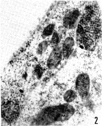
陽性率の減少傾向(英文)………・………・……一一…千馬 正敬,中村 剛,板倉 英世 25−28 ネッタイシマカおよびヒトスジシマカ(インドネシア株)の唾液腺におけるチクングニァ
ウイルス成熟の電子顕微鏡的研究(英文)………・…・…一Soedarto Soekiman,松村 武男 29−36 短 報
インドネシア由来のネッタイシマカ,およびヒトスジシマカにおけるデング3型ウイルスと チクングニアウイルス増殖の比較(英文)…一Soedarto Soek㎞an,小西 英二,松村 武男 37−41 会 報
昭和61年度第2回幹事会記録………一… 43−45 昭和61年度評議員会記録……一………・・ 45−46 第28回総会記録…… 46−47 昭和60年度会計決算書一一一 47 昭和62年度予算書…………一…一………一…・・…… 47 幹事選挙関係規則改正に至った経過要約……一…………曙一 47−48 斡事選挙関係規則改正案に対する説明一… 48−49
会則…………一 50−53
投稿規定・………一 54−55 会員名簿・…一・ 56−86
日熱医会誌
Japan.J.T.M.H. 日 本熱帯医学会
Japan. J. Trop. Med. Hyg., Vol. 15, No. 1, 1987, pp. 1 6 1
ECOLOGICAL STUDIES ON THE LUNG FLUKE, PARAGONIMUS OHIRAJ MIYAZAKI, 1939
II. INFECTION RATES WITH P. OHIRAr OF SNAILS AND RODENTS COLLECTED FROM THE IBI,
NAGARA AND KISO RIVERS IN THE TOKAI DISTRICT, CENTRAL JAPAN
KIKUO MATSUol AND KlYOSHI MAKJYA2
Received September 1 1986/Accepted January 8 1987
Abstract : In the area of the lbi, Nagara and Kiso rivers in the Tokai district, central Japan, a survey for the detection of Paraglonimus ohirai in snails and rodents was performed durirrg the period between October 1983 and October 1985. Snails, Assiminea parasitologica, positive for P. ohirai cercaria were found in 2 of 5 study sites. The over‑all infection rates were O. 41% at the sites on the lbi and 0.45% at those on the Kiso river. Two species of rodents, Rattus norvegicus and Apodemus speciosus, were collected, the infection rate for P. ohirai being 50% in the former and 0 fc, in the latter species. The results of the present study and our previous report (Matsuo and Makiya, 1985) dlsclosed the life cycle of P. ohirai in the areas along the lbi, Nagara and Kiso rivers; A. parasitologica is the Ist intermediate host, Sesarma dehaani and S. intermedia are the 2nd intermediate host, and R. norvegitcus is the final host.
INTRODUCTION
The frst report of this series revealed that 2 species of crabs, Sesarma dehaani and S.
intermedia, with Paragonimus ohirai were widely distributed along 3 rivers, the lbi, Nagara and Kiso, which flow into the Ise Bay at the boundary between the north‑eastern part of Mie Prefecture and the western part of Aichi Prefecture in the Tokai district, central Japan (Matsuo and Makiya 1985). Metacercariae were detected in the crabs collected at 17 out of 19 study sites which are situated about 5.5 to 13.0km up from the estuaries of the rivers. The infection rate was 2.7‑100% for S. dehaani and 0.9‑100% for S. intermedia.
The present report deals for the frst time with the infection rate of P. ohirai in snails and rodents serving the Ist intermediate and final hosts, respectively, collected from the study areas along the lbi, Nagara and Kiso rivers. Although lwata and Nagayoshi (1985) are of the opinion that P. ohirai is not an independent species, the authors here adopt the current theory that P.
ohirai is an independent species within the genus Paragonimus.
l Departrnent of Medical Zoology, School of Hygiene, Fujita‑Gakuen Health University, Toyoake, Aichi Prefecture 470‑11, Japan
2 Department of Medical Zoology, School of Medicine, University of Occupational and Enviroumental Heaith, Kitakyushu 807, Japan
SURVEY AREAS AND METHODS
The survey of the Ist intermediate host, Assiminea parasitologica, was carried out at 5 sites on the lbi, Nagara and Kiso rivers, shown in Figure 1, during 2 years from October 1983. The snails collected were crushed individually after measuring the shell length. Removing the broken sheu fragments, the specimens were checked for P. ohirai cercaria under a stereomicro‑
scope.
The final hosts, wild rodents, were captured with live traps set on the river‑banks along the waterway which are covered with communities of eulalia grass, reed and other grasses. The traps were set in the afternoon and rodents caught were gathered in the next morning. The rodents were dissected after identifying species and sexes and measuring body‑ and tail‑length.
Lungs and livers were carefully examiued for P. ohirai worms and feces were also checked for the eggs.
RESULTS AND DISCUssION
First intermediate host: Two species of snails, A. parasitologica and A. japonica were collected at 5 study sites shown in Figure I . Assiminea parasitologica inhabits damp areas on river‑banks covered with eularia grass and other weeds, and was collected from the surface of soil, deadwood and fallen leaves. Assiminea japonica, on the other hand, was mainly seen on the surface of wet soil and on stones on the bottom of the river and the lower part of reed stems, which were submerged at high tide.
The detection of P. ohirai cercaria was carried out for A. parasitologica which is known as the principal Ist intermediate host in other endemic areas of P. ohirai in Japan (Yokogawa et al.
1957; Yoshida and Miyamoto 1959; Miyamoto 1961). The results are summarized in Table 1 and Figure I . Parnglonimus ohirai cercariae, as shown in Figure 2, were detected in snails collected at B and E out of 5 study sites. Site B is located about 8 krn up from the estuary of the lbi River, where Route I crosses the river. The cercariae were found in 8 of 1,782 snails (infection rate = O. 45%), where the infection rate of the 2nd intermediate host crab with P. ohirai metacercariae was 90% for S. dehaani and 88. 9% for S. intermedia as reported in the frst part of this study. Site E is situated about 6 km up from the estuary of the Kiso River, where 10 out of 2,075 snails were positive for P. ohirai (infection rate =0.48%). The infection rate in crabs here was 69.3‑85. 7% in the previous report. At the other sites, all the snails examined were negative for P. ohirai cercariae. In conclusion, a total of 18 out of 5,305 snails examined were positive for P. ohirai (infection rate = 0.34%).
In addition to the dissection of snails, an experirnent on the cercarial emergence from snails was carried out with those collected irom Site E. The snails were individually submerged in water in small petri‑dish for I ‑2 hours and then P. ohirai cercariae emerging from the snails were counted. The cercarial emergence was detected in 8 out of 2,380 snails (emergence rate = O. 34%).
Yoshida and Miyamoto (1959) investigated the Ist intermediate host snails in the wide endemic area of P. ohirai along the Maruyama River, Hyogo Prefecture. The natural infection rate of P. ohirai cercaria was O. 048% for A. parasitologica, but negative for A. japonica and the experimental infection rate was 60. 8% for the former and 2.0% for the latter species. The
3
Table 1 Infection rate of snail, Assiminea parasitologica, with Paragonimus ohirai cercariae collected along the lbi, Nagara and Kiso rivers in Tokai district, central Japan
Collection site Date of survey No. of snails examined
No. of snails infected
(%)
A lbi River Apr. , 1984 190 o (o)
B lbi River Jan. , Jul. , 1984
Oct., 1985 1 , 782 8 (0.45)
c Nagara River Jan. , Jul. , 1984 l, 105 o (o)
D Kiso River Aug. , 1985 1 53 O (o)
E Kiso River Oct. , 1983
Jul. , Oct. , 1984 2,075 10 (O. 48)
Total 5, 305 18 (O. 34)
Figure l
D
ID
IID
1 23
1 : Ibi River
̲ 2: Nagara River
3: Kiso River
Route No. 1 D
B c Mehan Highway o
e
Route No. 23
Ise Bay
Sketch maps showing the lbi, Nagara and Kiso rivers where the survey on Paragonimus ohirai was carried out.
Results of examination of the Ist intermediate host. B and E: Sites where snails positive for the cercaria were collected. A, C and D: Sites where all snails examined were negative for the cercaria
Results of examination of the final host. Black dots: Sites where norway rats positive for the adult worm were collected. White dots: Sites where norway rats negative for the adult worm were collected. Minus marks: Sites where wood mice negative for the adult worm were collected
Results of examination of the 2nd intermediate host summarized in the previous report. Black dots: Sites where crabs positive for the metacercaria were collected. White dots: Sites where all crabs collected were negative for the metacercaria
Figure 2 A: Paragordmus ohirai cercariae recovered from the snail Assiminea para‑
sitologica collected from the lbi River. B: Adult worm of Paragonimus ohirai recovered from the rat Rattus norvegicus collected from the Nagara
River .
present survey on snails reveaied for the frst time that A. parasitologica serves as the Ist intermediate host of P. ohirai in the areas along the lbi and Kiso rivers and that the naturai infection rate for this snail (O.45( /0 at site B and 0.48% at site E) was 8 to 9 times higher than that in the Maruyama River area.
Final host: A total of 190 live traps were set at area8 5‑12, 7‑13 and 4‑8 km up from the estuaries of the lbi, Nagara and Kiso rivers, respectively, during 6 months from December 1983 to May 1984. As showa in Table 2 and Figure 1, a total of 18 norway rats (R . norvegicus) and 6 wood mice (A. speciosus} were captured during this period. Of these, 9 norway rats were positive for P. ohirai (infection rate = 50%), but all the wood mice were negative. Eight of the infected norway rats were identified as adults, in the lungs of which adult worms of P, ohirai were detected. Paragonimus ohirai eggs were also found from the feccs of all the adult
Table 2 Infection rate of rodents, Rattus norvegicus and Apodemus speciosus, with Paragord=
mus ohirai adult worms collected along the lbi, Nagara and Kiso rivers during six month trom December 1983
Collection
s ite
R attus norvegicus Ap(rdemus specios us No. exarnined No. positive
( / ) No. examined No. positive
(%) lbi River
Nagara River Kiso River
6 6 6*
4 (66. 7) 2 (33.3) 3 (50, o)
4 2 o
o o
Total 18 9 (50, o) 6 o
* including one young rat
5
8
g3
*o 6
,L)
4
;
2 e
,
D observed
e calculated
e ' ・ e I . . .
J L
o 2 4 6 8 Io 12 14 25 37
Number of adult worms
Figure 3 Frequency of Parnglonimus ohirai adult in Rattus norvegicus observed frequehcy agreed well with the negative binomial (k = 0.189, x2=1.214, p=0.271).
rodents. One rat was young and an irnmature worm of P. ohirai was obtained from the liver of this rat. The number of worms per positive rat was I ‑37, the average being 5. 06. The frequency distribution of the adult worms among the final host was regarded as overdispersed, and this agreed well with the negative binomial [(Bliss and Fisher, 1953; degree of overdisper‑
sion k=0. 189 and fitness p=0. 271 (Figure 3)]. This meant that many rats harboured a hrnited number of worms but that only a few individuals were infected with a large number of parasites.
This type of distribution is considered to be advantageous to the parasite, because most of the parasite‑carrying rats can survive longer and disserninate the eggs even if a few heavily‑infected hosts may die earlier. Further, when live traps were set, crab fragments were found around the lairs of the norway rats in the area. This suggests that the crabs are one of the major food sources of norway rats in the study area.
In other endemic areas of P. ohirai in Japan, the natural infection rate of the final host was reported as folbws: 7‑35% in weasels in the Maruyama River area, Hyogo Prefecture, 47% in weasels, 25 : o in badgers, 14% in wild boars, 8% in Japanese meadow mice (Microtus montebelli) and O% in wood mice in the lzu area, Shizuoka Prefecture.
Comparing our own with these reports, the present infection rate of norway rats (50%) is even higher than the highest rate for weasels in Shizuoka Prefecture (47%) and much higher than those of other final hosts including the Japanese meadow mouse and wood mouse. Norway rats feed not only on cereals but on fish, birds and insects, while wood mice have a mainly vegetation diet, such as the bark of young trees. It would therefore seem that the present difference in the natural infection rate between norway rats and wood mice is due to their differing food habits.
The present survey for rats revealed for the hrst time that the norway rat is one of the final hosts of P. ohirai in the areas along the 3 rivers, Ibi, Nagara and Kiso.
CONCLUSIONS
A parasitological survey of P. ohirai was carried out for the Ist intermediate host in the
areas along3hvers,the Ibi,Nagara and Kiso in the Tok由(五sthct,centralJapan. The fluke was con㎞ed to heav丑y㎞ect t上e sna丑ノ1.ρσ名硲訪oJo9∫oαand廿1e norway rat R.no甥磐∫6z硲血廿1e survey areas. Considered together with廿1e previous study on廿1e2nd k}termediate host,it
was c㎞ed jor the血』st t㎞e that P.ohゼ名痂in these areas inhabits the snaiL4.加昭s吻Joゆ08as lhe lst inte㎜ediate host,the crabs S.吻㎞伽nd S.∫n勧%8伽as the2nd and the ratR.
切nノεμo%s as廿1e f血al host.
REFERENCES
1) Bhss,C.1.a皿d Fisher,R A.(1953): Fitti119廿1e negative b血omial distribution to biological data Imd note on the ef6ective ntting of the negative binomial,Biometrics,9,176−200
2)Iwata,S.and Nagayoshi,K.(1985):The species name of P酩㎎o 伽%s in Japan,J.Kumme Med.
Assoc.,48,383−396(㎞Japanese)
3)Matsuo,K and Makiya,K(1985):Ecological studies on the lung nuk,Pα猶㎎oπ吻螂oみ加∫Miyazaki,
1939. 1.1価ection rate ofP.ohf耀∫metacercariae hl brackish water crabs co皿ected ffom the six rivers in the Tokai district,centraUapan,Jap.J.Trop、Med.Hyg.,13,307−313
4)Miyamoto,M(1961):Studies on Pα名㎎i㎝伽πs and Paragonjmiasis in the northem Disthct of Hyogo Prefecture. Part IL An ecological studies on Po猶㎎o 」 ¢z偲oゐ∫瓶J Myazaki,1939hl its ende血c area along the banks of the Maruyama River,J.Kyoto Pre£Univ.,69,1669−1683(in Japanese)
5)Yokogawa,M.,Yosh㎞ura,H.,Sano,M and Suzuki,S.(1957):P幽㎎o 伽%s oh加5in the southem Izu disthct,Tokyo Iji Shinshi,74,403−406(㎞Japanese)
6) Yoshida,Y.and蜥yamoto,M(1959): Studies on a fkst hltemediate host,ノ13s伽z初昭ρα名㏄訪o o蜘8 Kuroda,1958( 毛Pロ」%4初εJJα吻σf傭 of Yokogawa and Koyamaθ α乙non Gould),of Pα猶㎎o 伽%s o雇π房 Myaz面,1939,JPn.J.ParasitoL,8,122−129(血Japanese)
大平肺吸虫の生態学的研究
II 東海地方の揖斐川,長良川,木曽川流域産カイ ならびにネズミにおける本種の検出成績
松尾喜久男1・真喜屋 清2
1983年10月から1985年10月の間に,伊勢湾に注ぐ三重,愛知県下の揖斐川,長良川,木曽川の河口 周辺に生息するカイ,ネズミについて大平肺吸虫の検出を行った。計5地点で採集したムシヤドリカ
ワザンショウ計5,305個体を剖検し,そのうち,揖斐川,木曽川の各1地点から本種セルカリアを検出 した。この2地点における検出率はそれぞれ,0.45%,0.48%であった。ネズミについては,3河川 流域からドブネズミ計18頭,アカネズミ計6頭を捕獲して剖検した。ドブネズミでは8頭の成熟ネズ ミの肺から本種成虫計90個体,1頭の未成熟ネズミの肝から未熟虫体1個体を検出したが,アカネズ ミはすべて陰性であった。既報のカニの成績ならびに今回のカイ,ネズミの調査結果から,3河川河 口流域の広大な大平肺吸虫分布地において,本種の生活史に第1中間宿主としてムシヤドリカワザン ショウ,第2中問宿主としてクロベンケイ,ベンケイガニ,終宿主としてドブネズミが関与している ことが初めて明らかになった。
1藤田学園保健衛生大学衛生学部医動物学教室 2産業医科大学医動物学教室
Japan. J. Trop. Med. Hyg. , Vol 15 No 1 1987 pp 7 15 7
LEISHMANIASIS IN DIFFERENT ALTITUDES ON ANDEAN SLOPE OF ECUADOR
YOSHIHISA HASHIGUCH11, EDUARDO A. GOMEZ LANDIRES2
VICENTA VERA DE CoRONEL2, TATSIJYUKI MIMOR13 AND MASATO KAWABATA4
Received November 1 1 1986 / Accepted February 15 1987
Abstract : An epidemiological survey was performed in a leishrnaniasis‑endemic area aiong highway which was established about 15 years ago on the Andean slope of Ecuador; the area ranged from 300 m to 1,500 m above sea level. In general survey, 64 (14.3%) of the 446 subjects examined were positive for leishmanial signs. In order to know leishmanial infections in relation to the altitudes of dwemng sites of subjects, analysis was made on 224 children with 5 to 15 years of age. At 4 different sites with 500 m, 1,000 m, 1,300 m and 1,500 m above sea level, the infection rates of the subjects from the individual sites were 17.4, 18. 8, 5. 6 and 8.8%, respectively. A statistically significant difference was recognized between the altitudes, 00‑
1,000 m and 1,300‑1,500 m (0.01<p<0.05, x2=5.314), but not between 500 m and 1.000 m and between 1,300 m and 1,500 m. Leishmanial infections of the children who came irom forest and highway areas were compared in each altitude. But no significant difference was found between forest and highway dewellers at any study sites.
INTRODUCTION
In Ecuador, transmission of American cutaneous and mucocutaneous leishmaniasis occurs in rural populations living in bilateral regions of the Andes mountains from the lowlands to highlands up to the elevation of 2,000 m. The disease is widely spread in most provinces and is a considerable public health problem in the country. In the endemic areas, however, little epidemiological study has been done on the community base, and no control measure has been applied to reduce or interrupt the transmission of the disease. For a future control, it would be necessary to clarify the epidemiological features in each endemic area of lowlands and highlands.
New World cutaneous or mucocutaneous leishmaniasis is more difficult to control than is those of Old World, since it is principally a disease of wild mammals in the dense forest, and numerous reservoir hosts are arboreal; thus, in most endemic areas, reservoir‑vector control is ahnost impossible (Marinkelle, 1980). At present, the only alternative measure for the control
1 Department of Parasitology, Kochi Medical School, Nankoku City, Kochi 781‑51, Japan
2 Departamento de Parasitologia, Instituto Nacional de Higiene y Medicina Tropical, Apartado 3961, Gua‑
yaquil, Ecuador. S. A.
3 Department of Parasitic Diseases, Kumamoto University School of Medicine, Honjo, Kumamoto 860, Ja pan
4 Department of Clinical Pathology, School of Medicine, Nihon University, Ohtaniguchi, Tokyo 173, Japan
in most parts of the neotropics seems to be evacuation of the entire human population from potentially dangerous areas, but such measure is inconceivable because of political, socioecono‑
mic and logistic reasons (Marinkelle, 1980). Under such circumstance, it would be worthwhle to evaluate the effect of environmental changes in relation to the transmission of leishmaniasis.
The present paper deals with the resuslt of an epidemiological survey performed in different altitudes of leishmaniasis‑endemic areas with 300 m to 1,500 m above sea level. In the area, migration of inhabitants occurred from forest area to the viciuity of highway which was con‑
structed on the Andean slope about 15 years ago. The leishmanial infections, therefore, were also compared between forest and highway dwellers, in order to know the effect of change in the life mode of the inhabitants on the transmission.
MATERIALS AND METHODS Study area
The study site is located in the Departrnent of Cafiar on the south east of Ecuador, 2'30' West longitude, and located on the Pacific slope of the Andes, ranging irom 300 m to I , 500 m above sea level. Two vinages, Ocaffa and Javin, in the above area are situated about 70 km from Guayaquil City and established as agricultural cornmunities along highway to Cuenca City.
A simplified sketch of the area is shown in Figure 1.
The paved highway with 10 meters in width was constructed about 15 years ago in the area and it changed a mode of viuager's life, including agricultural systems. Before construction of the highway, bananas and yucas were the main agricultural products in densely forested areas, but they were replaced by sugar canes cultivated in largely deforested areas after the highway construction. The highway supported not only the movement of villagers but also transporta‑
tion of their agricultural products to major cities, such as Cuenca and Guayaquil. Thus, the highway construction made a great enviroumental change of the endemic area of leishmaniasis.
Still, however, there were some surviving patches of natural dense forest which would provide the breeding sites for vector sandflies and reservoir hosts of leishmaniasis. Such lintited patches are distributed in the one side of the highway, while on the other side there was a continuous undisturbed dense forest through lowlands to highlands along highway (Photos. I , 2 and 3).
The majority of dweWngs in the study area were built along highway, but the remainings were in forest areas. There was no livestock, but the people raised the dogs, cats, pigs, guinea pigs and domestic fowls. Wild mammals (sloths, arrnadilbs, opossums, rats and mice) and 2 species of man‑biting sandflies, Lutzomyia trapidoi and Lu. hartmanni, were found in the area (Hashiguchi et al., 1985a, b, c).
Epidemiological exam inations
In the inhabitants, the survey was performed by house visit, while it was done in children with 5 to 15 years of age by visiting 4 study sites (schools), La Delicia (A, 500 m above sea level), Ocafia (B, 1,000 m), Las Copas (C, 1,300 m) and Javin (D, 1,500 m). All the subjects were interviewed about their life history, such as occupation, cultivation, migration and contact history with sandfiies, then examined clirLically by well‑experienced physician (E . A.G.L. and V. V. C.) to find ulcers (active lesions) and scars (cured lesions) of leishmaniasis. When they had active lesions, tissue samples were taken from the margin of ulcers for microscopic examina‑
i li
9
t
Z : Highway dweller . : :,: '.. '; '
I : Forest dweller
" : C""':.:: .
,. ,',
": ' i:: , : '.'; , ・・": L I : : "
' ' . ' . '.:.:;:' ': prlmary forest "
l l
,
l' ' I
t ' I ・‑
,
, l
1
/
,
t C
, ,
" " .
. . ' '
,
,
.,
l
, l
l
, t ,
. l
"'1 "'
l" :
1500m above sea level
:: .e D
e‑
,
1'
Highway
l
eee
dr
300m above sea .ievel
,
/
1 km
/
"dP /
ez . ̲ A ,. ' s "
e ., /
",,ld::
..p ,,
, , ,
l l
/
,
J ,
,
'・'・・:: 7:・ . '
: ' :・ ,:'4 7 ;7); , //1 : C
e ':,
.
d "":1
'::
////////. ' .
'
'
Figure 1 A simplified sketch of the study sites in the Department of Cafiar, Ecuador. A, La Delicia (500 m above sea level); B, Ocaiia (1,000 m); C, Las Copas (1,300 m); D, Javin (1,500 m).
Pho,to .
Photo.
l 2
Showing 4 houses (arrows) along highway in the study site (highway dwellers).
Showing feld of sugar ca e cultivation and remainiug patchee of natural (primary) Arrows on the above side show 3 hou8es in forest area (farest dwellers).
f ore st .
11
tions. The location, size, onset and duration of lesions were recorded.
With regard to the subjects with scars, the diagnosis was determined only clinically, i.e., mainly based on the type and localization of the scars, duration of the lesions and contact history with sandflies; the chronic leishmanial ulcers resulted in a thin, depigmented scar (Photo. 4).
The viuagers have been calling leishmaniasis as llaga de montaha and sandflies, manta blanca in Spanish, respectively.
The causative agent of these cutaneous and mucocutaneous leishmaniasis in Ecuador has been considered Leishmania braziliensis s.1. , based mainly on the clinical manifestations, be‑
havior and localization of the organisms in sandfly vectors and growih in cultures.
RESULTS
In the inhabitants from whole area, the positive rates with leishJnanial signs were arranged by age and sex in Table I . In a total of 446 subjects examined, 64 (14. 3%) were found to be positive for leishmanial signs. No marked difference was recognized between both sexes in the positive rates.
In Table 2, Ieishnianial infections of 224 children with age 5 to 15 years in the total ex‑
aminees were arranged by the altitudes of their dweurng sites. The data revealed a statistically significant difference between altitudes, 500‑1,000 m and 1,300‑1,500 m above sea level (0.01<p<0.05, x2=5.314), but not between 500 m and 1,000 m and between 1,300 m and 1,500 m. The result, therefore, suggested that the intensity of transmission was markedly influenced by the altitudes of dweWng sites in the endemic area.
To evaluate the intensity of transmission, the above 224 children were reanalyzed between forest and highway areas (Table 3). They were divided into the fonowing two groups; 1) forest dwellers who have or had experience living in forest area, that is, those who settled down or
Table I Leishrnanial infections among 446 inhabitants arranged by age and sex in the Depart‑
ment of Caitar, Ecuador
Male Female Total
Age No. examined + * No exammed + No exanuned + %
‑10 11‑20 21‑30 31‑
110 77 16 19
12(4) 14(3) 4(1) 2
10. 9 18. 2 25. O 10. 5
1 09
70 19 26
12(3) 12(1)
3 5
ll.O
17. l
15.8
19. 2
219 147 35 45
24(7) 26(4) 7(1) 7
11.0 17.7
20. O 15. 6
Total 222 32(8) 14.4 224 32(4) 14.3 446 64(12) 14.3
* Positives with leishmanial scars or ulcers; the number in parentheses shows positives with active lesions (ulcers).
Photo. 3
Photo. 4
A house constucted in the vicinity of highway (1,000 m above sea leVel), but surrounded by dense forest (arrow in Figure 1). A considerable number of sandfiy was couected around the house.
All the persons of this family, 8 in total, had already suffered from leishmaniasis, showing the typical scars.
A typical leishnranial scar (arrow) found on the foreann of a 34‑year‑old female who lives in the house shown in Photo. 3.
Table 2 Leishrnanial infections of 224 chldren, ing sites in the Department of Cafiar,
5‑15 years old, Ecuador
arranged by altitude of dwell‑
Altitude*
(in meters) Schools** No. examined Positives with leish‑***
manial signs (%) 500
1, OOO 1, 300 1 , 500
A C D
46 85 36 57
8 (17.4) 16 (18.8) 2 (5.6) 5 (8.8)
*
**
***
Altitude above sea level.
A, La Delicia; B, Ocafia; C, Las Copas; D, Javin.
Statistically significant difference between the altitudes, 500‑ 1,000 m and 1,300‑ 1,500 m (0.01 < p < 0.05, x2= 5.314), but not between 500 m and 1,000 m and between 1,300 m and 1,500 m.
Table 3 Leishmanial ini;ections of 224 children, 5‑ 15 years old, arranged by sites in forest and highway areas in the Departrnent of Cafiar, Ecuador
their dwelling
Children
from Schools* No. examined Positives 'with leish‑
manial signs (%)
Forest
A B C D
27 34 36 ll
6 (22.2) 5 (14.7) 2 (5.6) 1 (9.1)
Total 1 08 14 (13.0)
Highway
A C D
19 51 O 46
6 (31.6) 11 (21.6) 4 (8.7)
Total ll6 21 (18.1)
Total 224 31 (13.8)
* A, La Delicia; B, Ocaiia; C, Las Copas; D, Javin.
lived in the past in forest area, 2) highway dwellers who were born at or imrnigrated from non‑endemic area to highway area. No statisticauy significant difference was recognized be‑
tween forest and highway dwellers in each study site of A, B, C and D.
The localization and number of lesions were examined in 52 subjects with leishmanial scars.
Of 1 10 scars found, 40. 9% were in the face, 30.0% in the upper extremities, 26. 4% in the lower extremities and 2.7% in the trunk. The majority of scars measured less than 15mm in dia‑
meter. Only 12 persons (2. 7% of the total examinees), 8 males and 4 females, had I to 8 ulcers in the cheek, ear and upper or lower extremities. In interviews, the duration of ulcer ranged from I month to 2 years; the lesions measured between 4 mrn and 32 mm in diameter.
13
DlscussloN
In our previous study in the Pacific slope of the Andes in Ecuador, 15. 8% of the examinees were positive for active leishmanial lesions and 60%, for leishmanial scars (Hashiguchi et al. , 1984). The present study showed rather low intensity of the transmission, in the Department of Cafiar, Ecuador.
In the examination at 4 sites with different altitudes, the prevalences were higher at 500 m (17.4%) and 1,000m (18.8%) above sea level than 1,300 m (5.6%) and 1,500 m (8.8%). The fact is quite noticeable in connection with the infection of sandfiy vectors with leishmanial pro‑
mastigotes. In this area, Hashiguchi et al. (1985c) examined natural irLfections with the parasites in man‑biting species of sandflies, and reported that the infection rate of Lu. hartmanni was 5. 9% at 350‑600 m, 3. 8% at 950 m, 2.3% at 1,200‑1,500 m and O% at 2,000 m, while the other sandfiy, Lu. trapidoi, was positive for the parasites at only one site of 350‑600 m (8. 1%), showing a markedly reduced number of fly catches at higher sites. These results indicated that leishmanial transmission in the Andean slope was greatly infiuenced by the altitudes of dwemng sites, and also that the intensity of transmission would be very low at higher lands of 1,300 m or over.
By the data analysis of 224 chldren, the prevalence of leishmaniasis was compared between forest and highway dwellers revealed no marked difference between two groups. The result, therefore, suggested that there might be no remarkable difference between forest and highway areas, in terms of the intensity of leishmaniasis transmission in the present endemic area. This might be due to the existence of prirnary forest along highway or the remaining patches of natural forest around dweuings, which would play a role as the breeding sites for the vector and reservoir of the disease. In Panama, an insular effect resulting from clearance of prirnary forest which surrounded a settlement, guarded properly the community against leishmanial infection prevalent in the nearby forest (Herrer et al. , 1976). To reduce the transmission in the present area, ,a iutther clearance of the forested areas would be necessary.
The localization of lesions in the subjects examined mostly agreed with that reported by Rodriguez and Aviles (1953) and Hashiguchi et al. (1984) from Pacific coastal regions of Ecuador, but greatiy difiered from that in Amazon regions of the country, where 60% of the lesions observed were in lower extremities (Amunarriz, 1982). This discrepancy might be caused by the difference of biting behavior of sandflies or clothing habits of inhabitants between the two regions.
ACKNOWLEDGEMENTS
We are most grateful to Drs. Hugo H. de Nully. M. Arzube R. , R. Lazo S., E. Gutierrez V.
and F. Parra Gil of the Direction of Instituto Nacional de Higiene y Medicina Tropical (INHMT), Ecuador for their support throughout this study. We owe a debt of gratitude to our colleagues, Dr. N. Urdiuares and Messrs. R. Sud and M. Leyion for their assistance in the field phase of the study. Thanks are also due to all the members of the Departamento de Parasitologia, INHMT for their support in laboratory works. We thank Dr. I. Tada of Kumamoto University and Dr.
T. Yoshimura of University of Occupational and Environmental Health for critical reading of the manuscript. Finally, we are much indebted to Drs. N. Ishida (Tohoku University), S. Hayashi (National Institute of Health, Japan), N. Suzuki and H. Osaki (Kochi Medical School) for their
encouragement throughout the study. This study was supported by the Ministry of Public Health, Republic of Ecuador and the Japan International Cooperation Agency (JICA) and Over‑
seas Scientific Research Program of the Ministry of Education, Science and Culture, Japan (No.
6104 1059).
REFERENCES
1) Amunarriz, M. U. (1982) : Leishmaniasis, Salud y enfermedad patologia tropical en la region amazonica, 71‑88, Edicion CICAME, Ecuador
2) Hashiguchi, Y. , Gomez, E. A . L. and Coronel, V. V. (1984) : An epidemiological study of leishrnaniasis in a plantation "Cooperativa 23 de Febrero" newly established in Ecuador, Jpn. J. Parasit., 33, 393‑401 3) Hashiguchi, Y. , Gomez, E. A. L., Coronel, V. V. , Mimori, T. and Kawabata, M. (1985a) : Leishmania
isolated from wild marnmals caught in endemic areas of leishmaniasis in Ecuador, Trans. Roy. Soc. Trop.
Med. Hyg., 79, 120‑121
4) Hashiguchi, Y., Gomez, E. A. L. , Coronel, V. V. , Mimori, T. and Kawabata, M. (1985b) : Biting activ‑
ity of two anthropophilic species of sandfiies, Lutzomyia, in an endemic area of leishmaniasis in Ecuador, Ann. Trop. Med. Parasit., 79, 533‑538
5) Hashiguchi, Y., Gomez, E. A. L., Coronel, V. V. , Mimori, T. and Kawabata, M. (1985c) : Natural inL fection with promastigotes in man‑biting species of sandflies in endemic areas of leishrnaniasis in Ecuador, Arn. J. Trop. Med. Hyg. , 34, 440‑446
6) Herrer, A. , Christensen, H. A. and Beumer, R. J. (1976) : Epidemiological pattems of cutaneous leish‑
maniasis in Panama, II. Incidental occurrence of case in non‑endemic settlements, Ann. Trop. Med.
Parasit., 70, 67‑71
7) Marinkelle, C. J. (1980) : The control of leishmaniasis, Bull. Wld. Hlt. Org. , 58, 807‑818
8) Rodnguez, J. D. M. and Aviles, F. N. (1953) : Algunas observaciones sobre leishmaniasis cutaneo‑
mucosa en el Ecuador, Rev. Ecuat. Hig. Med. Trop., 10, 35‑58
15
エクアドル国アンデス斜面の高度差による リーシュマニア症浸淫
橋口 義久1・EDuARDoA.GoMEzLANDIREs2・VIcENTAVERADECoRoNEL2 三森 龍之3・川端 真人4
エクアドル国のアンデス斜面低地から高地(海抜300m−1,500m)にかけて居住する住民につい て,リーシュマニア症の罹患状況を調べた。この流行地には約15年前にハイウェイ(道幅約10m)が 建設され,住民の居住環境ならびに生活様式に大きな変化が認められた。
一般住民446名について検査したところ,64名(14,3%)は本症によると考えられる治癒病変,また は皮膚潰瘍を保有していた。
居住地の高度差による住民のリーシュマニア症罹患状況を知るため,被検者のうち5−15歳の学童 224名を対象に,4地点(A,海抜500m;B,1,000m;C,1,300m;D,1,500m)において,居住地区 別の罹患を比較した。その結果,学童のリーシュマニア症罹患率は,海抜500m地点で17.4%,
1,000mで18、8%,1,300mで5.6%,1,500mで8,8%となり,500m−1,000mの地域と1,300m−
1,500mの地域との間には,統計学的に有意の差を認めた(0.01<p<0.05,Z2=5.314)。このこと は,アンデス斜面のリーシュマニア症流行地の比較的低い地域(1,000m以下)では,本症の罹患率 が高くなるが,より高い地域では罹患率は低くなることを示唆している。
一方,ハイウェイ沿いと山間部との間で,本症罹患率の差異を検討するため,上記224名の学童を,
その居住地の状況によって次の2群に分類した。1)山間部に定住または過去に一時期居住した者,2)
ハイウェイ沿いで出生または非流行地から移住した者。上記2群問での学童の罹患率を見る上で,高 度差による影響を除去するため,各地区ごとの山間部住民とハイウェイ住民との比較を行った。その 結果,4地点のいずれにおいても両群間に有意な差を認めなかった。したがって,本調査地において
は,ハイウェイが建設され,環境変化や住民の移動がみられたものの,道路沿いの原生林や人家,お よび農耕地周辺に原生林の一部が残存し,これがサシチョウバエや保虫宿主の供給源の役割を果たし ているものと判断された。
1高知医科大学寄生虫学教室 2エクアドル国熱帯医学研究所寄生虫学部 3熊本大学医学部寄生虫病学教室 4日本大学医学部臨床病理学教室
CHANGES OF BLOOD OXYGEN AFFlNITY IN FANSIDAR‑TREATED MICE INFECTED
WITH PLASMODIUM BERGHEI
ATSUSHI HIOKI AND HIROSHI OHTOMO Received November 10 1986/Accepted February 15 1987
Abstract : Changes of blood oxygen affinity in mice infected with Plasmodium berghei and treated with Fansidar (20 mg/kg body weight sulfadoxine and I mg/kg body weight pyrimetha‑
mine orany) were observed. When 5‑weeks‑old male mice were inoculated intraperitoneally with l07 of P. berghei‑infected red cells and treated with Fansidar on day 5 after inoculation, 67%
of the anirnals sulvived. Parasitemia decreased and abnormal values of glycolytic intermediates began to recover following Fansidar adrninistration. However, hemoglobin content still de‑
creased on day 2 of the treatnent, and methemoglobin iraction increased on days 1, 2, 3 and 4.
Blood oxygen affinity increased markedly on days 2 and 3. These findings suggested aggrava‑
tion of the host's hypoxic state on day 2 of the treatment.
INTRODUCTION
Malaria is relatively easy to cure if treated properly on the basis of early diagnosis.
However, delay in the onset of treaiment, especially for falciparum malaria, causes a higher level of parasitemia, severe complications and sometirnes death of the host. Furthermore, death of patients with markedly decreased parasitemia following antimalarial treatment have been re‑
ported (Devakul et al. , 1966; Stone et al. , 1972), but its mechanism is not yet understood.
The pathophysiology of falciparum malaria is considerably complex. Most organs are re‑
ported to be involved in this infection, and many factors contributing to dysfunction of these organs have been enumerated (WHO, 1980; Bruce‑Chwatt, 1980). Tissue hypoxia is thought to be one of the main causes of severe complications (Hall, 1977). We have already reported that the decrease in blood oxygen affinity in the course of Plasmodium berghei infection in mice may be a compensatory mechanism for tissue hypoxia resulting from malaria (Ohtomo et al. ,
l 982).
In this study, we intended to clarify the effect of treatment on blood oxygen affinity in P.
berghei infected mice. We used Fansidar̲ as an antimalarial drug because it is effective in a single dose and has clear action of only blocking the 2 sequential stages of the pathway leading to tetrahydrofolate (Donno, 1974; Weidekamm et al. , 1982).
MATERIALS AND METHODS Male mice of ddY strain, 5 weeks old, were used in this study.
NK65, has been maintained in our laboratory by injecting a dilute
Plasmodium berghei, strain suspension of infected blood
Department of Parasitology, Gifti University School of Medicine, 40 Tsnkasa‑machi, Gifti 500, Japan
18
into mice from the corresponding strain every week. Blood was conected from an infected mouse by cardiac puncture using heparin as an anticoagulant, diluted with saline to yield I x l08 infected red cells /ml, and O. I ml of this suspension was injected intraperitoneally. The time interval between exsanguination and inoculation was less than I hour. Mice were kept at 24‑
26'C and 40‑60% humidity and exposed to a photoperiod of 12 h light and 12 h darkness.
Under these conditions, mice died 6‑7 days after inoculation.
In the initial trials, desigued to evaluate the time of treaiment, 3 groups of 6 mice each were infected. Fansidar (F. Hoffrnann‑La Roche, Switzerland) was dissolved in distilled water and administered orally (1 mg pydmethamine and 20 mg sulfadoxine / kg body weight) on day 4 aiter inoculation to group 1, day 5 to group 2, and day 6 to group 3, respectively (Ferraroni and Speer, 1982). The course of the infection was followed by determining parasitemia and reticulo‑
cyie count daily frst by new methylene blue‑ followed by Giemsa‑stained blood smears from the
tail vein.
In the present study, the mice were inoculated with P. berghei and treated with Fansidar on day 5 of infection. Eleven mice each were anesthetized with pentobarbital sodium (50 mg/kg body weight intraperitoneally) on days O, 1, 2, 3, 4, 5 or 6 after Fansidar treatment, and whole blood was collected into heparinized syringes from the carotid artery withirL 5 minutes. Para‑
sitemia was determined on Giemsa‑stained thin blood filrn. Total hemoglobin concentration was measured by the cyanmethemoglobin method and methemoglobin fraction according to the KCN addition method (International Committee for Standardization in Haematology, 1978; Zwart et al. ,
1981). Hematocrit was defermined by means of a microhematocrit centrifuge. Oxygen equilibrium curve (OEO of whole blood was measured at 37'C on a Hem‑O‑Scan analyzer (Aminco Co. , USA), and blood oxygen affinity was expressed as the half‑saturation pressure values at actual pH in vivo (P50 act pH). The Hill‑coefficient n was obtained by linear regression analysis of log [y/(1 ‑ y)] vs. Iog P02, where y is 02 saturation for the data points of the OEC (02 saturation=30, 35, 40, 45, 50, 55, 60, 65 and 70 f ). Whole blood pH was measured im‑
mediately at 37'C with a glass electrode. 2, 3‑Diphosphoglycerate concentration was deter‑
mined according to a modified kit (No. 148334; Boehringer Mamheim GmbH, F. R. Germany) of the enzymatic end‑point method. Blood adenosine triphosphate (ATP) concentration was deter‑
mined by means of the luciferase reaction with a CHEM‑GLOW ( ninco Co.) (Chapman et al. , 1971). Concentrations of blood glucose, pyruvate and lactate were determined according to a modified kit (Nos. 124028, 124982 and 124842; Boehringer Marmheim GmbH).
RESULTS
Fansidar treatment on day 6 of Plasmodium berghei inoculation resulted in 100% mortality in mice 1‑2 days after treatment. Sixty‑seven percent of mice treated on day 5 and 100% of those on day 4 survived more than 10 days after treaiment (Table 1). Parasitemia decreased fouowing treatment compared with increasing in untreated subjects. Simultaneously, polychro‑
matophilic erythrocyies in the peripheral blood began to increase and significant reticulocylosis was observed from day 2 after treaiment (p < O. 05). When Fansidar was given on day 5, most of the parasites appeared to have degenerated on day 2 of treatment, and parasitemia became undetectable on day 4. Reticulocyie count reached its peak on day 5 after treatment, although uninfected controls remained low (Table 1).
Parasitemia of mice sacrificed for measurement (Table 2) were much the same as above.
Table 1 Time course of changes in parasitemia and reticulocylosis of mice treated with Fansidar 4 and 5 days after Plasmodium berghei inoculation
Day of Fansidar treatment
Days after Fansidar treatment
O 1 3 4 5 6 8
4 days after inoculation
No. of sutviving mice Parasitemia (%) Reticulocyie (%)
26. 4 7. 5 6 6
5.0 +1.6 O. 5 3. 4 :!:0.2 +0.6
6 6 1.7 O 0.5 +0
6. O lO. O
1.3 +1.3 6 o
+0
23. 7 d: 6. o
6
0 O
28. 5 :b 4. 2
6 O
+0
19. 6 d: 2. 9
6
+0 O 15.9 d:1.3
6
+0 O 11.7
2. O
5 days after inoculation
No. of surviving mice Parasitemia (%) Reticulocyie (%)
62. 7 41. 7 6 4
:1:2.2 +5.8 O. 4 4. 1 :1:0.1 0.5
4 4 4
10. 7 2. 4 O 2.9 +1.0 0
15. 6 17. 9 29. l 1.6 3・3 4.1
4 O
+0
35. 2 6・ 4
4 O
+0
31.0
3. 5
4
+0 O
20. 9 4. 2
4 O d:O
8. 9 O. 2
Uninf ected controls
No. of mice
Reticulocyie (%) 6 6
2.8 2.9
d:0.1 0.2
6 6 6 6 6 6
2. 6 2. 7 2. 9 2. 7 2. 6 2. 7 0.2 0.3 0.3 0.3 0.2 0.3
6
3. 1 O. 4
Unilufected and Fansidar treated controls
No. of mice Reticulocyle (%)
6 6 2.9 3.2 0.4 0.3
6 6 6 6 6 6
2. 7 3. 2 2. 6 2. 9 2. 7 2. 9 0.3 0.3 0.3 0.3 0.2 0.4
6
2. 9 O. 4
Values are means SE of sulviving mice.
Table 2 Hematological data of mice infected with P.
infection
berghei and treated with Fansidar on day 5 of
Uninfected controls Days after Fansidar treatnent Fansidar
treatment treatm nt O No 1 2 3 4 5 6
Parasitemia (%) Hb (g/dl) Ht (%)
MCHC (%)
12. 8 O. 2 40. 5 O. 7
31.7
O. 2
12. 7 O. 2 40. 8 O. 6
31.2
O. 3
65. 5 2.6
7. 2 O. 3 25. 7 1. l 27. 9 O. 6
43. 7 +̲5.0 7.4 O. 4 25. 4 1. 1 29. 2 O. 5
12. 6 1.4 4. 6 + O. 2 16. 9 O. 8 27. 4 + O. 7
1.5 O. 7 5. 7 O. 7 20. 6 :!:1.8 27. 2 + 1.0
O +̲O
6・ 5 O・ 3
25. 6 + 1.4 25. 7 O. 5
O
+0
7. 6 O. 6
28.4 1.8
26・ 9 O. 7
O
0
9・ 9 O. 3 36. 6 + O. 7 27. l O. 5
Values are means SE for 1 1 sacrificed mice each.
MCHC, mean corpuscul r hemoglobin concentration.
Hb, hemoglobin; Ht, hematocrit;
Hemoglobin content and hematocrit decreased on day 2 of treatrnent and then increased from day 3 as summarized in Table 2. No significant changes were seen in the parameters of the uninfected controls after the administration of Fansidar. Methemoglobin fraction elevated on days I (9.3+̲ 1.0%, mean SE, n=6), 2 (13.8 3.2%), 3 (9.6 2.9%) and 4 (13.6 2.3%) after treatment compared to days O (5.4 0.9%), 5 (5.9+̲ 1.8%) and 6 (4.3 1.5( o), and uninfected controls and uninfected‑Fansidar‑treated ones showed 0.8 0. 1% and 0.9 0. l% respectively.