第19巻第4号 1991年12月15日
内 容
原 著
Immmogenicity of slowly sedimenting antigen of Japanese encephalitis virus envelope glycoprotein isolated from infected culture fluids
・・Golam Masud Mohammad Shameem 321−330
フィラリア感染好適宿主としてのMongolian gerbi1の基礎的検討 一血液学的特性に関する被毛色間の比較一(英文)
一清水 眞澄,七戸 和博,月舘 説子,藤田紘一郎 331−338 Mebendazoleによる糞線虫症治療に関する臨床的研究(英文)
一志喜屋孝伸,上地 博之,斎藤 厚,安里 龍二 339−346 AIDS以外の免疫抑制患者におけるニューモシスチスカリニ肺炎の管理(英文)
・・勝又 達哉,河野 茂,古賀 宏延,吉富 祐子,
松田 治子,光武耕太郎,東山 康仁,宮崎義継,
原耕平 347−355
ジャワ島産アシマダラブユ亜属の2新種について(英文)
一高岡 宏行,Upik Kesumawati Hadi357−370 皮膚リーシュマニア症の治療一Promastigote型原虫に対するアンチモン剤と
Metronidazoleの薬効の比較
…・矢後文子
会報・記録
日本熱帯医学会会員への手紙一 会員消息…一
投稿規定…一……
編集部だより 一…
日本医学会だより
371−385
387−388 388 389−390 391−394 395−396
日本熱帯医学会
第19巻 第4号 平成3年12月15日 印刷 平成3年12月15日 発行
編集者 板 倉 英 喜
印刷所昭和堂印刷
諌早市長野町1007−2(⑰854)
智0957−22−6000 本雑誌の刊行にあたりその費用の一部を文部省科学研究費補助金
(研究成果公開促進費)によった。
Jpn. J. Trop. Med. Hyg., Vol. 19 No. 4 1991, pp. 321 330 321
IMMUNOGENICITY OF SLOWLY SEDIMENTING ANTIGEN OF JAPANESE ENCEPHALITIS VIRUS ENVELOPE GLYCOPROTElN ISOLATED
FROM INFECTED CULTURE FLUIDS
GOLAM MASUD MOHAMMAD SHAMEEM
Received July 12 1991/Accepted September 2 1991
Abstract: Slowly sedimenting antigen (SE) and the rapidly sedimenting antigen (RE) , associated with envelope glycoprotein E were prepared from the concentrated infected culture fluids of Japanese encephalitis (JE) virus. Mice were immunized by each antigen before and after inactivation of virus infectivity by ultraviolet (UV) irradiation.
The immunogenicity of the antigens determined by the plaque reduction neutralization test (PRNT), indirect ELISA and hemagglutination‑inhibition (HD test, indicated that SE was almost equally immunogenic as RE, and that the PRNT epitopes on both antigens were more immunogenic before UV‑inactivation than after the inactivation.
INTRODUCTION
JE is an acute viral encephalitis and is a serious public health problem in many Asian countries (Umenai et al., 1985). The etiologic agent, JE virus, belongs to the Flaviviridae family (Westaway et al., 1985) comprising about 70 viruses (Calisher et al., 1989), of which many are pathogens for humans and domestic animals (Monath, 1986) . Current JE vaccine has been developed as formalin‑inactivated and highly purified virion from infected mouse brains and has successfully been used for JE control in Japan, Republic of Korea, and some other countries (Hammon et al.. 1971; Huang, 1982; Oya, 1988; Igarashi, 1988) . Besides rapidly sedimenting viru antigens associated with complete virion, slowly sedimenting hemagglutinin or soluble complement‑fixing antigen (SCF) of fiaviviruses have been de‑
scribed in the infected mouse brain homogenates or in culture fluids by sucrose gradient sedimentation (Igarashi et al., 1963; Kitaoka and Nishimura, 1965; Smith et al.. 1970;
Shameem et al., 1989). However the immunogenicity of SE has not well been documented.
In order to clarify the immunogenicity of SE and RE, we examined anti‑JE titer of the sera from mice immunized with SE or RE before and after UV‑inactivation.
MATERIALS AND METHODS
Cells and virus
Aedes albopictus, clone C6/36, cells (Igarashi, 1978) were grown at 28'C and BHK21 cells Department of Virology, Institute of Tropical Medicine, Nagasaki University,
12‑4 Sakamoto‑machi, Nagasaki 852, Japan
at 37'C in Roux bottles with 750 cm* volume. The cell growth medium was Eagle's minimum essential medium supplemented with 0.2 mmol/1 each of 7 nonessential amino acids (Eagle, 1959) and 10% heat‑inactivated fetal calf serum (FCS) . The origin of a wild strain JE virus, JaOArS982, was described by Hori et al. (1986) , and seed virus was prepared in C6/36 cells at 28'C.
Preparation of slowly sedimenting and rapidly sedimenting antigens of JE virus
The SE and RE were isolated by sucrose gradient sedimentation of concentrated infected culture fluids as described in our previous communication (Shameem et al., 1989) . In this experiment specimens were harvested 36 hr after infection for both cell lines, but incubated at 28'C for C6/36 and 37'C for BHK21 cells, respectively.
Inactivation of the infective virus by UV‑irradiation
Immunogens (2.5 ml) were spread on a petri dish (60 mm) and exposed to 2 UV bulbs (20 W) at a distance of 42 cm for 5 minutes. After the exposure, infectivity of the virus was undetectable (Sachiko Matsuo, personal communication) .
Immunization of mice
BALB / c mice were immunized by 4 intraperitoneal injections at I week interval (0.1 ml/mouse/dose) , using either UV irradiated or unirradiated SE and/or RE fractions, which were mixed with an equal volume of Freund's complete adjuvant for the first injection or incomplete adjuvant for the subsequent injections. One week after the last injection, mice were individually bled and serum was separated for further test.
Plaque reduction neutralization test (PRNT or N)
This procedure was carried out as described by Hashimoto et al. (1971) with some modification. Serial two fold serum dilutions beginning from l:lO were made using the diluent of 5% FCS in Eagle's medium and mixed with an equal volume of the seed virus diluted to yield 100‑200 PFU/0.1 ml. For virus control the working dilution of the virus was mixed with an equal volume of the diluent. The mixtures were incubated for 90 min at 37'C in humidified atmosphere containing 5% C02. At the end of the incubation, the mixture was inoculated to the monolayer of BHK21 cells (0.1 ml/welD grown on 24‑well polystyrene tissue culture plate. Adsorption was carried out at 37'C for 2 hr and the cells were covered with 1.25% methyl cellulose in the maintenance medium (cell growth medium from which serum concentration was reduced to 2%). Plates were incubated for 5 days in the same condition as above and cells were fixed with cold methanol, stained with 0.1% trypan blue in 0.9% NaCl at room temperature for I hr to reveal the plaques. The N was scored as positive when more than 50% plaque reduction was observed compared with the negative control of virus‑diluent mixture. Reciprocal of the highest dilution of the test serum with positive plaque reduction was considered as N titer.
Sandwich ELISA
The modified procedure of Voller et al. (1976) was described in our previous communica‑
tion (Shameem et al., 1989).
323
Indirect micro ELISA
This was carried out according to Voller et al. (1976) using purified JE antigen (100 pgl mt). ELISA titer of test specimens was calculated b, comparing the OD with those by serially diluted standard positive serum as described before (Igarashi et al.. 1981; Morita et al., 1982) . Standard anti‑JE mouse serum was prepared by repeated intraperitoneal inocula‑
tion of purified JE virus (Srivastava et al., 1987).
Hemagglutination‑inhibition (HI) test
The procedure was carried out according to Clarke blood cells in virus adjusting diluent (VAD) at pH 6.6.
and Casals (1985) using goose red
Statistical analysis
Student's t test was performed according to the standard procedure.
RESULTS
Mouse immunogenicity of SE and RE before UV‑inactivation
In the initial experiment, mice were immunized with SE or RE fractions of both C6/36 and BHK21 cells without UV‑inactivation. The antibody titer of individual serum from immunized mice was shown in Table 1. When antibody titers raised by SE and RE were compared. SE immunogen from C6/36 cells produced less N and HI titers than RE from the same cells, and these differences were stati tically significant (Table 3) . While, both SE and
Table I Immunogenicity of SE and RE before UV‑irradiation, as shown by titers in log for N. ELISA and HI
Serum
No. Immunogen N ELISA HI
Titer Mean Titer Mean Titer Mean 1‑1
l‑2 l‑3 l‑4 2‑1 2‑2 2‑3 2‑4 3‑l 3‑2 4‑1 4‑2 4‑3 4‑4 4‑5
C6/36 SE
C6/36 RE
BHK21 SE BHK21 RE
1 . 30 l . 60 1 . 90 l . 90 2 . 20
2.51
1 . 90 2 . 20 l . 90 2 . 20 2 . 20 2 . 20 2 . 20 l . 90 2 . 20
l . 68 O . 29
(GMT 48)
2.20d:0.25 (GMT 158)
2 . 05 O . 21
(GMT I12)
2 . 08 i O . 16
(GMT 120)
3 . 85 4 , 72 4 . 04 4 . 49 4 , 16 4 , 65 4 . 22 4 , 14 4 , 49 4 . 48 4 . 06 4 , 48 4 . 72 3 , 89 3 , 96
4.28i0.40
(GMT 19 , 054)
4 . 29:!: O . 24 (GMT 19 , 498)
4.49:k0.01
(GMT 30 , 902) 4 . 22 d: O . 38 (GMT 16 , 595)
l . 90 1 . 90 1 . 90 2 , 51 2 . 56 2 . 51 2 . 51 3 . 11 2 . 20 2 . 51 2 . 81 2 . 51 2 . 51 3 , 11 2 . 51
2.05 0.31 (GMT I12)
2 . 66 O . 30
(GMT 457)
2 . 36 O . 22
(GMT 229) 2.69 0.27 (GMT 490)
Note: GMT =Geometric mean titer
Table 2 Immunogenicity of SE and RE before UV‑irradiation, as shown by titer ratio of ELISA/N and ELISA/HI
Serum ELISA/N ELISA/HI
Immunogen
N o. Titer Mean GMT Titer Mean GMT
1‑l 1‑2 1‑3 1‑4 2‑1 2‑2 2‑3 2‑4 3‑l 3‑2 4‑1 4‑2 4‑3 4‑4 4‑5
C6/36 SE
C6/36 RE
BHK21 SE BHK21 RE
2 . 55 3 . 12 2 , 14 2 . 59 1 . 96 2 , 14 2 , 32 1 . 94 2 . 59 2 . 28 1 . 86 2 . 26 2 . 52 1 . 99 1 . 76
2 . 60 d: o . 40
2.09 0 17
2.44 :0.22 2.07 0 31
398
123
272
119
1 . 95 2 . 82 2 . 14 1 . 98 l . 65 1 . 14 1 . 71 1 . 03 2 . 29 1 . 97 .1 . 25 1 . 97 2 . 21 O . 78 1 . 45
2.
1 22 o .
.38:!:O.
2.
1 13i O .
. 53 O .
41
35
23
57 166
24
134
34
Table 3 Statistical analysis on N, ELISA and HI titer and titer ratio difference.s raised by SE and RE before UV‑irradiation
Immunogens Iog titer and
titer ratio P value SE and RE of C6/36
SE and RE BHK21
of
N
ELISA HI ELISA/N ELISA/HI
N ELISA HI ELISA/N ELISA/HI
o.
o.
o.
o.
02< P <0‑
> O ・ 10
02< P <0‑
>0・10 02< P <0‑
>0.10 02< P <0.
> O . 10
>0.10
> o . 10
05 05 05 05
RE from C6/36 cells produced similar ELISA titers. In the case of immunogen from BHK21 cells, both SE and RE produced similar N and HI titers, while ELISA titer produced by RE was less than the titer by SE and the difference was statistically significant (Table 3). The antibody titers produced by immunogens from C6/36 and BHK21 cells were almost the same except that N titer by SE antigen from C6/36 cells was lower than the titer by other antigens (Table 1). Since immunogens of SE and RE from C6/36 cells and SE from BHK21' cells
325
possessed comparable ELISA titers as shown in Table 4, the results may indicate low immunogenicity of SE from C6/36 cells to induce N antibodies. Lower ELISA titer by RE from BHK21 cells may be explained by its slightly less ELISA titer (Table 4) . The antibody titers were compared by their ratios (ELISA/N and ELISA/HD , and the results were shown in Table 2. The SE from C6/36 cells showed higher ELISA/HI ratio than RE from the same cells with statistically significant difference, while other ratios did not show significant difference between SE and RE. In this series of experiment a high mortality rate of mice around 40% was observed probably due to encephalitis caused by residual infective virus in the immunogens (data not shown) . Therefore, it was conceivable that the antibody titers and their ratios in Tables I and 2 might reflect the effect of viral antigens produced in mice after virus replication, besides immunogenicity of inoculated specimens. Therefore, subsequent experiments were performed by immunogens which had been inactivated by UV‑irradiation.
Mouse immunogenicity of SE and RE after UV‑irradiation
Each immunogens of SE and RE from C6/36 and BHK21 cells were irradiated by UV as described in the Materials and Methods, and ELISA titers of these immunogens were shown in Table 4. The ELISA titer of SE from both C6/36 and BHK21 cells was reduced around 78%, while the titer reduction of RE was less (around 42‑43%) .
The antibody titers of individual mouse serum raised by UV‑inactivated immunogens were shown in Table 5. Only the HI titer produced by RE from BHK21 cells was higher than the titer by SE from the same cell, and the difference was statistically significant (Table 7) . The titer ratio (ELISA/N and ELISA/HD of the individual mouse serum was shown in Table 6. In this case, only the ELISA/HI produced by SE from C6/36 cells was higher than the titer by RE from the same cells, and the difference was statistically significant.
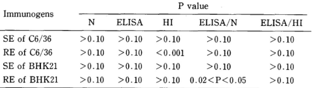
Comparison of the immunogenicity before and after UV‑irradiation
The effect of UV‑irradiation on the immunogen was evaluated by comparing the antibody titers (N, ELISA, HD as well as their ratios (ELISA/N, ELISA/HD raised by each immunogen before and after UV‑irradiation. The statistically significant difference was observed only for the reduction in HI titer produced by RE from C6/36 cells and the increase in ELISA/N ratio produced by RE from BHK21 cells (Table 8). Other immunogenicities were apparently not affected by UV‑irradiation.
Table 4 ELISA titer of immunogens before and after UV‑
irradiation
ELISA titer of immunogen % Reduction
Immunogen before UV after UV in ELISA titer
C6/36 C6/36
BHK21 BHK21
SE RE SE RE
232 206 229 186
50 116 50 107
78 . 45 43 . 69 78 . 15 42 . 48
Table 5 Immunogenicity of SE and RE after UV irradiation, titers in log for N, ELISA and HI
as shown by
Serum
No Immunogen Titer N ELISA HI
Mean Titer Mean T iter Mean 1‑l
1‑2 1‑3 l‑4 l‑5 2‑l 2‑2 2‑3 2‑4 3‑1 3‑2 3‑3 3‑4 3‑5 4‑1 4‑2 4‑3 4‑4
C6/36 SE
C6/36 RE
BHK21 SE
BHK21 RE
2 . 20 l . OO 1 . OO 1 . 90 l . 60 2 . 20 2 . 20 1 . 90 1 . 60 2 . 20 1 . 60 1 . 30 2 . 20 1 . 90 1 . 90 l . 90 2 . 20 1 . 60
l.54: O
(GMT
1 . 98 O
(GMT
1.84d:O
(GMT
1.90i:O
(GMT
. 54
35)
. 29
95)
. 39
69)
. 24
79)
4 . 58 4 . 58
4.11
4 . 48 4 . 35 4 . 29 4 . 23 4 . 51 4 . 58 4 . 64
4.51
4 . 61 4 . 35 4 . 08 4 . 35 4 . 58 4 . 44 4 . 61
4.42 0.20 (GMT 26,302)
4.40 0.
(GMT 25
17 118)
4 . 43i O . 23
(GMT 26,915)
4 . 50i O . 12
(GMT 31,622)
1 . 90 1 . 90 l . 90 2 . 20 1 . 90 2 . 51 1 . 90 2 . 20
2.51
2 . 51 1 . 90 2 . 20 2 . 20 2 . 20 2 . 51 3 . Il 2 . 20 2 . 81
l.96 0
(GMT
. 13
91)
2 . 28 O . 29
(GMT 191)
2.20 0.22 (GMT 158)
2 . 66 O . 39
(GMT 457)
DISCUSSION
From the results of our immunological studies it appears that the SE of JE virus is immunogenic in mice to induce N, ELISA and HI antibodies, and the immunogenicity of both antigens, SE and RE, were almost comparable. Although UV‑irradiation totally inactivated infectivity and partially destroyed ELISA antigenicities in both SE and RE, they still retained their immunogenicities. The different UV‑susceptibility of ELISA antigenicity between SE and RE from both C6/36 and BHK21 cells may be explained by different target size of the particle. The RE is the complete virion and possesses larger size and greater number of repetitive epitope units than SE, which is smaller size with less number of repetitive units. It is reasonable to imagine that RE with larger number of repetitive ELISA antigenic epitopes is more resistant to UV‑irradiation than SE. Kimura‑Kuroda and Yasui (1983) reported that at least five topographically distinct antigenic determinants including N, HI, and ELISA epitopes, were present on E protein and the epitopes of HI and N were separated from each other by monoclonal antibodies.
Recently, Mason et al. (1991) reported that the JE vaccinia virus recombinants possess‑
ing JE virus CDNA inserts produced particulate form of antigen containing M (membrane) and E proteins of JE virus. The antigen migrated in sucrose gradients slower than the complete virion, similar to the SE, and induce high level of anti‑JE N antibodies and conferred protection against lethal JE virus infection.
327 Table 6 Immunogenicity of SE and RE after UV‑irradiation, as shown by
titer ratio of ELISA/N and ELISA/HI Serum
No. Immunogen ELISA/N ELISA/HI
Titer Mean GMT T iter Mean GMT
1‑l l‑2 l‑3 1‑4 1‑5 2‑1 2‑2 2‑3 2‑4 3‑l 3‑2 3‑3 3‑4 3‑5 4‑l 4‑2 4‑3 4‑4
C6/36 SE
C6/36 RE
BHK21 SE
BHK21 RE
2 . 38 2 . 88 i O . 47 3 . 58
3.11
2 . 58 2 . 75
2 . 09 2 . 45i O . 46 2 . 03
2 . 61 2 . 98
2 . 44 2 . 60i O . 50 2 . 91
3 . 31 2 . 15 2 . 18
2 . 45 2 . 59 O . 32 2 . 68
2 . 24 3 . Ol
758
281
398
389
2 . 68 2 . 68 2 . 21 2 . 28 2 . 45 1 . 78 2 . 33 2 . 31 2 . 07 2 . 13 2 . 61 2 . 41 2 . 15 l . 88 1 . 84 l . 47 2 . 24 1 . 80
2.
2.
2
l
46 0 .21
12 :!: O .
.23 0
. 84 O
25
. 28
. 31
288
131
170
69
Table 7 Statistical analysis on N, ELISA and HI titer and titer ratio differences raised by SE and RE after UV=irradiation Immunogens log titer and
titer ratio P value SE and RE of C6/36
SE and RE of BHK21
N ELISA HI ELISA/N ELISA/HI
N ELISA HI ELISA/N ELISA/HI
o.
O.
> O . 10
> O . 10
> O . lO
> O . 10
02< P <0.
> O . lO
> O . 10
02< P <0.
> O . 10
>0.05
05
05
Table 8 Statistical analysis on the difference in antibody titers and titer ratios obtained by immunogens before and after UV‑irradiation
P value Immunogens
N ELISA HI ELISA/N ELISA/HI
SE of C6/36 RE of C6/36 SE of BHK21 RE of BHK21
> O . 10
>0.10
> O . 10
> O . 10
>0・10
>0・10
> O , lO
> O ・ 10
> O ・ 10
< O . OO1
> O . 10
> O ・ 10
> O ・ 10
> O . 10
> O . 10 O . 02 < P < O . 05
> O . 10
>0.lO
>0.10
>0.10
At present, inactivated JE vaccine has been manufactured from infected mouse brain homogenates, by purifying formaline‑inactivated complete virion using primarily
ultracentrifugation (Takaku et al., 1968) . Till now, Iittle or no attention has been paid for the recovery of SE during JE vaccine preparation, and most of the SE would have been lost in the supernatant of ultracentrifugation. It would be worthwhile to consider the im‑
munogenicity of SE in the crude vaccine preparation before ultracentrifugation in order to recover more immunogens.
Some flaviviruses have been shown to produce slowly sedimenting antigen along with rapidly sedimenting antigen or complete virion in the mouse brain homogenates or in tissue culture system (Igarashi et al., 1963; Smith et al., 1970; Shameem et al., 1989). This information has permitted us to predict that some form of 'incomplete virion' is synthesized in the early period of replication of JE virus. In the case of dengue type 2 virus, another important member of flavivirus, Cardiff et al. (1971) argued that most of the incomplete virus or top component was associated with several unrelated phenomena, viz. defective interfering particles, adsorbing interfering particles, pleomorphic aggregates of coat structures, ma‑
cromolecular capsid precursors and some naturally occurring particles which morphological‑
ly resemble infectious form except that they did not contain no core structure. Alternatively, SE may represent a protein synthesized during the course of viral replication which might be released into the medium, besides complete virion, sharing several biological properties possessed by envelope glycoprotein E.
ACKNOWLEDGMENTS
I would like to express my heart felt thanks to Professor Akira lgarashi for his pains‑
taking support, keen interest and critical reading and comments of the manuscript. A part of this work was supported by a Grant‑in‑aid for Scientific Research from the Ministry of Education, Science and Culture of Japan, No. 61480161 in the year of 1989‑90. The animal experiment was performed in the Animal Research Center for Infectious Tropical Diseases, Institute of Tropical Medicine, Nagasaki University. Special thanks are due to Drs. K.
Morita and S. Matsuo and all ,the staff members of this department for their co‑operation through out the whole period of my.research work.
329
REFERENCES
1 ) Calisher, C.H., Karabatsos, N., Dalrymple, J.M., Shope, R.E., Porterfield, J.S., Westaway, E.G.
and Brandt, W.E. (1989): Antigenic relationships between fiaviviruses as determined by cross neutralization tests with polyclonal antisera, J. Gen. Virol., 70, 37‑43
2 ) Cardiff. R.D., Brandt, W.E., McCloud, T.G., Sapiro, D. and Russell, P.K. (1971): Immunological and biophysical separation of dengue‑2 antigens, J. Virol., 7, 15‑23
3 ) Clarke, D.H. and Casals, J. (1958): Techniques for hemagglutination and hemagglutination‑
inhibition with arthropod‑borne viruses, Am. J. Trop. Med. Hyg., 7, 561‑573
4 ) Eagle. H. (1959) : Amino acid mefabolism in mammalian cell cultures. Science, 130, 432‑437 5 ) Hammon, W. McD., Kitaoka, M. and Downs, W.G. (eds.) (1971): Immunization for Japanese
encephalitis. Igaku‑shoin. Tokyo
6 ) Hashimoto, N., Yamada, K. and Kanamitsu, M. (1971): A microtiter method for assay of neutralizing antibodies against group B arboviruses, Virus, 21, 55‑59
7 ) Hori, H., Morita, K. and lgarashi, A. (1986): Oligonucleotide fingerprint analysis of Japanese encephalitis virus strains isolated in Japan and Thailand, Acta Virol., 30, 353‑359
8 ) Huang, C.H. (1982): Studies of Japanese encephalitis in China, Adv. Virus Res., 27, 71‑101 9 ) Igarashi, A., Kitano, H. and Fukai, K. (1963): Heterogenicity in hemagglutinating agent of
Japanese B encephalitis virus, Biken J., 6, 25‑26
10) Igarashi, A. (1978): Isolation of Singh's Aedes albopictus cell clone sensitive to dengue and chikungunya viruses. J. Gen. Virol., 40, 531‑544
11) Igarashi, A., Bundo, K., Matsuo, S. and Lin, W.J. (1981): Enzyme‑linked immunosorbent assay (ELISA) on Japanese encephalitis virus. I. Basic condition of the assay of human immuno‑
globulin, Trop. Med., 23, 49‑53
12) Igarashi, A. (1988) : Development of the second generation Japanese encephalitis (JE) vaccine, South East Asian J. Trop. Med. Publ. Hlth., 9, 493‑500
13) Kitaoka, M. and Nishimura, C. (1985): Infectious, hemagglutinating, and complement‑fixing components in suckling mouse brains infected with Japanese encephalitis virus, Jpn. J. Med.
Sci. Biol., 18, 177‑187
14) Kuroda‑Kimura, J. and Yasui, K. (1983) : Topographical analysis of antigenic determinants on envelope glycoprotein V3 (E) of Japanese encephalitis virus, using monoclonal antibodies, J.
Virol., 45, 124‑132
15) Mason, P.W., Pincus, S., Fournier, M.J., Mason. T.L.. Shope. R.E. and Paoletti, E. (1991):
Japanese encephalitis virus‑vaccinia recombinants produce particulate forms of the structural membrane proteins and induce high levels of protection against lethal JEV infection, Virology, 180, 294‑305
16) Morita, K., Bundo, K. and lgarashi, A. (1982): Enzyme‑linked immunosorbent assay (ELISA) on Japanese encephalitis virus. IV. A computer system to calculate ELISA end point titer from ELISA‑OD at a single dilution of test sera. Trop. Med., 24, 131‑137
17) Oya, A. (1988): Japanese encephalitis vaccine, Acta Paediatr. Jpn., 30, 175‑184
18) Shameem, G.M.M., Morita, K., Bundo‑Morita, K. and lgarashi, A. (1989) : Production of slowly sedimenting and rapidly sedimenting components associated with Japanese encephalitis virus envelope glycoprotein E in infected cell culture fluids, Trop. Med., 31, 111‑123
19) Smith, T.J., Brandt, W.E., Swanson, J.L.. McCown, J.M. and 'Buescher, E.L. (1970): Physical and biological properties of dengue‑2 virus and associated antigens, J. Virol., 5, 524‑532 20) Srivastava, A.K., Aira, Y., Mori, C., Kobayashi, Y. and lgarashi, A. (1987): Antigenicity of
Japanese encephalitis virus envelope glycoprotein V3 (E) and its cyanogen bromide cleaved fragments examined by monoclonal antibodies and Western blotting. Arch. Virol., 96, 97‑107 21) Takaku, K., Yamashita, T.. Osanai, T., Yoshida, I., Kato, M., Goda, H., Takagi, M., Hirota,
22)
23)
T., Amano, T., Fukai, K., Kunita, N.. Inoue, K., Shoji, K., Igarashi, A. and Ito, T. (1968):
Japanese encephalitis purified vaccine, Biken J., Il, 25‑39
Umenai, T., Krzysko, R., Bektimirov, A. and Assaad, F.A. (1985) : Japanese encephalitis:
current world wide status, Bull. WHO, 63, 625‑631
Voller, A., Bidwell. O. and Bartlet, A. (1976): Microplate enzyme immunoassay for the immunodiagnosis of viral infections. pp. 506‑512. In N.R. Friedman (ed.) Manual for Clinical Immunology, American Society for Microbiology, Washington, D.C.
Jpn. J. Trop. Med Hyg Vol 19 No 4 1991, pp. 331‑338 331
BASIC STUDIES ON THE MONGOLIAN GERBIL AS A SUSCEPTIBLE HOST TO FILARIAL INFECTION;
COMPARATIVE STUDIES ON HEMATOLOGICAL FEATURES BETWEEN THE WILD‑COLORED
GERBIL AND THE COAT COLOR MUTANTS
MASUMI SHIMIZU , KAZUHIRO SHICHINOHE*, SETSUKO TSUKIDATE' AND KOICHIRO FUJITA'
Received August 16 1991/Accepted October 9 1991
Abstract: Hematological baseline parameters of the Mongolian gerbils were inves‑
tigated to be compared between the wild‑colored gerbil (agouti type) and the other coat color mutants such as white spotted‑agouti, albino, black and white spotted‑black type.
Erythrocyte counts of the agouti type were higher than those of the coat color mutants.
But, there was no significant difference. A frequent occurrence of polychromasia and basophilic stippling in circulating erythrocytes was known to be a particular feature of the Mongolian gerbil among laboratory animals. The polychromasia and the basophilic stippling were proved to be present in the coat color mutants in the same degree as the agouti type. As to sex dimorphism in erythrocytic values, it was only hemoglobin concentration that was observed significant sex‑related difference in all coat color gerbils.
In leukocytic values, quantitative sex‑related difference was not seen in this experiment.
A presence of basophils in peripheral blood was observed on all blood films of all coat color gerbils. These results confirmed that the coat color mutants of gerbils had the same unique hematological characteristics as an agouti type.
INTRODUCTION
The Mongolian gerbil (Meriones unguiculatus) has been recognized an useful rodent having considerable potential for laboratory studies, especially in the field of parasitology, as a susceptible host to filarial infection. The gerbil, ordinary used in laboratories has a wild coat color called an agouti type. We obtained some new coat color mutants from the gerbil and reported a comparative study on biological aspects among these mutants (Shimizu et al..
1990) . Some hematological data of agouti gerbils have been reported and it was ascertained that the blood of this species manifested some interesting characteristics not generally observed in other rodents (Mays, 1969; Dillon and Glomski, 1975a; Smith et al.. 1976; Termer and Glomski, 1978) . But, according to the coat color mutant, only a limited data has been available that the white spotted‑agouti type of mutants is with a slight anemia (Waring et
l
2
Department of Laboratory Animal Science, Nippon Medical School, 1‑1‑5 Sendagi, Bunkyo‑
ku, Tokyo 113, Japan
Department of Medical Zoology, Faculty of Medicine, Tokyo Medical and Dental University, 1‑5‑45 Yushima, Bunkyo‑ku, Tokyo ll3, Japan
al., 1978) . Hematological baseline parameters of the coat color mutants of gerbils were studied basically in order to analyze the susceptibility of gerbils to filarial infection. A comparative study on the blood picture among coat color mutants was carried out in the present work and showed some interesting hematological features of the coat color mutants of the gerbils as well as an agouti type.
MATERIALS AND METHODS
The animals were bred under a conventional condition and housed 5 adults of the same sex in one cage. The room temperature and the humidity were maintained at 24: 2'C and 60 d: 5%, respectively. Ten of 3 to 5‑month‑old gerbils, such as agouti, white spotted‑agouti, albino, black and white spotted‑black, of both sexes were used in this experiment. Samples of blood were prepared from the retro‑orbital venous plexu of the ether‑anesthetized gerbils by means of hepalinized capillary pipettes. All samples were collected between 0900 and lIOO hours at 1‑month intervals from the animals. Total erythrocyte and leukocyte counts were performed using hemocytometer under a light microscope. Hematocrit determinations were made by means of microcentrifugation and hemoglobin concentrations were determined spectrophotometrically by the cyanmethohemoglobin technique. Dry film blood smears were stained with May‑Grunwald and Giemsa stain and differential counts were identified at least 100 Ieukocytes per an animal. Blood smears for reticulocyte counts were prepared by using Brecher's new methylene blue stain. One thousand of erythrocytes were examined and the number of cells containing a reticulurn were recorded. The numbers of basophilic stippled erythrocytes, polychromatophilic cells and cells with Howell‑Jolly bodies were also counted while examining 1,000 erythrocytes in the smears stained with May‑Grunwald and Giemsa
stain.
Table 1 Total gerbils
erythrocyte counts and related values in each coat color mutant of Mongolian
Coat color Sex
Erythrocytes
( x 10'1/d )
Hemoglobin Hematocrit Mean corpuscularMean corpuscularMean corpuscular
volume hemoglobin hemoglobin
(g/dl) (fl) (pg) concentration(o/o) (o/o)
Agouti
White spotted‑
agouti Albino
Black
White spotted‑
black
Male Female Male Female Male Female Male Female Male Female
8.8+0.5 8.6+0.8 8.6 1.0*
7,7:!:0,6
8.6+0,9 8.0d:0,5 8.2+0.7 8,0+0.9 8.2+0.7 7,9+0.4
16.4d:0.4t 14.9 0.8
15 . 7d: O . 6t
14.9d:0.6 15.7 0.6*
14.9d:0.7 15.1 0.4*
14.6d:0.5 15.6d:1.1' 14.6d:0.8
48 . 6 I . Ot
46.6+1.2
47 . 9d: I . 7t
45.7+0.8
47 . 9 i 2 . Ot
44.7+1,5 45.2+1.1 45.8+2.3 45.1+1.4 45. 1+1.8
52.0:i:4.6 54 . 6d: 5 . O
56.3+5.7 59.5d:3.9 56.1 5.8 56.2d:2.4
55 . 7d:: 5 . 3
57.9 9.0 55.4d:5.0 57.Id:3.3
18 . 6d: 1
17,5 1 18.5d:l 19.5 1
18 . 5d: 1 18 . 9dl 1
18.6 l 18.4i2
19. 1 l
18 . 6:!: l .o .6 .8 .5 .9 .8 .6 .4 .8 .2
33.7 1.0 32.2d:2.0 32.9 0.7 32.7il.2
32 . 9 I . 6
33.5 2,2
33 .8d: I .6 32 . Oi I . 6
34,6d:1.6 32,5 0.9 difference between male and female
* t test, p<0.05 t t test, p<0.01
n = 10, mean standard deviation
333 Table 2 Incidence of erythrocytes with reticulum, basophilic stippling, polychromasia
and Howell‑Jolly body in each coat color mutant of Mongolian gerbils
Coat color Sex Reticulocytes Basophilic stippled cel]s Polychromatophilic ce]Is Cells with Howell‑Jolly (/1,000 RBC) (/1,000 RBC) (/1,000 RBC) bodies (/1,000 RBC) Agouti
White spotted‑
agouti Albino
Black
White spotted‑
black
Male Female Male Female Male Female Male Female Male Female
27.4 11.9 28.Id: 6.3
37 . 9 10 , 2
31.2i:13.0 37.4 7.8
34 . 6d: 13 . 1
38.0d: 9.0 30.2d: 5.4 34.5d: 7.7 36.0d: 8.3
8.6+0.4 12.8 2,9 6.1+1.8 9.0d:3.7 14,0+4.6 ll*Od:3.l 14,4dll,7 12.5:!:4.0 13.3d:4.5 14.3:!:4,2
6.5d:0.3 6.5+2.2 3.6i0.6 5.9d:2.0 6.8d:1.8 5.3d:2.7 9.0d:1.9 6.7+2.6 6.9d:2.8 6.5+2.6
l.5+0.7 1.0+1.0 0.6+0.5 O.6+0.5 1.1+1.0 1.5+1.2 1.3+1.2 0.7+0.7 1.3+0.9 1.1+0.7 n= 10, meand:standard deviation
RESULT
The mean values and standard deviations for total erythrocyte counts as well as related data in the peripheral blood of each color mutant of Mongolian gerbils are presented in Table 1. The average erythrocyte counts for male gerbils were always higher than those for females in all color gerbils; the average counts for males were (8.2‑8.8) x 106/pl as compared to (7.7‑8.6) x 106/pl for females. However, the significant difference between sexes was proved only in the white‑spotted agouti type of gerbil among all coat color gerbils. The agouti type of male Was observed to have the highest value of total erythrocyte counts, but there is no significant difference among all coat color gerbils. No coat color‑related significant difference in the hemoglobin content was observed, but sex‑related difference was also evident in all coat color gerbils. The same tendency was seen in the hematocrit values of three coat color gerbils.
Table 2 shows the incidence of erythrocytes with cytologic features in each coat color mutant of gerbils. The average reticulocyte counts were (27.4‑38.0)/1,000 erythrocytes (RBO for males and (28.1‑36.0)/1,000 RBC for females and sex‑related difference was not seen in this case. That value of reticulocyte was lowest in the agouti type conversely to total erythrocyte counts. Polychromatophilic and stippled red blood cells were demonstrated frequently in all blood films in all coat color gerbils (Photos. 1, 2). The average counts of basophilic stippled cells were (6.1‑14.4)/1,000 RBC for both males and females and those of polychromatophilic cells were (3.6‑9.0) /1,000 RBC. It was observed in all blood films that one red blood cell had both polychromasia and stippling (Photo. 1). The erythrocytes with Howell‑Jolly bodies were also observed (0.5‑1.5)/1,000 RBC in all color gerbils (Photos. 2,
3) .
Total numbers of cirbulating leukocytes and differentials in each coat color mutant of Mongolian gerbils are shown in Table 3. Total circulating leukocytes were (12.4‑15.6) x 1031 pl for males and (14.2‑16.6) x 103/pl for females. In the circulating leukocyte counts, both sex‑related and coat color‑related differences were not seen in this study. The lymphocyte was observed most prevalent in all coat color gerbils of both sexes. Although the number of basophils and eosinophils accounted for only small propotion in the peripheral blood, it was
.' ** ==* . ," j. , ':=' ; .*;',*..""・'.:.;.'** ' ̲*, ; + ・ ・ '.
Photo. I Polychromatophilic and basophilic stippled erythrocytes near a lymphocyte on a blood film of a Mongolian gerbil. May‑Grunwald and Giemsa stain.
S
Photo. 2 An erythrocyte with polychromasia and with a Howell‑Jolly body on a blood film of a Mongolian gerbil. May‑Grunwald and Giemsa stain.
335
i{ =d '; { ;' r:* !i :' *' s' :*ip
mj} ;
"
Photol 3 A Howell‑Jolly body in an erythrocyte on a blood film of a Mongolian gerbill May‑Grunwald and Giemsa stainl
Table 3 Total leukocyte counts and differentials in each coat color mutant of Mongolian gerbils Coat color Sex Total leukocytes
( x 10'1pl)
Basophils (%)
Eoslnophils (%)
Neutrophils (%)
Lymphocytes (%)
Monocytes (%)
Agouti Male
Female White spotted‑ Male
agoutl Female Albino Male
Female
Black Male
Female White spotted‑ Male
black Female
13.8+3.6 14.8 3.8 15.6 4.9 15.9 5,8 15.5 3.6 14.8+4.8 12.4+2.6 16.6+5.6 15.6 2.9 14.2+4.4
o.
o.
o.
o.
o.
o.
o.
o.
o.
o.
3+0.
2 0.
3 0.
1+0.
4+0.
2+0.
2+0.
l+0.
l+0.
1+0.
5
5 3 5 6
3 3 3
0.8 0.6
0.3:!I0.5 1.2:t0.8 0.8+0.6 1.3 0.7 l.5 1.5 0.4+0.5 0.4 0.7 0.1iO.3 0.2+0.4
25 . 23 . 23 . 23 .
20 .
21.
30 . 26 . 30 . 32 .
0 6.5 8 6.9 7+g.9 6+5.3 3+3.6 8 4,0 0 9.3 0+8.6 7+6.9 5+9.6
70.6+7.0 72.6 7.0 70.9 9.2 73.6i5.6 75.5+3,8 74.5+4.2 67.9+8.8 71.1+8.7 66.8 6.7 64.8+9.2
3.3 1.4 3.1+1.6 3.9 1.2 1.9+0.9 2.5+1.6 2.0 1.4 l.5 0.7 2.4+1.3 2.3+0.8 2.4+1.4 n = 10, mean standard deviation
confirmed that these cells were present in the all blood films in all color mutants of gerbils.
DISCUSSION
In this paper, we investigated hematological profiles in 4 kinds of the coat color mutants of gerbils and compared them to the agouti type. There was only one past report on a hamatological characteristics in a coat color mutant of a gerbil (Waring et al., 1978). They noted that total erythrocyie counts of spotted gerbils were significantly lower than those of
an agouti type indicating the presence of a slight anemia. From their description about the location of white spots and about breeding data of the mutant, it seemed that their spotted gerbil was the same mutant as our white spotted‑agouti type. But, their result of erythrocyte counts was different from ours. The total erythrocyte counts of agouti type were observed also higher than the other coat color gerbils in our experiment, however, there was no significant difference and an anemia of white spotted‑agouti gerbils was not observed. The total erythrocyte counts of agouti type in our experiment were comparable to those of the other investigators (Ruhren, 1965; Mays, 1969; Dillon and Glomski, 1975a; Termer and Glomski, 1978) , but, those of Waring et al. (1978) were about a half level of them.
As their own particular feature of the blood of the Mongolian gerbil, it was known the frequent occurrence of polychromasia and basophilic stippling in circulating erythrocytes throughout the life span of agouti gerbils (Ruhren, 1965; Dillon and Glomski, 1975a; Smith et al.. 1976; Termer and Glomski, 1978) . In this study, it was proved that those cells were also present in the blood of the coat color mutants of gerbils as the agouti type. Those cytologic features were considered a property of immaturity of erythrocytes (Smith et al., 1976) . High incidence of those cells refiected that immature red cells emerged to the peripheral blood because of. a brief life span of an erythrocyte (Dillon and Glomski, 1975b) . Although gerbils have been known to have very wide sensitivity to various parasites compared with the other laboratory rodents, the cause of a high sensitivity was not investigated. Since a marked difference of the gerbil from the other rodents was the continuous presence of stippled and polychromatophilic cells in the adult gerbils as far as a hematological measurement, the brief span of blood cells may participate in a wide sensitivity of this animal.
Certain numbers of reticulocytes were also observed. Although there was no statistical difference, it was lowest in the agouti gerbil conversely to total erythrocyte counts.
As to sex dimorphism in total erythrocyte counts and related values, there have been some discussions. Male predominance of hemoglobin concentration, hematocrit, mean corpuscular volume or mean corpuscular hemoglobin was reported by some investigators (Mays, 1969; Dillon and Glomski, 1975a; Termer and Glomski, 1978) . In this experiment, only hemoglobin concentration showed statistical sex‑related difference in all coat color gerbils.
The possible existence of quantitative sex‑related difference in the leukocytes has been a feature described for this rodent (Mays, 1969; Dillon and Glomski, 1975a). But, in this experiment, sex‑related difference was not observed in total leukocyte counts in all coat color gerbils. This finding supported the result of the study by Termer and Glomski (1978) . The predominant cells in the circulating leukocytes of the gerbils were lymphocytes, a characteristic common to other laboratory rodents (Wolford, 1986). On the other hand, a presence of basophils in peripheral blood was a specific characteristics of the gerbil and it was not observed in mice or rats. At the point of existence of both basophils in peripheral blood and mast cells in connective tissues or mucosal tunics of the gerbil (data not shown), this animal can be said more resemble to human being than mice or rats. The gerbil may be useful for analysis of the infection kinetics to various parasites, because the animal has both mast cells in connective tissues and a comparatively larger amount of basophils in peripheral blood.
Finally, this study confirmed that the coat color mutants had the same unique hematological characteristics as the wild‑colored gerbil. Further detail hematological and other physiological experiment concerning this animal will help us to know why the gerbil is
337 very sensitive to various parasite infections.
REFERENCES
l ) Dillon, W.G. and Glomski, C.A. (1975a): The Mongolian gerbil: qualitative and quantitative aspects of the cellular blood picture, Lab. Anim., 9, 283‑287
2 ) Dillon, W.G. and Glomski, C.A. (1975b) : Erythrocyte survival in the Mongolian gerbil, J. Nucl.
Med., 16, 682‑684
3 ) Mays, A.Jr. (1969): Baseline hematological and blood biochemical parameters of the Mon‑
golian gerbil (Meriones unguiculatus). Lab. Anim. Care, 19, 838‑842
4 ) Ruhren, R. (1965): Normal values for hemoglobin concentration and cellular elements in the blood of Mongolian gerbils, Lab. Anim. Care, 15, 313‑320
5 ) Shimizu, M., Shichinohe, K., Tsukidate, S. and Fujita, K. (1990): Basic studies on the Mongolian gerbil as a susceptible host to filarial infection; comparative studies on growth and reproduction among coat color mutants and genetic analysis of coat colors, Japan. J. Trop.
Med. Hyg., 18, 301‑310
6 ) Smith, R.A., Termer, E.A. and Glomski, C.A. (1976): Erythrocyte basophilic stippling in the Mongolian gerbil, Lab. Anim., 10, 379‑383
7 ) Termer, E.A. and Glomski, C.A. (1978): The cellular blood picture of the Mongolian gerbil throughout the first year of life; a longitudinal study, Exp. Hemat., 6, 499‑504
8 ) Waring, A.D., Poole, T.W. and Perper, T. (1978): White spotting in the Mongolian gerbil, J.
Heredity, 69, 347‑349
9 ) Wolford, S.T., Schroer, R.A., Gohs, F.X., Gallo, P.P., Brodeck, M., Falk, H.B. and Ruhren, R.
(1986): Reference range data base for serum chemistry and hematology values in laboratory animals, J. Toxicol. Environm. Health, 18, 161‑188
フィラリア感染好適宿主としてのMongolian gerbilの基礎的検討 一血液学的特性に関する被毛色間の比較一
清水 眞澄1・七戸 和博1・月舘 説子2・藤田紘一郎2
Mongolian gerbi1(スナネズミ)の被毛色突然変異体(white spotted−agouti,albino,black,
whitespotted−black)の血液学的特性について,野生色(agouti)のものと比較検討した。赤血 球数は統計学的な有意差はなかったが,agoutiが最も多かった。他の実験動物には見られないス
ナネズミ独特の性質として,多染性や好塩基斑点を持つ赤血球が多いことが知られているが,そ れらが被毛色突然変異体の末梢血中にも多数出現することが観察された。赤血球関連の検査値に おいてすべての被毛色で有意な性差が見られたのは,ヘモグロビン濃度のみであった。白血球数 には性差が確認されなかった。白血球中で最も多くの割合を占めたのは,各被毛色ともリンパ球 であった。すべての被毛色の各個体の末梢血中に好塩基球の存在が確認された。
以上のように被毛色突然変異体は,agoutiと同様にスナネズミ独自の血液学的性質を持ってい ることがわかった。
日本医科大学実験動物管理室(〒113東京都文京区千駄木1−1−5)
東京医科歯科大学医学部医動物学教室(〒113東京都文京区湯島1−5−45)