in vivo Endothelial Function by Measurement of
Flow-Mediated Dilation in Rats
著者
芹澤 賢一
year
2014
その他のタイトル
ニコランジルは酸化ストレ抑制を介して血管内皮機
能を改善する ; 血流依存性血管拡張反応測定によ
るラットの in vivo 血管内皮機能評価
学位授与大学
筑波大学 (University of Tsukuba)
学位授与年度
2013
報告番号
12102甲第6913号
URL
http://hdl.handle.net/2241/00123624
Nicorandil Improves Endothelial Function via
Reduction of Oxidative Stress;
Evaluation of in vivo Endothelial Function by
Measurement of Flow-Mediated Dilation in Rats
January 2014
Nicorandil Improves Endothelial Function via
Reduction of Oxidative Stress;
Evaluation of
in vivo
Endothelial Function by
Measurement of Flow-Mediated Dilation in Rats
A Dissertation Submitted to
the Graduate School of Life and Environmental Sciences,
the University of Tsukuba
in Partial Fulfillment of the Requirements
for the Degree of Doctor of Philosophy
(Doctoral Program in Biological Sciences)
i
Table of Contents
Abstract --- 1
Abbreviations --- 4
General Introduction --- 6
Chapter 1
Studies of effect of nicorandil on diabetic endothelial dysfunction
in rat
1-1. Introduction --- 10
1-2. Materials and Methods --- 12
1-3. Results --- 18
1-4. Discussion --- 30
Chapter 2
Studies of effect of nicorandil on paclitaxel-induced endothelial
dysfunction in rat
2-1. Introduction --- 35
2-2. Materials and Methods --- 37
2-3. Results --- 42
2-4. Discussion --- 59
General Discussion --- 63
Acknowledgements --- 69
References --- 70
Prior Publications --- 84
1
Abstract
Endothelial dysfunction is a powerful surrogate marker of cardiovascular risk. Flow-mediated dilation (FMD) is an indicator of endothelial function and is identified as an independent predictor of future cardiovascular events in clinical settings. Nicorandil, an anti-angina agent with ATP-sensitive potassium channel (KATP channel) opening and nitrate-like activity, has been shown to improve the prognoses of patients with stable angina pectoris via several mechanisms, including endothelial protective effects. Although patients with angina have many risks for endothelial dysfunction, such as diabetes and drug-eluting stent, it has not been also fully understood about the risks that nicorandil is effective against. The purpose of this study is to evaluate the endothelial protection provided by nicorandil against two risk factors, diabetes and paclitaxel. Paclitaxel was used for drug-eluting stent. Furthermore, I tried to evaluate the endothelial function by FMD.
In chapter 1, I examined the effect of nicorandil on diabetic endothelial dysfunction. Male Sprague-Dawley rats were intraperitoneally injected with streptozotocin (STZ) to induce diabetes. Nicorandil and tempol were administered in drinking water for one week, starting 3 weeks after STZ injection. Endothelial function was evaluated by measuring FMD in the femoral arteries of anaesthetized rats. Femoral arteries were harvested and protein expression was analyzed by western blotting. High glucose-induced reactive oxygen species (ROS) production was measured in cultured human coronary artery endothelial cells (HCAECs). I could observe the reduction of
2
FMD in diabetic rats, which was improved by nicorandil or tempol. NADPH oxidase and endothelial nitric oxide synthase (eNOS) uncoupling were increased in femoral arteries of diabetic rats, which were inhibited by nicorandil. High glucose-induced ROS production in HCAECs was inhibited by nicorandil, N-omega-nitro-L-arginine methyl ester (L-NAME) or apocynin.
In chapter 2, I examined the effect of nicorandil on paclitaxel-induced endothelial dysfunction. An osmotic pump was implanted subcutaneously into a small pouch between the shoulder blades of rats to continuously infuse paclitaxel. Nicorandil, diazoxide, isosorbide dinitrate (ISDN) or tempol were administered for 1 week from just after implantation of the osmotic pump. FMD was measured at 1 week after implantation of the osmotic pump. Femoral arteries were harvested for real-time PCR. ROS production by paclitaxel was measured in HCAECs and in vivo in the mice. I could observe the reduction of FMD by paclitaxel, and nicorandil, diazoxide and tempol, but not ISDN, improved the reduction of FMD. NADPH oxidase was increased in femoral arteries by paclitaxel, which was inhibited by nicorandil. Paclitaxel-induced ROS production in HCAECs was inhibited by nicorandil. This effect was reduced by 5-HD, but not ODQ. ROS production was also observed in vivo mice treated with paclitaxel, which could be prevented by nicorandil.
In the present study, I demonstrated that nicorandil improved the reduction of FMD through its anti-oxidative effect in diabetic and paclitaxel-treated rats. Moreover, the anti-oxidative effect of nicorandil
3
might be exerted via KATP channel opening. Therefore, nicorandil may improve the prognoses of angina patients with concomitant diabetes and drug-eluting stent treatment by protecting their endothelial function.
4
Abbreviations
ADMA: asymmetric dimethylarginine BH4: tetrahydrobiopterin
DAF-2DA: Diaminofluorescein-2 diacetate DCF: 2’, 7’-dichlorofluorescein
DES: drug-eluting stent
EDHF: endothelium-derived hyperpolarization factor eNOS: endothelial nitric oxide synthase
FMD: flow-mediated dilation GCH-1: GTP cyclohydrolase 1
HCAECs: human coronary artery endothelial cells H2DCF-DA: 2', 7'-dichlorodihydrofluorescein diacetate ISDN: isosorbide dinitrate
KATP channel: ATP-sensitive potassium channel L-NAME: N-omega-nitro-L-arginine methyl ester NF-B: nuclear factor-kappa B
5
NOx: nitrate and nitrite NTG: nitroglycerin
ODQ: 1H-[1,2,4] oxadiazolo [4,3-a]quinoxalin-1-one PAI-1: plasminogen activator inhibitor
PES: paclitaxel-eluting stent PKC: protein kinase C
PVDF: polyvinylidene difluoride ROS: reactive oxygen species STZ: streptozotocin
Tempol: 4-hydroxy-2,2,6,6-tetramethylpiperidine-N-oxyl 5-HD: 5-hydroxydecanoate
6
General Introduction
It has become evident that the endothelium plays a crucial role in regulating vascular tone and structure. Common conditions that predispose to atherosclerosis, such as diabetes, hypertension, hypercholesterolemia, are associated with endothelial dysfunction. They may be linked to proinflammatory and prothrombotic endothelial phenotypes (Landmesser et al., 2004). Alterations in endothelial function play a pivotal role in the development and progression of atherosclerosis and its clinical complications. Therefore, endothelial dysfunction is a powerful surrogate marker of cardiovascular risk, and the severity of endothelial dysfunction correlates with a patient’s risk for experiencing an initial or recurrent cardiovascular event (Widlansky et al., 2003).
Clinically, endothelial function generally has been evaluated by endothelium-dependent relaxation. Flow-mediated dilation (FMD) is an established method for assessing endothelial function (Celermajer et al., 1992). It is determined from the dilation of conduit arteries following temporary ischemia and reperfusion by release of a forearm-occluding cuff and. Temporary ischemia induces dilation of the downstream microvasculature, then reperfusion increases blood flow (i.e., reactive hyperemia) and wall shear stress (Kelm, 2002; Pyke and Tschakovsky, 2005). These lead to a rapid activation of endothelial nitric oxide synthase (eNOS) that increases formation of nitric oxide (NO), leading to vasodilation (Rassaf et al., 2006). Endothelial function can be evaluated noninvasively using this technique. FMD also has been used to evaluate endothelial function in dogs
7
and pigs (Jones et al., 2011; Weber et al., 2011). Recently, Heiss et al. reported a method to measure endothelial function by FMD in rats (Heiss et al., 2008).
Nicorandil is an antiangina agent with ATP-sensitive potassium channel (KATP channel) opening and nitrate-like activity. Nicorandil has been shown to improve the prognoses of patients with stable angina pectoris by preconditioning-like cardioprotective effects in the IONA study and the JCAD study (Horinaka et al., 2010; IONA study group, 2002). In addition to the preconditioning-like cardioprotective effects, some recent reports demonstrated that nicorandil can exert endothelial protective effects. Long-term administration of nicorandil significantly improved endothelial function in patients with ischemic heart disease or with cardiovascular risk factors, as evaluated by FMD in forearm arteries (Ishibashi et al., 2008; Sekiya et al., 2005). In swine heart, nicorandil reduced myocardial no-reflow after ischemia reperfusion by protecting endothelial function (Zhao et al., 2006). In human umbilical vein endothelial cells, nicorandil inhibited apoptosis induced by serum starvation by inhibiting production of reactive oxygen species (ROS) (Date et al., 2005). Therefore, it is believed that nicorandil can improve the prognoses of patients with angina due, in part, to protecting endothelial function. However, the endothelial protective mechanisms of nicorandil have not been fully understood. Moreover, the risk factors of endothelial dysfunction against which nicorandil is effective are not clear.
8
dysfunction, such as diabetes, hypertension, hypercholesterolemia, smoking, aging and obesity (Celermajer et al., 1992; Sorensen et al., 1994; Steinberg et al., 1996; Widlansky et al., 2003). In addition to these traditional risk factors, drug-eluting stent (DES) recently have been considered as another potential cause of endothelial dysfunction. DES, which is used for percutaneous coronary intervention, is coated with anti-proliferative compounds, such as paclitaxel to prevent in-stent restenosis. While DES dramatically reduce in-stent restenosis (Stone et al., 2004), the potent anti-proliferative agents have raised concerns regarding endothelial dysfunction (Shin et al., 2007).
Accordingly, the purpose of this study was to elucidate the effects and mechanisms on the endothelial protective effect of nicorandil against two risk factors. In the chapter 1, I examined the effect of nicorandil on diabetic endothelial dysfunction in rats. In the chapter 2, I examined the effect of nicorandil on paclitaxel-induced endothelial dysfunction in rats. In these studies, I tried to evaluate the endothelial function by measurement of FMD in living rats.
9
Chapter 1
Studies of effect of nicorandil
10
1-1. Introduction
Diabetes mellitus is regarded as an independent major risk factor for the development of cardiovascular disease, since long-term survival and freedom from cardiac events were reduced in diabetic coronary angioplasty patients (Kasai et al., 2008; Kip et al., 1996; The Japanese Coronary Artery Disease (JCAD) Study Investigators, 2006). Endothelial dysfunction plays a central role in diabetic vascular diseases (Creager et al., 2003). A common mechanism underlying this endothelial dysfunction could involve increased production of ROS in vascular tissue (Gryglewski et al., 1986). High glucose greatly increases endothelial superoxide production (Cai et al., 2005), leading to an eNOS uncoupling state, followed by lower production of NO and more ROS production, which acts to quench NO (d’Uscio et al., 2003; Landmesser et al., 2003; Laursen et al., 2001; Tiefenbacher et al., 2000; Vasquez-Vivar et al., 2002). Reduced NO availability attenuate its beneficial vascular effects such as vasodilation, regulation of vascular smooth muscle proliferation and expression of cellular adhesion molecules that are involved in initiating atherosclerotic plaque formation (Bian et al., 2008). Therefore, increased ROS production with diabetes has been speculated to reduce endothelial NO availability, leading then to endothelial dysfunction (Landmesser et al., 2004; Nakagawa et al., 2011).
Nicorandil is an anti-angina agent that opens KATP channels and acts like nitrate compounds. It reportedly improves prognoses in patients with angina pectoris via preconditioning effects (IONA study group, 2002). This beneficial effect of nicorandil is exerted across a broad range of patients with
11
stable angina, for example in diabetic patients (IONA study group, 2004). In general, however, the preconditioning effect is abolished in diabetic animals (Miki et al., 2009; Katakam et al., 2007) and humans (Ishihara et al., 2001). Therefore, it is believed that the cardioprotective effect of nicorandil is mediated by the other mechanisms. Recently, nicorandil has been shown to exert endothelial protective effects in both clinical settings and animal studies. Therefore, I hypothesized that nicorandil can prevent diabetic endothelial dysfunction.
In the present study, I investigated the protective effect of nicorandil on endothelial function in rats with diabetes induced by streptozotocin (STZ). Endothelial function was evaluated by measuring FMD in femoral arteries using a high-resolution ultrasound system under in vivo conditions in which blood flow, many humoral factors and nerve activity were maintained. The mechanism underlying the protective action of nicorandil also was investigated in relation to ROS production in the endothelium, both in vivo and in vitro.
12
1-2. Materials and Methods
AnimalsMale Sprague-Dawley rats (Charles River Japan, Yokohama, Japan, 6 weeks old, 200-240 g) were used in all experiments. All rats were fed ordinary laboratory chow and allowed free access to water under a constant light and dark cycle of 12 h. Diabetes was induced by intraperitoneal administration of STZ (40 mg/kg, Wako Pure Chemicals Co., Osaka, Japan) once a day for 3 days. Glucose concentrations were measured one week after STZ administration. Diabetes was considered to have been induced when the
glucose level was higher than 250 mg/dL. Nicorandil
[N-(2-hydroxyethyl)nicotinamide nitrate ester] was synthesized in the Chugai Organic Chemistry Laboratory. Nicorandil (15 mg/kg/day) and 4-hydroxy-2,2,6,6- tetramethylpiperidine-N-oxyl (tempol, 20 mg/kg/day, Sigma-Aldrich, St. Louis, MO, USA) were administered in drinking water for one week, starting 3 weeks after STZ administration.
All animal procedures were conducted in accordance with Chugai Pharmaceutical's ethical guidelines for animal care, and all experimental protocols were approved by the Animal Care Committee of the institution and conformed to the Guide for the Care and Use of Laboratory Animals published by the US National institutes of Health.
Measurement of FMD
Blood pressure (tail-cuff method, BP-98A Softron, Tokyo, Japan) and blood glucose were measured four weeks after STZ administration, and the
13
rats were anaesthetized with thiobutabarbital with constant monitoring of rectal temperature. A catheter was inserted into the jugular vein to administer drugs. The animals were kept stable with a heated sheet and warming lamps.
FMD measurement in rats was described previously (Heiss et al., 2008). The diameter of femoral arteries and Doppler flow were measured using a high-resolution ultrasound system (Vevo 770, VisualSonics, Toronto, Canada). The femoral artery was visualized with a 30- or 40-MHz transducer. After identifying the femoral artery by its characteristic flow pattern, the position of the probe was optimized to clearly show the vessel wall/lumen interfaces and then fixed throughout the investigations. Experiments were started over an equilibration period of 15 min and when body core temperature was stable (37 1C).
Ultrasound measurements of diameter and Doppler-flow were obtained from longitudinal sections of the femoral artery before and after 5 min of hindlimb ischemia. Ischemia and reperfusion of the hindlimb were achieved with a snare occluder positioned upstream from the site to be visualized, around the common iliac artery, through a trans-abdominal access. The snare occluder consisted of a 5-0 nylon surgical suture around the artery that was passed through a 4 cm PE-200 tube, and the skin was closed with surgical clips. Hindlimb ischemia was created by pulling on the filament through the tube and clamping it.
Baseline recordings were taken after an equilibration period, and then the common iliac artery was occluded with the snare occluder. Flow arrest
14
was confirmed by abrogation of the Doppler signal. After 5 min of ischemia, the hindlimb was reperfused by releasing the occluder. The changes in flow velocity and the diameter of the femoral artery were monitored at 0, 0.5, 1, 2 and 3 min after reperfusion. In this study, FMD was defined as the peak change in femoral artery diameter measured at 1 min after reperfusion. The clinical guideline to assess FMD defined the peak change in brachial artery diameter as an endothelial function, which occurred about 1 min after reperfusion (Corretti et al., 2002).
To evaluate endothelium-independent vasodilation, nitroglycerin (NTG) was administered to the same rats after FMD measurement, with 10 min breaks between measurements. After obtaining baseline recordings of diameter, NTG (5 g/kg) was administered intravenously via the jugular vein catheter. Changes in femoral artery diameter were monitored at 10, 30, 60 and 120 sec after administration.
Western blot analysis
Femoral arteries were harvested and frozen in liquid N2 immediately after isolation and stored in a -80C freezer until protein expression was measured by Western blotting. The femoral artery was homogenized in a homogenization buffer composed of 25 mM Tris-HCl (pH 7.4), 1 mM dithiolthreitol, 25 mM sodium fluoride, 1 mM sodium orthovanadate, protease inhibitor cocktail tablet, phosphatase inhibitor cocktail and 1% Triton X-100. The homogenates were centrifuged at 14,000 g for 20 min at 4C. Supernatants were collected, and protein concentrations were
15
determined using a BCA Protein Assay Kit (Thermo Scientific, Woltham, USA). Equal amounts of protein extracts were separated on 10% SDS-polyacrylamide gel and immobilised on polyvinylidene difluoride (PVDF) membranes (Millipore, Billerica, USA). To investigate the eNOS uncoupling state, low-temperature SDS-PAGE was performed to detect the dimer and monomer of eNOS. Briefly, protein extracts were mixed with sample buffer (without -mercaptoethanol), and non-boiled samples were separated on SDS-PAGE. The membranes were blocked in a PVDF Blocking Reagent (TOYOBO, Osaka, Japan), and incubated with anti-p47phox, anti-eNOS (Santa Cruz Biotechnology, Santa Cruz, USA) or anti-GTP cyclohydrolase 1 (GCH-1) antibodies (Abnova, Taipei, Taiwan). After washing, the membranes were incubated with anti-rabbit or anti-goat IgG conjugated with horseradish peroxidise (Santa Cruz Biotechnology). Immunoreactive signals were visualized with the SuperSignal West Dura Extended Duration Substrate (Thermo Scientific), and detected using a ChemiDoc XRS system (Bio-Rad Laboratories, Hercules, USA). Each protein signal was normalized to -actin expression from the same sample.
Measurement of serum NOx
To determine NO excretion, total nitrate and nitrite (NOx) concentrations were measured by the Griess method with a Total Nitric Oxide Assay Kit (Enzo Life Sciences, Farmingdale, USA). Serum samples were diluted 1:2 into reaction buffer and ultra-filtered through a 10,000 molecular weight cut-off filter. Samples were read at 540 nm with a
16
Microplate Reader (Molecular Devices, Sunnyvale, USA).
Cell culture and treatment
Normal human coronary artery endothelial cells (HCAECs) were purchased from Lonza Inc. (Walkersville, MD, USA). HCAECs at passages 3-5 were cultured with EBM-2 medium supplemented with 5% fetal bovine serum (Lonza). To measure production of ROS, the cells were seeded onto plastic dishes (1105 cells/2 mL/dish) and cultured as monolayers in a 5% CO2 humidified incubator at 37C overnight. After incubation, the HCAECs were exposed to normal (5.6 mM) or high glucose (35.6 mM) for 24 h. Nicorandil (100 M), apocynin (100 M, Calbiochem, Darmstadt, Germany) or N-omega-nitro-L-arginine methyl ester (L-NAME, 300 M, St Louis, Sigma-Aldrich) were added over the same period.
Measurement of ROS production using a fluorescent probe in HCAECs The production of ROS was monitored using the fluorescent probe 2', 7'-dichlorodihydrofluorescein diacetate (H2DCF-DA) (Invitrogen, Carlsbad, USA). Briefly, cultured cells were incubated with 10 M H2DCF-DA for 45 min in a 37C incubator, and 2’, 7’-dichlorofluorescein (DCF) fluorescence was then quantified by confocal microscopy (Zeiss Axiovert 200) from at least 20 randomly-selected cells/dish, using three dishes for each experimental condition.
17
Statistical analyses
All data are expressed as mean SE. The n values refer to the number of individual animals on which experiments were performed. The statistical significance of differences was determined using Tukey's Multiple Comparison Test. Probability values less than 0.05 were considered significant. Statistical analyses were performed using SAS version 8.2 software (SAS Institute, Cary, USA).
18
1-3. Results
AnimalsBody weight, blood glucose, blood pressure and heart rate are shown in Table 1. Body weight increased in each group of rats for 4 weeks after STZ administration, although the increase in the STZ rats was smaller than in normal rats. Blood glucose levels in STZ rats were significantly higher than in normal rats. Systolic blood pressure was slightly increased, while heart rate decreased significantly in STZ rats. Nicorandil appeared not to influence any of these parameters in STZ rats.
19
Table 1. Body weight, blood glucose, blood pressure and heart rate at 4 weeks after STZ administration normal STZ STZ+nicorandil N 7 6 6 Body weight (0 wks, g) 213.2 ± 1.6 212.2 ± 3.4 217.1 ± 4.3 Body weight (4 wks, g) 417.8 ± 8.7 299.4 ± 15.4* 327.7 ± 9.8* Blood glucose (mg/dL) 96.7 ± 1.7 392.3 ± 17.4* 424.5 ± 16.1* Systolic blood pressure
(mmHg) 121.8 ± 1.8 135.4 ± 5.9 133.5 ± 3.2 Heart rate (bpm) 359.4 ± 11.9 312.4 ± 14.5* 334.5 ± 11.4
20
FMD in STZ-induced diabetic rats
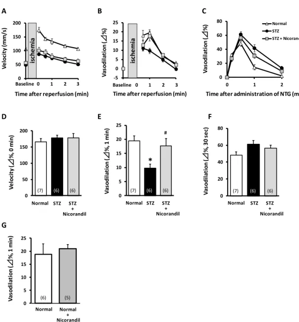
Endothelial function was evaluated by the measurement of FMD in rat femoral arteries. Reperfusion after 5-min of hindlimb ischemia instantly increased flow velocity (i.e. reactive hyperaemia) compared with baseline femoral artery flow, followed by a rapid decay to baseline values at around 3 min (Fig. 1A). The increase in flow velocity was associated with a delayed increase in femoral arterial vasodilation that peaked at 1 min after reperfusion (Fig. 1B). This delayed vasodilation in the femoral artery was defined as FMD. In STZ rats, peak velocity in the femoral artery was lower than that in normal rats (normal, 106.7 5.0; STZ, 49.5 2.5 mm/s; n = 6-7, Fig. 1A) at baseline (i.e. before ischemia). However, hyperaemia, expressed as the % change in peak velocity just after reperfusion, did not differ between normal and STZ rats (Fig. 1D). FMD was lower in STZ rats compared with normal rats (normal, 19.5 1.7; STZ, 9.7 1.4%; n = 6-7, Fig. 1B, E). In evaluating endothelium-independent vasodilatory potency, intravenous administration of NTG led to vasodilation of the femoral artery that peaked at 30 sec (Fig. 1C). NTG-induced vasodilation was similar in normal and STZ rats (Fig. 1C, F).
Nicorandil (15 mg/kg/day) was administered in drinking water for one week starting 3 weeks after STZ administration to evaluate its effects on endothelial function. Nicorandil restored the reduced FMD in STZ rats to almost same level as in normal rats (STZ + Nicorandil, 17.7 2.6%; n = 6, Fig. 1B, E), whereas nicorandil affected neither NTG-induced vasodilation nor flow velocity. In normal rats, nicorandil produced no significant changes in
21
FMD (normal, 18.8 4.0; nicorandil, 20.9 1.7%; n = 5-6, Fig. 1G). These results demonstrated that nicorandil could ameliorate endothelial dysfunction in STZ rats, without changing flow velocity or vascular smooth muscle function.
22
Figure 1. Effect of nicorandil on endothelial dysfunction induced in rats by STZ
Ultrasound measurements were performed at 4 weeks after STZ administration. Time courses of velocity (A), FMD (B) and NTG-induced vasodilation (C) in rat femoral arteries were measured after 5 min ischemia. Percent change in velocity (Δ%, D) and NTG-induced vasodilation (F) were similar among groups. FMD peaked at 1 min after reperfusion and then decreased in diabetic rats, showing it was improved by nicorandil (E). Nicorandil showed no significant changes in FMD of normal rats (G). *p<0.05 vs normal, #p<0.05 vs STZ (n = 5-7). The numeral in each column is the number of animals.
Baseline 0
Time after reperfusion (min)
2 3 1 * # Baseline 0 1 2 3 Normal STZ STZ + Nicorandil 0
Time after administration of NTG (min) 2 1 A D B E C F -5 0 5 10 15 20 25 V as o d il at io n ( ⊿ %) 0 5 10 15 20 25 V as o d il at io n ( ⊿ % , 1 m in ) 0 50 100 150 200 V e lo ci ty (m m /s )
Time after reperfusion (min)
0 50 100 150 200 V e lo ci ty (⊿ % , 0 m in ) 0 20 40 60 80 V as o d il at io n ( ⊿ % , 3 0 s e c) is ch e m ia is ch e m ia Normal STZ STZ + Nicorandil Normal STZ STZ + Nicorandil 0 20 40 60 80 V as o d il at io n ( ⊿ %) NormalSTZ STZ + Nicorandil 0 5 10 15 20 25 V as o d il at io n ( ⊿ % , 1 m in ) Normal Normal + Nicorandil G (7) (6) (6) (7) (6) (6) (7) (6) (6) (6) (5)
23
Involvement of oxidative stress in decreased FMD in STZ-induced diabetic rats
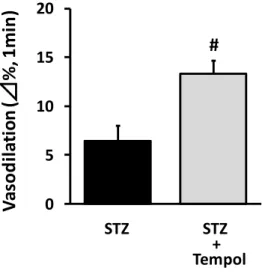
The effect of tempol was investigated to clarify how oxidative stress was involved in endothelial dysfunction in rats treated with STZ. Tempol (a radical scavenger) significantly reversed the decrease in FMD (STZ, 6.5 1.5; STZ + Tempol, 13.3 1.4%; n = 6, Fig. 2). Expression of NADPH oxidase was measured in femoral arteries of STZ rats because it is a potential source of vascular superoxide. In STZ rats, the p47phox protein (a major NADPH oxidase component) increased in the femoral artery. Nicorandil prevented an increase in expression of p47phox (Fig. 3A). Expression of total eNOS was measured in the rat femoral artery to examine how it is associated with endothelial dysfunction. Unexpectedly, total eNOS expression increased in STZ rat femoral arteries, and the increase was prevented by nicorandil (Fig. 3B). However, this increase in total eNOS expression did not raise serum NOx levels in STZ rats, and nicorandil also showed no significant changes (Fig. 4). In oxidative state, reducing tetrahydrobiopterin (BH4) causes eNOS uncoupling state, resulting in production of superoxide by eNOS monomer (Landmesser et al., 2003). In this study, the eNOS dimer/monomer ratio decreased (Fig. 3C), and expression of GCH-I (a synthetase of BH4) also decreased in STZ rat femoral arteries (Fig. 3D). Nicorandil restored the eNOS dimer/monomer ratio and GCH-1 expression to normal values (Fig. 3C, D).
24
Figure2. Effect of tempol on endothelial dysfunction induced in rats by STZ
Ultrasound measurements were performed 4 weeks after STZ administration. Data indicate the % changes in femoral diameter at 1 min after reperfusion. Tempol lessened the decrease in FMD in diabetic rats. #p<0.05 vs STZ (n = 6).
# 0 5 10 15 20 V as o d il at io n ( ⊿ % , 1 mi n ) STZ STZ + Tempol
25
Figure 3. Effects of nicorandil on p47phox, total eNOS and GCH-1 expression, and eNOS uncoupling in rat femoral arteries
Femoral arteries were harvested from diabetic rats at 4 weeks after STZ administration. Nicorandil decreased the increase in p47phox (A) and total eNOS proteins (B) in the
arteries. Nicorandil increased the decrease in the eNOS dimer/monomer ratio (C) and GCH-1 protein (D) in the arteries. *p<0.05 vs normal, #p<0.05 vs STZ (n = 6-8). The numeral in each column means the number of animals.
0 2 4 6 8 10 12 eN O S d im er /m o n o m er ra ti o A 0 0.5 1 1.5 2 2.5 3 R e la ti ve p 4 7 p h o xe xp re ss io n
*
# p47phox -actin 0 0.5 1 1.5 2 2.5 R e la ti ve e N O S p ro te in e xp re ss io n*
# Total eNOS -actin B Normal STZ Normal STZ STZ + Nicorandil STZ + Nicorandil 0 0.2 0.4 0.6 0.8 1 1.2 R el at iv e G C H -1 p rot ei n e xpr es si on Dimer eNOS Monomer eNOS GCH-1 -actin STZ + Nicorandil STZ Normal STZ + Nicorandil STZ Normal*
#*
# C D (6) (7) (8) (6) (7) (8) (6) (7) (8) (6) (6) (8)26
Figure4. Effect of nicorandil on serum NOx levels in STZ rats
NO excretion was assessed from serum NOx concentration measured by the Griess method. Serum NOx concentrations were not changed among the groups (n = 6-8). The numeral in each column is the number of animals.
0 5 10 15 20
Se
ru
m
N
O
x
(
M)
Normal STZ STZ + Nicorandil (6) (8) (7)27
ROS production in HCAECs induced by high glucose
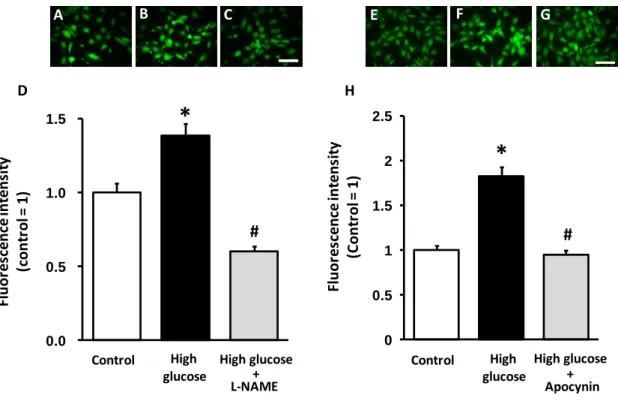
To clarify the possibility that eNOS itself might serve as a source of superoxide in the diabetic state, I examined the influence of the NOS inhibitor L-NAME on production of ROS induced by high glucose in HCAECs. High glucose increased ROS production indicated as DCF fluorescence in HCAECs. Interestingly, L-NAME decreased high glucose-induced ROS production in HCAECs (Fig. 5A-D), suggesting that eNOS is an important superoxide source in high glucose conditions. Furthermore, apocynin, an NADPH oxidase inhibitor, also decreased high glucose-induced ROS production in HCAECs (Fig. 5E-H). Therefore, NADPH oxidase is also another important superoxide source in high glucose conditions. Moreover, the anti-oxidative effect of nicorandil was confirmed because nicorandil improved expression of p47phox and eNOS uncoupling state in the femoral arteries of STZ rats (Fig. 3A, C). Nicorandil also prevented high glucose-induced ROS production in HCAECs (Fig. 6A-D). Taken together, these results suggest that nicorandil could exert an anti-oxidative effect by normalizing NADPH oxidase and eNOS uncoupling state in endothelial cells.
28
Figure 5. Effect of L-NAME and apocynin on ROS production in HCAECs treated with high glucose
HCAECs were exposed to high glucose (35.6 mM) for 24 h, and L-NAME (300 M) or apocynin (100 M) for the same period. These representative images show DCF fluorescence in control (A, E), high-glucose (B, F) and high-glucose + L-NAME (C) or + apocynin (G). High glucose treatment increased ROS production, which could be prevented by L-NAME (D) or apocynin (H). *p<0.05 vs control, #p<0.05 vs high-glucose (20 cells/dish from 2-3 dishes). Scale bar = 100 m.
0.0 0.5 1.0 1.5 Flu or es ce n ce in te n si ty (con tr ol = 1) Control High glucose High glucose + L-NAME
*
# B D C A 0 0.5 1 1.5 2 2.5 Fl u o res cence in tens it y (C o n tr o l = 1 ) Control High glucose High glucose + Apocynin*
# F H G E29
Figure 6. Effect of nicorandil on ROS production induced by high-glucose treatment in HCAECs
HCAECs were exposed to high glucose (35.6 mM) for 24 h, and nicorandil (100 M) for the same period. Representative images show DCF fluorescence in control (A), high-glucose treatment (B) and high-glucose + nicorandil (C). High-glucose treatment increased ROS production, which was prevented by nicorandil (D). *p<0.05 vs control, #p<0.05 vs high-glucose (20 cells/dish from 3 dishes). Scale bar = 100 m.
0.0 0.5 1.0 1.5 2.0 2.5
Fl
u
o
res
cence
in
tens
it
y
(co
n
tr
o
l =
1
)
*
#
B
C
A
D
Control High glucose High glucose + Nicorandil30
1-4. Discussion
The present study demonstrated that nicorandil protected STZ rats from endothelial dysfunction assessed by the FMD reduction without affecting endothelium-independent vasodilation. Because earlier studies showed that endothelial dysfunction in diabetes is caused by increased ROS (Gryglewski et al., 1986), there should be 2 possibilities for mechanisms underlying endothelial protection. Nicorandil could inhibit the expression and activity of NADPH oxidase, a major source of ROS (Griendling et al., 2000; Muller and Morawietz, 2009). Or, because monomer state of eNOS is a source of ROS but not NO depending on the BH4 level (Landmesser et al., 2003), nicorandil could reduce ROS production by inhibiting the monomerization of eNOS through increased expression of BH4 synthase.
These results suggested that nicorandil improved endothelial function in STZ rats through an antioxidative effect exerted by normalizing p47phox and eNOS uncoupling. Nicorandil may offer a novel therapy for diabetes complications by targeting endothelial dysfunction caused by ROS.
Endothelial dysfunction and NADPH oxidase in diabetes
Endothelial dysfunction is attributable to endothelial ROS production derived from vascular NADPH oxidase, which is an important source of superoxide (Griendling et al., 2000; Muller and Morawietz, 2009). The present results generally agree with earlier reports that endothelial-dependent relaxation of arteries is impaired by NADPH oxidase increasing ROS production in both type I (Hink et al., 2001; Ojaimi et al.,
31
2010) and type II diabetic models (Gupte et al., 2010; Mishra et al., 2010). This study demonstrated that tempol, a radical scavenger, improved FMD of STZ rats (Fig. 2), and that more NADPH oxidase subunit p47phox was expressed in femoral arteries (Fig. 3A). As well, apocynin significantly decreased high glucose-induced ROS production in HCAECs (Fig. 5H); indicating that endothelial dysfunction in STZ rats was caused by the oxidative stress produced by NADPH oxidase. Moreover, nicorandil inhibited expression of NADPH oxidase in STZ rat femoral arteries (Fig. 3A), and also prevented high glucose-induced ROS production in HCAECs (Fig. 6). These results suggest that nicorandil ameliorates endothelial dysfunction by decreasing NADPH oxidase which inhibited production of ROS. This agreed with a previous report that nicorandil can inhibit production of ROS by NADPH oxidase activation in response to hypoxia-reoxygenation in human umbilical vein endothelial cells (Tajima et al., 2008).
An increase in ROS production caused by NADPH oxidase in diabetic vessels is at least partially mediated by protein kinase C (PKC) and nuclear factor-kappa B (NF-B) (Mariappan et al., 2010; Ohshiro et al., 2006). Because nicorandil exerts inhibitory effects on PKC activity (Iliodromitis et al., 2008) and NF-B activation (Kawamura et al., 2005), nicorandil might prevent the increase in NADPH oxidase by inhibiting PKC or NF-B in diabetic rats. Moreover, it has been suggested that mitochondrial ROS may activate vascular NADPH oxidase through a PKC-dependent process (Wenzel et al., 2008A). Eguchi et al suggested that nicorandil inhibits
32
ROS-induced ROS release in the mitochondria of endothelial cells (Eguchi et al., 2009). Therefore, nicorandil may inhibit NADPH oxidase by protecting mitochondria.
Contribution of eNOS to ROS production in diabetes
eNOS can produce NO or superoxide. eNOS is uncoupled when BH4, an essential cofactor of NOS, is reduced in oxidative states (Landmesser et al., 2003). As a result, production of superoxide is increased by the eNOS monomer; whereas, in the presence of abundant BH4 the eNOS dimer produces mainly NO (Landmesser et al., 2004). In the diabetic state, eNOS is believed to serve as another source of ROS (Hink et al., 2001; Wenzel et al., 2008C). High glucose can induce an eNOS uncoupling state due to BH4 deficiency (Cai et al., 2005), leading to decrease in NO production and increase in superoxide production (Hink et al., 2001; Satoh et al., 2008). In fact, there are clinical and experimental reports that describe administration of BH4 to improve endothelial function (Heitzer et al., 2000; Pieper, 1997). In the present study, the expression of GCH-1, a major enzyme for BH4 synthesis, was decreased in STZ rats (Fig. 3D). This was consistent with the decrease in eNOS dimer/monomer ratio (Fig. 3C). These results suggest that ROS production in STZ rats could be derived from an uncoupled state of eNOS. In fact, the NOS inhibitor L-NAME completely inhibited production of ROS induced by high glucose treatment in HCAECs (Fig. 5D), suggesting that ROS production in STZ rats was derived mainly from eNOS.
33
and activity of eNOS. This occurs not only in normal rat hearts (Horinaka et al., 2001) but also in several disease models such as salt-sensitive Dahl rats (Horinaka et al., 2006; Xu et al., 2005), myocardial infarcted rats (Horinaka et al., 2004) and monocrotaline-induced pulmonary hypertensive rats (Hongo et al., 2005). Increased release of NO by nicorandil was correlated with enhancement of eNOS phosphorylation in cultured rat cardiac fibroblasts (Liou et al., 2011). The increase in eNOS expression is related to the improvement of symptoms. On the other hand, the present study demonstrated that nicorandil decreased the increase in total eNOS expression in STZ rats (Fig. 3B) and improved endothelial function. To our knowledge, this is the first demonstration that nicorandil inhibits the increase in expression of eNOS in diabetic rats. There is a possibility that nicorandil inhibited the expression of eNOS. In diabetic rats, hyperglycemia increased superoxide production by activating mitochondrial electron transport chain (Nishikawa et al., 2000), which activated PKC, resulting in upregulation of eNOS in the endothelium (Creager et al., 2003; Hink et al., 2001). Because nicorandil and diazoxide can reduce the mitochondrial ROS production (Ozcan et al., 2002), the inhibition of mitochondrial ROS by nicorandil may serve to inhibit the increase in eNOS expression in STZ rats. Further investigation is required to clarify the mechanisms by which nicorandil regulates eNOS expression.
34
Chapter 2
Studies of effect of nicorandil
35
2-1. Introduction
It is well known that the endothelium plays an important role in the regulation of vascular tone via release of factors such as NO, prostaglandin I2, endothelium-derived hyperpolarization factor (EDHF), and endothelin. Endothelial dysfunction predicts adverse cardiac events in patients with coronary artery disease (Kitta et al., 2009). It is mainly caused by loss of NO that occurs from several pathological conditions, such as atherosclerosis, hypertension, and diabetes. Recently, DES has been considered as another potential cause of endothelial dysfunction, despite their successful use in the treatment of ischemic heart diseases by providing dramatic reduction in in-stent restenosis (Dawkins et al., 2005; Stone et al., 2005; Stone et al., 2004). Paclitaxel, one of the major drugs used in DES, is known to cause endothelial dysfunction in humans (Shin et al., 2007) and swine (Pendyala et al., 2009) because it induces apoptosis and cytotoxicity in human coronary artery endothelial cells (Wessely et al., 2007).
Nicorandil, an anti-anginal drug with KATP channel opening and nitrate-like activity, was shown in the IONA (IONA study group, 2002) and JCAD (Horinaka et al., 2010) studies to improve the prognoses of patients with ischemic heart disease by a pharmacological preconditioning effect (Horinaka, 2011). In both clinical settings and animal studies, it also has been reported that nicorandil reduced cardiac events partially by protecting endothelium. Nicorandil improved FMD (an indicator of endothelial function) in patients with ischemic heart disease (Sekiya et al., 2005) or with cardiovascular risk factors (Ishibashi et al., 2008). And it reduced myocardial
36
no-reflow in swine by protecting endothelial function (Zhao et al., 2006). In chapter 1, I demonstrated that nicorandil protected endothelial function in diabetic rats. However, there are no reports that nicorandil provides protection from endothelial dysfunction caused by paclitaxel.
In the next study, I investigated the protective effect of nicorandil on paclitaxel-induced endothelial dysfunction. I also sought the mechanisms underlying this protective effect in association with NO bioavailability in vascular endothelial cells.
37
2-2. Materials and Methods
ChemicalsNicorandil was synthesized in the Chugai Organic Chemistry Laboratory. Paclitaxel and the other reagents were analytical grade and purchased from Wako Pure Chemicals Co. Tempol, 5-hydroxydecanoate (5-HD) and 1H-[1,2,4] oxadiazolo [4,3-a]quinoxalin-1-one (ODQ), isosorbide dinitrate (ISDN) and diazoxide were obtained from Sigma-Aldrich. Nitroglycerin came from Nippon Kayaku (Tokyo, Japan) and acetylcholine was supplied by Daiichi-Sankyo (Tokyo, Japan).
Paclitaxel was dissolved in 50% dimethylformamide, 25% Cremophor EL, and 25% distilled water. Diazoxide and ISDN were suspended in 3% gum arabic solution. Nitroglycerin and acetylcholine were dissolved in, and diluted with, 0.9 % saline solution. All other drugs were dissolved in distilled water just before the experiment. The concentrations of paclitaxel used in this study were chosen on the basis of studies published previously (Kim et al., 2004; Wessely et al., 2007).
Animals
Male Sprague Dawley rats (250–300 g) were used. All rats were fed ordinary laboratory chow and allowed free access to water under a constant light and dark cycle of 12 h. The rats were anesthetized with an intraperitoneal injection of pentobarbital (50 mg/kg) and adequate anesthetic depth was monitored by pinching the toe. An osmotic pump (model 2ML1; Durect Corp., Cupertino, CA, USA) was implanted
38
subcutaneously into a small pouch between the shoulder blades to continuously infuse paclitaxel (0.5, 1.5, 5 mg/kg/day). Bupivacaine (5 mg/mL) was applied topically to the skin wound. For the vehicle group, the osmotic pump containing vehicle (50% dimethylformamide, 25% Cremophor EL, and 25% distilled water) was subcutaneously implanted. Nicorandil (15 mg/kg/day) and tempol (20 mg/kg/day) were dissolved in drinking water and administered for 1 week, from just after implantation of the osmotic pump. Diazoxide (15 mg/kg) and ISDN (15 mg/kg) were administered by gastric gavage once a day for 1 week from just after implantation. FMD was measured in rats 1 week after implantation of the osmotic pump.
All animal procedures were conducted in accordance with Chugai Pharmaceutical’s ethical guidelines for animal care, and all experimental protocols were approved by the Animal Care Committee of the institution and conform to the Guide for the Care and Use of Laboratory Animals published by the US National Institutes of Health. All animals were euthanized by exsanguination under anesthesia after experiments.
Measurements of endothelial function in live rats
FMD was measured in living rats as described in Chapter 1. In this study, vasodilation induced by acetylcholine was also evaluated as another indicator of endothelial function. Acetylcholine (3 ng) was injected into the aorta just proximal to the iliac bifurcation after femoral artery was visualized using a high-resolution ultrasound system in anesthetized rats. The change in diameter of femoral artery was monitored until 1 min after
39
injection.
Real-time PCR analysis in rat femoral arteries
After measurement of FMD, femoral arteries were harvested and stored in RNAlater solution (Ambion, Austin, TX, USA) until RNA isolation was performed. Tissues were homogenized with a Micro Smash homogenizer (Tomy Digital Biology, Tokyo, Japan), and total RNA was isolated using an RNeasy Fibrous Tissue kit (Qiagen, Dusseldorf, Germany). Real-time PCR was performed using TaqMan Gene Expression Assays for NADPH oxidase components p47phox and gp91phox in an ABI PRISM 7500 Sequence Detection System (Applied Biosystems, Carlsbad, CA, USA). Gene expression was normalized to the endogenous control, -actin.
Measurement of ROS in mouse testicular arteries
In vivo ROS production was monitored by the fluorescent probe, dihydrorhodamine 123 hydrochloride (Wako Pure Chemical) according to the method as previously described (Eguchi et al., 2009). Briefly, male ICR mice (10 to 11 weeks old) were anesthetized with pentobarbital (50 mg/kg, i.p.). Endothelial cells were observed by isolectin B4 labeled with FITC: an endothelial cell marker specific to mice (Vector Lab, Burlingame, CA, USA). After the testicular artery (approx. 200 μm in diameter) was carefully exposed, dihydrorhodamine 123 (2.5 mg/kg) was given intravenously, followed by intravenous administration of FITC-labeled isolectin B4 (1.25 mg/kg). The fluorescence intensity was observed by an ultrafast laser
40
confocal microscope equipped with a piezoelectric motor control unit, and was recorded as arbitrary units (au). Under pentobarbital anesthesia, mice were subcutaneously implanted osmotic pump (model 1007D; Durect Corp.) with body temperature kept stable and monitoring adequate anesthetic depth by pinching the toe. Paclitaxel (5 mg/kg/day), was infused continuously from the osmotic pump. Nicorandil (15 mg/kg/day) was administrated in drinking water from just after infusion of paclitaxel.
Measurements of ROS in HCAECs
To measure production of ROS, HCAECs were seeded onto plastic dishes (1×10⁵ cells/2 mL/dish) and cultured in monolayers in a 5% CO2 humidified incubator at 37°C. After overnight incubation, paclitaxel (10 ng/mL) was added to the culture medium for 24 h. Nicorandil (100 M), 5-HD (500 M), and ODQ (20 M) were added over the same period. The cellular level of ROS production was evaluated as described in Chapter 1.
Western blot analysis in HCAECs
Paclitaxel (10 ng/mL) was added to the culture medium for 24 h. Nicorandil (100 M) was added over the same period. Then, cells were harvested and used for western blotting. Equal amounts of protein extract were separated on 10% SDS-polyacrylamide gel and immobilized on PVDF membranes. The membranes were incubated with anti-eNOS (Santa Cruz Biotechnology), anti-phospho-eNOS (Cell Signaling Technology, Danvers, USA) and anti--actin antibodies. Immunoreactive signals were detected
41
using a ChemiDoc XRS system (Bio-Rad).
Measurements of NO in HCAECs
Intracellular NO was assessed with diaminofluorescein-2 diacetate (DAF-2DA; Sekisui medical, Tokyo, Japan) fluorescence probe (10 mol/L) (Wang et al., 2008). Briefly, HCAECs were incubated with PBS containing 10 M DAF-2 in the dark for 30 min at 37 °C, and then washed twice with PBS. Fluorescence intensity was measured by spectrofluorophotometry (SpectraMax Gemini XS (Molecular Devices, CA, USA) with excitation and emission wavelengths of 485 and 545 nm, respectively.
Statistical analyses
All data are expressed as mean ± SE. The n values refer to the number of individual animals in each experiment. The statistical significance of differences was determined by using the Tukey Multiple Comparison Test. Probability (P) values less than 0.05 were considered significant.
42
2-3. Results
Measurements of FMD in rats
Endothelial function was evaluated by measuring FMD in rat femoral arteries. The femoral arterial flow velocity was 121.2 10.3 mm/s at baseline in rats treated with vehicle (Fig. 7A). When the common iliac artery was occluded for 5min and reperfused, the flow velocity increased instantly (i.e. reactive hyperemia) to 176.1 3.7 mm/s (Fig. 7B). The flow velocity decayed rapidly to baseline values at around 3 min (Fig. 7C). The increase in flow velocity was associated with the delayed increase in femoral arterial vasodilation. The femoral arterial diameter increased from 0.40 0.01 mm (Fig 7D) to 0.48 0.01 (Fig 7E) at 1 min after reperfusion. The percentage increase in arterial diameter (vasodilation) peaked at 1 min (Fig. 7F). This delayed vasodilation is FMD.
43
Figure 7. Post-ischemic reactive hyperemia and vasodilation of the rat femoral artery
Original Doppler flow velocity tracings at baseline (A) and at onset of reperfusion after 5 min occlusion of the common iliac artery (B). (C) Time course of peak flow velocity after 5 min ischemia. Representative ultrasound image of the femoral artery at baseline (D) and 1 min after reperfusion (E). Longitudinal sections of the rat femoral artery were visualized with a high-resolution ultrasound probe placed on the upper thigh. (F) Time course of vasodilation (percentage increase in arterial diameter) after 5 min ischemia (n
= 5). Arrow indicates femoral artery. Scale bars = 1 mm.
C
100 120 140 160 180 200 P ea k vel o ci ty (mm/ s)Time after reperfusion(min)
5 mi n is ch emi a Baseline 0 1 2 3 Baseline Just after reperfusion (0 min)
-20 -10 0 10 20 V el ocit y (cm /s ) -20 -10 0 10 20 V el ocit y (cm /s )
B
A
F
D
Baseline -5 0 5 10 15 20 25 30 V as o d ila ti o n ( ⊿ %)Time after reperfusion (min)
E
1 min after reperfusion
5 mi n is ch emi a Baseline 0 1 2 3
44
Effects of nicorandil on reduced FMD in rats treated with paclitaxel
I evaluated the influences of paclitaxel and nicorandil on FMD of rat femoral artery. Continuous subcutaneous infusion of paclitaxel (0.5, 1.5, 5 mg/kg/day, 7 days) reduced FMD in a dose-dependent manner (Fig. 8A). Reactive hyperemia was also evaluated as an inducer of FMD in the femoral artery to confirm that stimulation to the endothelium was similar among the groups. As a result, reactive hyperemia was similar among the groups (Fig. 8B). NTG was administered to evaluate endothelium-independent vasodilation because it was possible that paclitaxel impaired vascular smooth muscle function which influenced FMD. NTG-induced vasodilation was similar among the groups (Fig. 8C). These results imply that paclitaxel impaired endothelial function without changing contractility in vascular smooth muscle (Control, 21.6 ± 3.2; Paclitaxel [5 mg/kg/day], 7.1 ± 1.7%, Fig. 8A). On the other hand, co-treatment with nicorandil prevented the decrease in FMD caused by paclitaxel (15.5 ± 2.5%) without changing flow velocity or NTG-induced vasodilation (Fig. 8B, C). In the current study I found that treatment with paclitaxel and nicorandil for 1 week did not affect body weight, blood pressure or heart rate relative to rats treated with vehicle (Table 2).
45
Figure 8. Effects of nicorandil on endothelial function impaired by paclitaxel as evaluated by FMD
(A) Dose-dependent impairment of FMD by paclitaxel (0.5, 1.5, 5.0 mg/kg/day; 1 week) and the effects of nicorandil (15 mg/kg/day; 1 week) on paclitaxel-induced impairment of FMD. Peak velocity (B) and NTG-induced vasodilation (C) were similar among groups. *P < 0.05 vs. control; #P < 0.05 vs. paclitaxel (5 mg/kg/day) (n = 5–9).
0 5 10 15 20 25 30 V as o d ila ti o n ( ⊿ % , 1 m in ) A FMD
*
*
# Control Paclitaxel Nicorandil 0.5 1.5 5.0 5.0 0 50 100 150 200 250 Pe ak v el o ci ty (m m /s , 0 m in ) Control Paclitaxel Nicorandil 0.5 1.5 5.0 5.0 0 5 10 15 20 25 30 35 V as o d ila ti o n ( ⊿ % , 3 0 se c) Control Paclitaxel Nicorandil 0.5 1.5 5.0 5.0B Reactive hyperemia C NTG-induced vasodilation
(5) (6) (5) (9) (7)
46
Table 2. Body weight, blood pressure and heart rate at 1 week after paclitaxel administration Vehicle Paclitaxel 0.5 1.5 5.0 5.0 + Nicorandil N 5 6 5 9 7 Body weight (g) 330.9 ± 13.9 337.3 ± 3.5 343.5 ± 6.6 322.0 ± 16.1 293.3 ± 14.6 Systolic blood pressure (mmHg) 134.9 ± 3.5 136.6 ± 5.0 121.8 ± 5.0 129.2 ± 2.5 122.7 ± 3.4 Heart rate (bpm) 362.6 ± 8.1 369.2 ± 8.5 378.3 ± 8.0 369.1 ± 6.3 394.0 ± 9.5
47
Effects of nicorandil on vasodilation induced by acetylcholine in rats treated with paclitaxel
Acetylcholine-induced vasodilation serves as an indicator of endothelial function because it can induce NO-dependent vasodilation by eNOS activation in endothelium. Therefore, I next evaluated the influence of paclitaxel and nicorandil on acetylcholine-induced vasodilation in living rats. In vehicle-treated rats, local administration of acetylcholine into the iliac artery led to femoral arterial vasodilation. Peak dilation was at 10 sec and rapidly decayed to baseline by approximately 60 sec (Fig. 9A). In rats treated with paclitaxel (5 mg/kg/day), acetylcholine induced significantly less dilation in the femoral artery (Control, 12.7 ± 2.1%; Paclitaxel, 3.9 ± 1.1%). On the other hand, nicorandil significantly prevented the decrease in acetylcholine-induced vasodilation by paclitaxel (Paclitaxel + Nicorandil, 12.5 ± 3.5%, Fig. 9B). These results were consistent with the FMD measurements (Fig. 8A), indicating that nicorandil surely prevented endothelial dysfunction caused by paclitaxel.
48
Figure 9. Effects of nicorandil on endothelial function impaired by paclitaxel as evaluated by acetylcholine injection
Acetylcholine was injected into the common iliac artery through an intra-aortic catheter. Time course (A) and mean percentage increase in arterial diameter at 10 sec after acetylcholine injection (B) was evaluated at the femoral artery. *P < 0.05 vs. control; #P
< 0.05 vs. paclitaxel (5 mg/kg/day) (n = 7–8). 0 5 10 15 20 V as o d ila ti o n ( ⊿ % , 1 0 s e c)
*
# B Control Paclitaxel (5.0) Nicorandil 0 5 10 15 20 V as o d ila ti o n ( ⊿ %)Time after administration (sec)
vehicle paclitaxel (5.0) paclitaxel (5.0) + nicorandil A 0 30 60 (7) (8) (7)
49
Effects of diazoxide, ISDN, and tempol on FMD in rats treated with paclitaxel
Because nicorandil is known to exert multiple actions, such as KATP channel opening and nitrate-like activity, it was important to determine which action was more important in endothelial protective effect of nicorandil. Diazoxide, a mitochondrial KATP channel opener, was used instead of nicorandil. As shown in Figure 10, diazoxide prevented significantly the decrease in FMD caused by paclitaxel, suggesting that opening mitochondrial KATP channels is important in endothelial protection. Conversely, ISDN, a nitrate which serves as an NO donor, had no effect (Fig. 10). These results suggest that mitochondrial KATP channel opening was more important than nitrate-like activity in endothelial protection. Furthermore, I also examined the involvement of oxidative stress in paclitaxel-induced endothelial dysfunction. Tempol, a ROS scavenger, also prevented the decrease in FMD of rats treated with paclitaxel (Fig. 10). This result could mean that paclitaxel caused endothelial dysfunction by oxidative stress.
50
Figure 10. Effects of diazoxide, ISDN and tempol on FMD impaired by paclitaxel
Diazoxide (15 mg/kg/day) and ISDN (15 mg/kg/day) were suspended in 3% gum arabic solution and administrated by gastric gavage once a day for 1 week. Tempol (20 mg/kg/day, p.o.) was administrated in drinking water for 1 week. *P < 0.05 vs. control; #P < 0.05 vs. paclitaxel (5 mg/kg/day) (n = 5–7).
0
5
1015
20
25V
aso
d
il
at
io
n
(
⊿
%
, 1
m
in
)
*
#
*
#
Control Paclitaxel (5.0) Tempol Diazoxide ISDN (5) (7) (6) (6) (7)51
Effects of nicorandil on gene expression of NADPH oxidase in femoral arteries of rats treated with paclitaxel
Because the involvement of oxidative stress in paclitaxel-endothelial dysfunction was indicated in Figure 10, I examined the effect of nicorandil on the expression of NADPH oxidase which is a major source of ROS. The mRNA expressions of p47phox and gp91phox, the major components of NADPH oxidase, increased significantly in femoral arteries isolated from rats treated with paclitaxel (Fig. 11). On the other hand, nicorandil prevented significantly the increase in p47phox expression (Fig. 11A), and also limited the increase in gp91phox expression (Fig. 11B, P = 0.09 vs. paclitaxel). These results suggest that nicorandil could prevent oxidative stress induced by paclitaxel through inhibiting NADPH oxidase.
52
Figure 11. Effects of nicorandil on changes in expression of p47phox and gp91phox mRNA induced by paclitaxel in rat femoral arteries
p47phox (A) and gp91phox (B) expression levels in femoral arteries were measured using
real-time PCR. Data are shown as subunit/-actin ratio. *P < 0.05 vs. control; #P < 0.05
vs. paclitaxel (5 mg/kg/day) (n = 8).
A
B
0 0.2 0.4 0.6 0.8 1 p 47 phox / -a ct in*
# 0 0.2 0.4 0.6 0.8 1 1.2 gp9 1 phox / -a ct in*
Control Paclitaxel (5.0) Control
Nicorandil
Paclitaxel (5.0) Nicorandil
53
Effects of nicorandil on production of ROS in testicular arteries of mice treated with paclitaxel
Because nicorandil prevented the increase in NADPH oxidase by paclitaxel, I examined the effect of nicorandil on paclitaxel-induced ROS production. As shown in Figure 12, the fluorescence intensity of dihydrorhodamine 123 hydrochloride in mice treated with paclitaxel was 1.6 times greater than in mice treated with vehicle. On the other hand, co-treatment with nicorandil significantly inhibited any increase in fluorescence intensity (control, 33.0 ± 0.5; paclitaxel, 52.2 ± 1.1; paclitaxel + nicorandil, 44.3 ± 1.0 au). This result suggests that nicorandil could prevent paclitaxel-induced ROS production.
54
Figure 12. Effect of nicorandil on oxidative stress in mice caused by paclitaxel
Production of reactive oxygen species in mouse testicular arteries by paclitaxel was measured using dihydrorhodamine 123 hydrochloride. Representative images show fluorescence in control (A), 7 days after administration of paclitaxel (B), and 7 days after nicorandil was co-administered with paclitaxel (C). Scale bar =25 μm. (D) Each column denotes mean fluorescence intensity from 3 mice. Paclitaxel especially increased the fluorescence intensity at the edge of the spindle-shaped vascular endothelial cells (B). *P < 0.05 vs. control; #P < 0.05 vs. paclitaxel (5 mg/kg/day).
*
#
A Control B Paclitaxel(5.0) C Paclitaxel (5.0) + Nicorandil
D 0 10 20 30 40 50 60 Fl u o re sc e n ce ( ar b it ra ry u n it s) Control Paclitaxel (5.0) Nicorandil
55
Effects of nicorandil on production of ROS, NO release and eNOS phosphorylation induced by paclitaxel in HCAECs
I also examined the effect of nicorandil on paclitaxel-induced ROS production in HCAECs. Paclitaxel (10ng/mL) significantly increased DCF fluorescence compared with control. Nicorandil, in turn, significantly inhibited the increase in DCF fluorescence by paclitaxel (Fig. 13). This result indicated that nicorandil prevented paclitaxel-induced ROS production. A further experiment determined whether KATP channel opening or nitrate-like activity was more important in the anti-oxidative effect of nicorandil. The anti-oxidative potential of nicorandil was abolished by co-treatment with 5-HD, a mitochondrial KATP channel inhibitor (Fig. 14A), but not by co-treatment with ODQ, a specific inhibitor of soluble guanylate cyclase (Fig. 14B). Treatment with 5-HD alone increased DCF fluorescence compared with control (Fig. 14A), whereas nicorandil or ODQ alone did not affect DCF fluorescence (Fig. 13, 14B). Opening mitochondrial KATP channels may be important for the anti-oxidative effect of nicorandil.
Next, I examined the influence of paclitaxel and nicorandil on NO production in HCAECs. Paclitaxel (10 ng/mL) significantly decreased DAF-2 fluorescence intensity compared with control. This result indicated that paclitaxel decreased NO production in HCAECs. Nicorandil tended to inhibit the decrease in DAF-2 fluorescence intensity induced by paclitaxel (Fig. 15A). Nicorandil alone had no effect on fluorescence intensity. On the other hand, there was no significant difference in the phosphorylation of eNOS protein among the groups in HCAECs (Fig. 15B).
56
Figure 13. Effects of nicorandil on oxidative stress induced by paclitaxel in HCAECs
The images show representative fluorescences in control (A), after 24 h treatment with paclitaxel (10 ng/mL) (B), paclitaxel + nicorandil (100 M) (C), and nicorandil alone (100
M) (D). Fold changes in DCF fluorescence intensity are provided as the normalized ratio to control (E). Intracellular production of reactive oxygen species was measured with confocal microscopy after 45 min DCF (10 M) label as described in the Methods. Nicorandil inhibited paclitaxel-induced fluorescence intensity in HCAECs. *P < 0.05 vs.
control; #P < 0.05 vs. paclitaxel. Each column denotes mean fluorescence intensity of 20 cells/dish from 3 dishes. Scale bar = 100 µm.