51:756 症例報告
初期には脊髄小脳変性症と診断され,オリーブ・橋・小脳病変が
高度であった進行性核上性麻痺の 1 剖検例
岩崎
靖
1)*森
恵子
1)伊藤 益美
1)三室 マヤ
2)吉田 眞理
2) 要旨:症例は死亡時 72 歳の女性である.63 歳時に歩行障害と構音障害で発症し,画像所見上は小脳・脳幹萎縮 がめだち,初期には脊髄小脳変性症と診断された.眼球運動障害,体幹に強い筋強剛,把握反射,頭部 MRI での中 脳被蓋萎縮所見が経過とともに出現し,発症 6 年後に進行性核上性麻痺と診断を変更した.病理学的には前頭葉萎 縮,脳幹被蓋萎縮に加え,オリーブ・橋・小脳系の変性が高度で,神経原線維変化と Gallyas 陽性!タウ陽性のグリ ア細胞内構造物,argyrophilic thread を広範かつ多量にみとめた.小脳・脳幹病変が強く,脊髄小脳変性症に類似し た臨床症候を呈する進行性核上性麻痺のサブタイプに属する症例と考えられた. (臨床神経 2011;51:756-760) Key words:進行性核上性麻痺,失調,脊髄小脳変性症,神経原線維変化,オリーブ・橋・小脳障害 はじめに進行性核上性麻痺(progressive supranuclear palsy;PSP) は精神症状,核上性眼球運動障害,錐体外路徴候などの様々な 臨床症候と脳幹被蓋萎縮などの画像所見を示す1)∼3).われわ れは歩行障害と構音障害で発症し,失調症状や画像所見上の 小脳・脳幹萎縮から初期には脊髄小脳変性症と診断されてい た PSP の 1 剖検例を経験した.末期には PSP の典型的な臨 床症候を呈したが,病理所見ではオリーブ・橋・小脳系の変 性が高度であった.本症例の臨床経過と病理所見を対比検討 し,文献的考察を加えて報告する. 症 例 患者:死亡時 72 歳の女性 主訴:嚥下障害,歩行障害 既往歴:虫垂炎手術 家族歴・生活歴:特記すべきことなし. 現病歴:1998 年(63 歳時)にふらつき,歩行障害で発症し た.しだいにろれつが回らなくなり,発症 3 年後に神経内科を 受診した.構音障害,失調性歩行,四肢協調運動障害,頭部 CT 画像にて小脳・脳幹萎縮と第 4 脳室の拡大を指摘され (Fig. 1a),脊髄小脳変性症(オリーブ橋小脳萎縮症)と診断さ れた.眼球運動障害や筋強剛は明らかでなかった.タルチレリ ン水和物の経口投与で加療されたが,症状は徐々に進行した. 発症 5 年後に行動異常や認知機能障害が出現し,交通事故を おこしたり,仕事上のトラブルが頻回となり退職した.嚥下障 害と歩行障害が進行し,自宅療養が困難となったため発症 6 年後に小山田記念温泉病院神経内科に入院した. 入院時所見と経過:体幹の筋強剛が強く,頸部後屈,上下方 向への核上性眼球運動障害,把握反射陽性,Babinski 徴候陽 性などの神経所見から PSP と臨床診断を変更した.明らかな 自律神経障害はみとめなかった.頭部 MRI 画像では第 3 脳室 と第 4 脳室の拡大,中脳被蓋萎縮,小脳萎縮をみとめるものの (Fig. 1b),橋の十字架徴候や被殻背外側の異常信号など多系 統萎縮症をうたがう所見はみとめなかった.脳血流シンチグ ラフィでは前頭葉と小脳の血流低下をみとめた.眼振や口蓋 ミオクローヌス,静止時振戦などの不随意運動はみとめな かった.発症 7 年後に胃瘻造設術,発症 8 年後に気管切開術が 施行されたが,突然の心停止により全経過約 9 年で死亡した. 経過中に膀胱直腸障害や起立性低血圧は明らかでなかった. 病理所見:脳重は 1,030g で,前頭葉萎縮(とくに弓隆面と 中心前回),淡蒼球と視床下核の褐色調萎縮をみとめた.小脳 では歯状核と白質の萎縮が強かった(Fig. 2a).脳幹では被蓋 萎縮に加え(Fig. 2b),橋底部の萎縮もめだち(Fig. 2c),黒質 と青斑核の褪色が高度だった.下オリーブ核の仮性肥大はみ とめなかった.組織学的には前頭葉(とくに前頭前野と中心前 回),淡蒼球と視床下核に神経細胞脱落とグリオーシスをみと め,小脳ではプルキンエ細胞の脱落と Bergmann グリアの増 生,Torpedo の形成(Fig. 2d),歯状核の神経細胞脱落とグリ オーシス,グルモース変性をみとめた.上小脳脚の変性も高度 * Corresponding author: 小山田記念温泉病院神経内科〔〒512―1111 三重県四日市市山田町 5538―1〕 1) 小山田記念温泉病院神経内科 2) 愛知医科大学加齢医科学研究所神経病理部門 (受付日:2011 年 3 月 15 日)
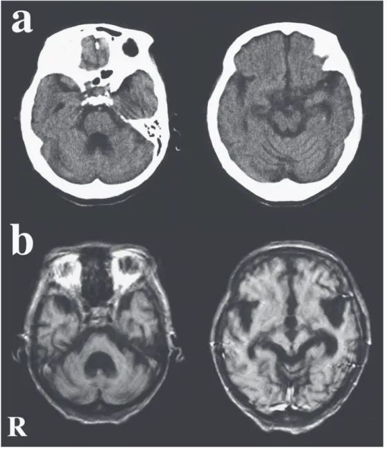
Fig. 1 Cranial CT and MRI findings.
a: CT, obtained 3 years after onset, shows mild atrophy of the cerebellum and brainstem and dilatation of the fourth ventricle.
b: T1 weighted images (TR 350ms, TE 17ms), obtained 9 years after onset, show moderate cerebellar atrophy and severe dilatation of the fourth ventricle. The tegmentum of the midbrain and pons shows severe atrophy. The third ventricle, inferior horns of the lateral ventricle, and Sylvian fissure are also dilatated.
(R: Right side) だった.黒質と青斑核は高度の神経細胞脱落を示し,動眼神経 核,橋核(Fig. 2e),下オリーブ核には中等度の神経細胞脱落 とグリオーシスをみとめた.Gallyas-Braak 染色および抗リン 酸化タウ抗体(AT-8;Innogenetics)をもちいた免疫染色で前 頭葉,基底核,脳幹,小脳白質には広範かつ多量に Gallyas 陽性!タウ陽性のグリア細胞内構造物(tuft-shaped astrocyte, coiled body)や argyrophilic thread をみとめ(Fig. 2f),プル キンエ細胞や Bergmann グリア,小脳歯状核神経細胞にもタ ウ陽性所見をみとめた(Fig. 2g).淡蒼球,視床下核,動眼神 経核,黒質,青斑核に Globose-type の神経原線維変化(neuro-fibrillary tangle;NFT)をみとめ,橋核,下オリーブ核にも 多数の Gallyas 陽性!タウ陽性の神経細胞をみとめた(Fig. 2 h,i).タウの沈着は変性の強いオリーブ・橋・小脳系にとく に強く,タウ陽性構造物は免疫組織学的には 3 リピートタウ を認識する抗体(RD3;Upstate Biotechnology)では陰性,4 リピートタウを認識する抗体(RD4;Upstate Biotechnology) では陽性であった.脊髄では前側索の萎縮と 平化をみとめ, 灰白質には Gallyas 陽性!タウ陽性の神経細胞,多数の
argy-臨床神経学 51巻10号(2011:10) 51:758
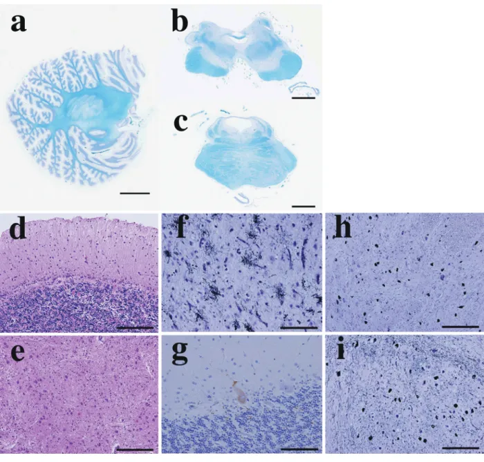
Fig. 2 Neuropathologic findings.
a: Macroscopic image of the sagittal section of the cerebellar hemisphere. The cerebellar white matter and dentate nucleus are atrophic. The cerebellar white matter also shows myelin pallor. b: Macroscopic image of the axial section of the midbrain. Tegmentum shows severe atrophy. c: Macroscopic image of the axial section of the pons. Not only the tegmentum but also the pontine base show atrophy. Degeneration of the superior cerebellar pedunculus is also observed.
d: The cerebellar cortex shows moderate Purkinje neuron loss with Bergmenn s gliosis and torpedoes.
e: The pontine nucleus shows moderate neuron loss with gliosis.
f: Numerous tuft-shaped astrocytes are observed. Coiled bodies and argyrophilic threads are also observed (frontal cortex).
g: A tau-positive Purkinje neuron is observed (cerebellar cortex).
h: The pontine nucleus shows numerous Gallyas-positive neurofibrillary tangles.
i: The inferior olivary nucleus also shows numerous Gallyas-positive neurofibrillary tangles. Numerous argyrophilic threads are also observed.
(a-c: Kl ver-Barrera staining; d, e: Hematoxylin-eosin staining, f, h, i: Gallyas-Braak silver staining, g: AT-8 immunostaining)
けて高度だった.海馬支脚から海馬傍回,内嗅領皮質には ar-gyrophilic grain と多数 の flame-shaped の NFT を み と め た
が,老人斑は明らかでなかった.抗リン酸化α―シヌクレイン
抗体(Santa Cruz Biotechnology)をもちいた免疫染色にて Lewy 小 体 や glial cytoplasmic inclusion な ど のα-synuclein pathology はみとめなかった. 考 察 Williams らは多数の PSP 剖検例の検討をおこない,早期か ら姿勢反射障害と易転倒性,垂直性眼球運動障害など典型的 な臨床像を呈する Richardson 症候群,非対称性の臨床症候と 振戦を呈し初期には L-Dopa 反応性パーキンソニズムを呈す る PSP-parkinsonism,すくみ足を主体とした純粋無動症を呈 するまれな pure akinesia with gait freezing などのサブタイ
プに PSP を分類することを提唱しているが3)4),本症例はいず れのタイプともことなる.本症例のように,病初期から失調 や小脳症状がめだち,画像所見上も小脳・脳幹萎縮を示し脊 髄小脳変性症に類似した症候を呈する症例が本邦で報告さ れ,これらの症例では神経病理学的に橋底部,小脳白質の病 変が強く,オリーブ・橋・小脳系に高度な変性をみとめてい る5)∼7).PSP における小脳性運動失調の存在については Steel らの原著においても記載され,それらの症例では歯状核変性 および上小脳脚の障害が高度であることがすでに指摘されて いる1).しかしながら NINDS-SPSP(National Institute of
Neu-rological Disorders and Stroke-the Society for Progressive Supranuclear Palsy)の PSP の臨床診断基準では「早期の著明 な小脳症状」は多系統萎縮症を鑑別するための除外項目とし てあげられている8). PSP の病変分布の強弱は症例によって差があり,大脳皮質 や基底核,小脳歯状核,橋核などの変性程度によって,病理学 的にもいくつかのサブタイプに分類される2).本症例の病変分 布は PSP としては典型的であったが,大脳皮質,小脳,脳幹 をふくめて全体的に萎縮と変性が強く,脊髄の病変も高度で あった.また本症例の Gallyas 陽性!タウ陽性構造物の出現量 は, われわれが以前に検討報告した PSP 剖検多数例の大脳, 基底核,脳幹および脊髄におけるこれらの出現量9)∼11)よりも 全体的に多かった. PSP の小脳・脳幹病変は,入力系の障害として下オリーブ 核と橋核の神経細胞脱落とグリオーシス,出力系の障害とし て小脳歯状核の神経細胞脱落とグルモース変性が観察され る1)2).本症例では小脳歯状核の変性が強く,小脳白質にも多 数の Gallyas 陽性!タウ陽性グリア細胞内構造物や argyro-philic thread が出現し,プルキンエ細胞の脱落もめだった.残 存するプルキンエ細胞や Bergmann グリア,歯状核神経細胞 にはタウ陽性所見をみとめた.オリーブ・橋・小脳系の変性 が強かったことは初期に脊髄小脳変性症(オリーブ橋小脳萎 縮症)と診断されていた臨床経過に対応すると思われた.しか しながら本症例のオリーブ・橋・小脳系の変性や小脳白質の 度は軽かった.Kanazawa らは本邦の PSP 22 剖検例を検討 し,プルキンエ細胞にタウ陽性封入体をともない,歯状核の神 経細胞脱落が高度な 3 症例の存在を強調し,これらの症例は Williams らの指摘するいずれのサブタイプにも分類できな いと報告している12).本症例には Kanazawa らが指摘した小 脳病変とオリーブ・橋・小脳系の変性が併存していると思わ れた. 本症例の臨床経過において,どの時点で PSP をうたがうこ とが可能であったかを後方視的に検討することも重要であ る.発症 3 年後の頭部 CT 画像を注意深く再検討すれば,橋は 底部より被蓋の萎縮が強く,この時点で PSP をうたがえた可 能性があると思われた.発症 5 年後に行動異常や認知機能障 害が加わった時点では PSP を鑑別する必要があった.錐体外 路症状が明らかとなった時点において自律神経障害がめだた ず,橋の十字架徴候や被殻の異常信号など多系統萎縮症をう たがう MRI 所見をみとめなかった点も重要であると思われ た. 小脳・脳幹病変が強く,とくにオリーブ・橋・小脳系の変 性がめだち,脊髄小脳変性症と臨床診断されやすいサブタイ プの存在は,PSP の臨床病理像を検討する上で重要であると 思われる.今後の症例の蓄積が期待される. 謝辞:本症例の病理解剖を施行していただいた小山田記念温泉 病院 病理検査科・永岡昌光先生に深謝いたします. 文 献
1)Steele JC, Richardson JC, Olszewski J. Progressive su-pranuclear palsy. A heterogeneous degeneration involv-ing the brain stem, basal ganglia and cerebellum with vertical gaze and pseudobulbar palsy, nuchal dystonia and dementia. Arch Neurol 1964;10:333-359.
2)吉 田 眞 理. PSP の 病 理. Clinical Neuroscience 2009;27:295-298.
3)Williams DR, de Silva R, Paviour DC, et al. Characteristics of two distinct clinical phenotype in pathologically proven progressive supranuclear palsy: Richardson s syndrome and PSP―parkinsonism. Brain 2005;128:1247-1258. 4)Williams DR, Holton JL, Strand K, et al. Pure akinesia
with gait freezing: a third clinical phenotype of proven progressive supranuclear palsy. Mov Disord 2007;22:2235-2241. 5) 場郁子, 齋藤由扶子, 安田武司ら. 小脳性運動失調の目 立った進行性核上性麻痺の 1 剖検例. 神経内科 2002;56: 230-233. 6)水谷智彦, 上倉 勲, 清水夏繒ら. モダンコンセプト CPC. 歩行障害・姿勢反射異常で発症し,経過中,小脳・橋の萎 縮,眼球運動障害を来たした 64 歳男性例. 栗原照幸, 田代 邦雄, 水野美邦, 編. モダンコンセプト神経内科 2. 東京: 医 学書院; 1992. p. 149-174. 7)森 秀生, 太田 聰, 田久保秀樹ら. オリーブ・橋・小脳系
臨床神経学 51巻10号(2011:10) 51:760
の変性を伴った進行性核上性麻痺(会).Neuropathology 1995;15 Suppl:31.
8)Litvan I, Agid Y, Calne D, et al. Clinical research criteria for the diagnosis of progressive supranuclear palsy ( Steele-Richardson-Olszewski syndrome ) : report of the NINDS-SPSP international workshop. Neurology 1996;47: 1-9.
9)Iwasaki Y, Yoshida M, Hattori M, et al. Distribution of tuft-shaped astrocytes in the cerebral cortex in progres-sive supranuclear palsy. Acta Neuropathol 2004;108:399-405.
10)Iwasaki Y, Yoshida M, Hashizume Y, et al. Widespread spinal cord involvement in progressive supranuclear palsy. Neuropathology 2007;27:331-340.
11)Hattori M, Hashizume Y, Yoshida M, et al. Distribution of astrocytic plaques in the corticobasal degeneration brain and comparison with tuft-shaped astrocytes in the pro-gressive supranuclear palsy brain. Acta Neuropathol 2003;106:143-149.
12)Kanazawa M, Shimohata T, Toyoshima Y, et al. Cerebel-lar involvement in progressive supranuclear palsy: a clini-copathological study. Mov Disord 2009;24:1312-1318.
Abstract
An autopsied case of progressive supranuclear palsy, initially diagnosed as spinocerebellar degeneration with severe olivopontocerebellar involvement
Yasushi Iwasaki, M.D.1)
, Keiko Mori, M.D.1)
, Masumi Ito, M.D.1)
,
Maya Mimuro, M.D.2)and Mari Yoshida, M.D.2)
1)
Department of Neurology, Oyamada Memorial Spa Hospital
2)
Department of Neuropathology, Institute for Medical Science of Aging, Aichi Medical University
A Japanese female patient presented with dysarthria and gait disturbance with ataxia at the age of 63. She was initially diagnosed with spinocerebellar degeneration because a head CT showed atrophy of the cerebellum and brainstem, and dilatation of the fourth ventricle. Symptoms including abnormal behavior, dementia, vertical gaze palsy, dysphagia, retrocollis, axial rigidity, grasp reflex and positive Babinski s sign were gradually observed. Tegmental atrophy of the brainstem and dilatation of the third ventricle were apparent on images. The diagnosis was modified to progressive supranuclear palsy (PSP), 6 years after the onset of symptoms. Gastrostomy and tra-cheotomy were performed 7 and 8 years after onset, respectively, and the patient died one year later.
At autopsy the brain weighed 1,030 g and showed atrophy of the frontal lobe and cerebellum. The brainstem was also atrophic, particularly in the tegmentum and pontine base. Neurofibrillary tangles, mainly globose-type, were widespread in the subcortical structure, particularly in the globus pallidus, subthalamic nucleus, nucleus of the oculomotor nerve, substantia nigra, locus ceruleus, pontine nucleus, and the inferior olivary nucleus. Numer-ous glial fibrillary tangles and argyrophilic threads were also observed particularly in the frontal lobe, basal gan-glia, brainstem and cerebellar white matter. The cerebellar dentate nucleus showed neuron loss with grumose de-generation and the Purkinje neuron layer showed neuron loss and Bergmann s gliosis with torpedoes. Tau posi-tive inclusions in the Purkinje neurons, Bergmann glias and dentate nucleus neurons were observed. Pathological findings were consistent with the diagnosis of PSP but the olivopontocerebellar involvement and the quantity of Gallyas-positive!tau-positive structures were generally more severe than in typical PSP cases.
According to these clinicopathologic findings and a review of the literature, we speculate on the existence of a PSP subtype with severe olivopontocerebellar involvement that tends to be clinically misdiagnosed as spinocere-bellar degeneration.
(Clin Neurol 2011;51:756-760)
Key words: progressive supranuclear palsy, ataxia, spinocerebellar degeneration, neurofibrillary tangle, olivopontocere-bellar involvement