Received June 2,2016;Accepted August 19,2016
Gr owt h hor mone def i ci ency as s oci at ed wi t h s hor t s t at ur e i n a ver y l ow bi r t h wei ght i nf ant af t er t r eat ment f or
s us pect ed i nt r acr ani al caver nous hemangi oma
Norihisa Wada,Shunsuke Minakata,Yuhei Konishi,Tomohiro Inoue,Mitsuru Okada and Tsukasa Takemura
Department of Pediatrics,Kindai University Faculty of Medicine
Abstract
A female infant born at 30 weeks of gestation and weighing 1,276 g developed a suspected intracranial cavernous hemangioma invading the sella turcica at the age of one month.She received high-dose steroid treatment with 5 mg/ kg/d of prednisolone,resulting in a reduction in the size of the hemangioma,and follow-up care began after discharge.At the age of six,it was noted that the girlʼs growth in height was insuf- ficient. Head magnetic resonance imaging (MRI)showed pituitary atrophy,and the results of a loading test led to a diagnosis of growth hormone deficiency resulting in a short stature.
However,the administration of growth hormone
brought her height up to the average by the age of 11. The patientʼs intracranial lesion during the newborn period,which was suspected to be a cavernous hemangioma,may have contributed to her pituitary atrophy.The findings of this case suggest that infants who develop a space-oc- cupying lesion in the sella turcica must be kept under long-term observation in view of the pos- sible occurrence of a defective pituitary func- tion.
Key words:growth hormone deficiency,short stature,very low birth weight infant,intra- cranial cavernous hemangioma
Introduction
Cavernous hemangiomas are benign vascular malformations of relatively low frequency,and congenital intracranial cavernous hemangiomas are particularly rare.웋 Clinical manifestations most often associated with intracranial cavern- ous hemangiomas include:seizures, hemorr- hagic syndrome,and a mass lesion effect with hydrocephalus or focal neurological deficits.워 We report a case of growth hormone deficiency resulting in a short stature after treatment for suspected intracranial cavernous hemangioma.
To our knowledge,no reports have been publi- shed on intracranial cavernous hemangioma in relation to a growth hormone deficiency-as- sociated short stature.
Case Report
The patient was a female appropriate-for-ges- tational-age infant born via emergency C-section at 30 weeks and 1 day,with a weight of 1,276 g and a height of 37 cm.The emergency C-section was performed because of the difficulty in managing the motherʼs hypertension induced by pregnancy,which had led to a loss of variability as indicated by cardiotocographic findings. Apgar scores were 6 at 1 and 7 at 5 minutes.An artificial pulmonary surfactant was administered for the respiratory distress syndrome,mechanical ventilation was continued for 3 days,and oxygen was administered for 7 days.By day 7 of life,a hemangioma-like erythema had appeared in the posterior cervical region, which gradually extended to the right and became raised. A
dermatologist at our institution diagnosed this as possible cavernous hemangioma.On day 27 of life,right optic disk enlargement was found during the first ophthalmologic examination, suggesting optic nerve compression. However, no abnormal findings were seen on the ocular fundus.Head computed tomography(CT)per- formed on day 38 of life revealed a right orbital tumor compressing the optic nerve.This tumor was found to have invaded the intracranial space on the contrast enhanced CT images taken on day 47 of life.These images also showed that the contrast effect presented by the intracranial tumor was similar to that of the subcutaneous mass in the right posterior cervical region,which provided a reasonable ground to suspect that this tumor was a hemangioma(Fig.1). Also,head MRI on day 46 of life detected a high-intensity signal in the cervical mass, which was radiologically determined to be consistent with the diagnosis of cavernous hemangioma.Based on these images,cutaneous findings,and follow- up observation,the patientʼs condition was clinically diagnosed as a possible intracranial cavernous hemangioma. In view of the site of the tumor as well as the patientʼs general physi- cal condition including her constitution,treat- ment with high-dose prednisolone was initiated on day 49 of life.We administered 5 mg/kg/d of
prednisolone for the first 6 weeks,after which the dose was tapered off over a period of 6 weeks before the administration was discontinued on day 134 of life.
On day 93 of life,the therapeutic efficacy was evaluated by means of contrast enhanced CT (Fig.2),which showed a tumor with contrast effect extending from the right orbit to cavernous sinus that had invaded the suprasellar region.
However,although the growth of the infant did not allow us to make an exact comparison with the same slices obtained on day 93 of life,our
Fig.1 Contrast-enhanced CT performed on day 47 of life, showing the invasion of a right orbital tumor into the intracranial space.The contrast effect presented by the intracranial tumor was similar to that of the subcutaneous mass in the right posterior cervical region.
Fig.3 Contrast-enhanced CT obtained on day 124 of life, showing tumor shrinkage.
Fig.2 Contrast-enhanced CT performed on day 93 of life, showing invasion of the tumor into the sella turcica.
radiologist diagnosed tumor shrinkage based on contrast enhanced CT on day 124 of life,which was 10 days before treatment discontinuation (Fig.3). The images obtained on day 146 showed no sign of relapse. For the cervical hemangioma, we performed cryocoagulation using liquid nitrogen initially on day 95 of life followed by two more times. As a result,it macroscopically reduced in size.
At the corrected age of 4 months,the patientʼs head circumference was of average size,and head MRI confirmed that myelination was sufficient for the age. No obvious abnormalities were identified in the pituitary gland or hypoth- alamus,and,although we did not run any endo- crinological tests while she was hospitalized, neonatal screening tests performed during the early neonatal period and prior to discharge showed no thyroid-stimulating hormone(TSH) abnormality.After discharge on day 189 of life (corrected age:4 months and 15 days)with a weight of 4,840 g,she was followed up as an outpatient.Although she could only sense light with her right eye due to optic nerve compression caused by the tumor,her neurological develop- ment was normal.However,her height develop- ment gradually slowed down and had become well below average by the age of 6. She was hospitalized for further examination,and the bone age of her left hand joint bones was evaluated at 2 years and 6 months.The insulin- like growth factor-1(IGF-1)value was markedly
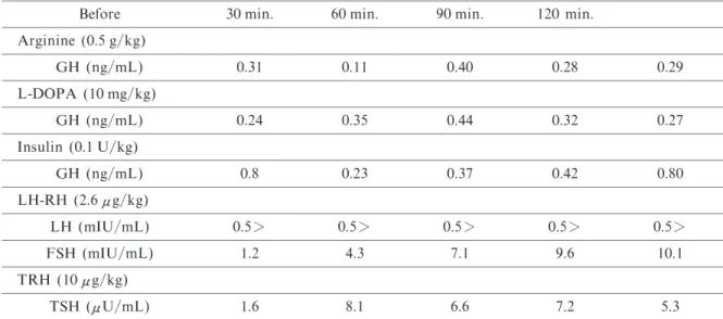
low,at 35 ng/mL(median value for Japanese 6- year-old females:147 ng/mL). A growth hor- mone(GH)secretion loading test indicated a marked secretion deficit (Table). While a thyrotropin-releasing hormone loading test showed a slightly insufficient response of thy- roid-stimulating hormone,it did not indicate pituitary dysfunction.Furthermore,responses of the luteinizing and follicle-stimulating hormones on a luteinizing hormone release hormone load- ing test showed no obvious anomalies for a prepubertal child.However,head MRI showed pituitary atrophy(Fig.4).We started the admin- istration of GH (0.16 mg/kg/week)when she was 6 years and 3 months old and 100.8 cm tall (-2.9 SD),which led to improvement in height development during the following years.When her height was 144.2 cm(-0.2 SD)at the age of 11 years and 3 months,the treatment was completed (Fig.5).Her first menstruation started at the age of 11 years and 9 months,and the development of secondary sexual characteristics and psychomotor development were normal. She attended regular classes at an elementary school.
Discussion
Although we were unable to establish a defi- nite pathological diagnosis for the intracranial lesion in this case,findings based on head images were consistent with cavernous hemangioma.
The differential diagnosis of vascular lesions
Table Results of hormone stimulation tests
Before 30 min. 60 min. 90 min. 120 min.
Arginine(0.5 g/kg)
GH (ng/mL) 0.31 0.11 0.40 0.28 0.29 L-DOPA (10 mg/kg)
GH (ng/mL) 0.24 0.35 0.44 0.32 0.27 Insulin(0.1 U/kg)
GH (ng/mL) 0.8 0.23 0.37 0.42 0.80 LH-RH (2.6μg/kg)
LH (mIU/mL) 0.5> 0.5> 0.5> 0.5> 0.5> FSH (mIU/mL) 1.2 4.3 7.1 9.6 10.1 TRH (10μg/kg)
TSH (μU/mL) 1.6 8.1 6.6 7.2 5.3 GH:growth hormone,L-DOPA:levodopa,LH-RH:luteinizing hormone releasing hormone,LH:luteiniz-
ing hormone,FSH:follicle-stimulating hormone,TRH:thyrotropin releasing hormone,TSH:thyroid stimu- lating hormone
involving the neuraxis in infancy includes hemangioma,hemangioblastoma,vascular mal- formation,and other neonatal tumors,such as soft-tissue sarcoma. The presence of radiologically indistinguishable extracranial tumor and concordant changes in size of the extracranial and intracranial lesions should sug- gest neuraxial hemangioma.웍That the contrast effect presented by the intracranial tumor was similar to that of the subcutaneous mass in the right posterior cervical region suggests that the intracranial tumor in this case was a cavernous hemangioma as was the case with the cutaneous
lesion. On the other hand,just because the intracranial tumor and the cervical mass reduced in size concordantly with each other does not necessarily mean that not only the cervical mass but the intracranial lesion also was cavernous in nature,considering that cryocoagulation had been performed on the cervical mass.However, since intracranial hemangiomas are often ac- companied by cutaneous lesions as in this case, and cases without cutaneous lesions have rarely been reported,웎the intracranial lesion in this case was considered most likely to be a cavern- ous hemangioma.Cavernous hemangiomas are histologically identical to what is categorized as infantile hemangiomas.웍Spontaneous regression of extracranial cavernous hemangiomas during childhood is common,while congenital intra- cranial cavernous hemangiomas can exhibit a progressive reduction in size after birth.웏In the case presented here,however,immediate treat- ment was considered necessary because the tumor was pressing on the optic nerve,indicating a marked likelihood of the manifestation of other symptoms.The use of steroids and interfer- on is reportedly effective for the treatment of cavernous hemangiomas.원웦웑Moreover,transcath- eter coil embolization has been used in some cases of neonatal hepatic cavernous heman- giomas with favorable results.웒While surgical excision is considered to be the most reliable therapy for intracranial cavernous hemangiomas in particular,워stereotactic radiosurgery for the management of cerebral cavernous malforma- tions remains controversial.웓 Taking into account the general physical condition of our patient,including her constitution,it was deter- mined that any surgical or invasive therapies would be too risky,and so we opted for high- dose steroid treatment,as described in the report by Sadan et al.웋월 This treatment resulted,as expected,in the shrinkage of the tumor without any major complications.Although we did not foresee any pituitary disorders after the heman- gioma treatment,we nevertheless periodically tracked and monitored our patientʼs growth and development because her weight at birth was very low.As a result of these follow-ups,she was found to have growth hormone deficiency ac- companied by a short stature at the age of six.
While this type of deficiency is almost always of idiopathic origin,there have also been cases with other causes,including organic,hereditary, and congenital.One of the organic causes of GH
Fig.5 Comparison of a standard growth chart for girlsʼ stature with the patientʼs growth chart(●).Period of growth hormone administration is indicated by (←GH→).
Fig.4 Sagittal T1-weighted head MRI,showing pituitary atrophy(arrow).
deficiency accompanied by a short stature is pituitary stalk transection associated with per- inatal abnormalities such as breech delivery and neonatal asphyxia,웋웋and,in recent years,cases of GH deficiency after traumatic brain injuries have also been receiving marked attention.웋워
GH deficiency in our case was thought to have been caused by pituitary atrophy. Although morphological variation of the pituitary gland is not necessarily accompanied by functional dis- orders,when a circulatory disorder due to some external factor causes degeneration or necrosis of the anterior pituitary,its function can be impair- ed depending on the severity of the degeneration or necrosis.웋웍As mentioned above,some cases of pituitary atrophy are known to occur in associa- tion with pituitary stalk transection due to per- inatal disorders,such as abnormal delivery or neonatal asphyxia,웋워ischemia,웋웎and cranial radi- ation.웋웏 Our patient was not born by breech delivery,but given that she was a very low birth weight infant with mild neonatal asphyxia,the possibility remains that the pituitary atrophy was indeed a result of perinatal abnormality.On the other hand,the incidence rate of GH defi- ciency is not particularly high in preterm infants, and our patientʼs perinatal period followed a course typical for a very low birth weight infant except for the onset of the hemangioma and the subsequent high-dose steroid therapy.The fact that the hemangioma was found to have invaded the sella turcica suggests that this was the cause of the pituitary atrophy.While it is difficult to establish at what degree of tumor invasion destruction of the pituitary gland occurs based on images,the pituitary gland can become flattened even by spinal fluid infiltrating the sella turcica,which,in turn,causes endocrine disruption.웋원Moreover,a suprasellar tumor can cause irreversible pituitary dysfunction.웋웑 A diagnosis of GH deficiency accompanied by a short stature is typically established after the patient has reached the age of three to four,when GH is thought to exert its greatest effect on height growth,and yet it becomes evident that the increase in height has slowed down.It can thus be assumed that our patientʼs growth had been sustained until she turned 3 or 4 by factors other than GH,such as nutrition and thyroid hormones.This is consistent with our supposi- tion that the tumorous compression impaired her pituitary gland in an irreversible fashion during early infancy.Although a high dose of steroids
was administered,no reports,as far as we can determine,have associated steroid administra- tion with pituitary atrophy or a manifestation of GH deficiency after a long period of time.Also, while the incidence of a short stature in small for gestational age infants has been drawing much attention in recent years,웋웒it is not likely the case here since our patient was an infant with an appropriate stature for her gestational age. Moreover,a short stature in small for gestational age infants is usually not accompanied by GH deficiency.
Hypopituitarism after traumatic brain injuries, which has also attracted attention in recent years, is considered to be caused by various factors including mechanical damage to the hypoth- alamic-pituitary system,웋웓and this kind of hypopituitarism can progress with age.워월 If a similar type of mechanical damage had been involved in the GH deficiency in our case,vari- ous symptoms may therefore occur in the future, necessitating life-long follow-up not only of her GH secretory state but of her overall pituitary function as well.
The authors declare no financial conflict of interest.
References
1.Ng BH,et al.(2006)Familial cerebral cavernous haemangioma diagnosed in an infant with a rapidly growing cerebral lesion. Australas Radiol 50:583‑ 590
2.Azam M,et al.(2009)Intracranial cavernous heman- gioma and seizures in a newborn infant.J Pediatr 155:
298‑298
3.Viswanathan V,et al.(2009)Infantile hemangiomas involving the neuraxis:clinical and imaging findings. Am J Neuroradiol 30:1005‑1013
4.Jalloh I,et al.(2014)Giant intracranial hemangioma in a neonate.Acta Neurochir 156:1151‑1154 5.Hayashi S,et al.(2004)Congenital cavernous an-
gioma exhibits a progressive decrease in size after birth.
Childs Nerv Syst 20:199‑203
6.Dourmishev LA (2005) Craniofacial cavernous hemangioma:successful treatment with methylpred- nisolone.Acta Dermatovenerol Alp Pannonica Adriat 14:49‑52
7.Castello MA,et al(1997)Successful management with interferon alpha-2a after prednisone therapy fail- ure in an infant with a giant cavernous hemangioma.
Med Pediatr Oncol 28:213‑215
8.Kretschmar O,et al.(2008)Interventional treatment of a symptomatic neonatal hepatic cavernous heman- gioma using the Amplatzer vascular plug.Cardiovasc Intervent Radiol 31:411‑414
9.Nagy G,et al.(2013)Stereotactic radiosurgery of
intracranial cavernous malformations.Neurosurg Clin N Am 24:575‑589
10.Sadan N,et al.(1996)Treatment of hemangiomas of infants with high doses of prednisone.J Pediatr 128:
141‑146
11.Ioachimescu A.G.et al.(2012)The pituitary stalk transection syndrome:multifaceted presentation in adulthood.Pituitary 15:405‑411
12.Fernandez-Rodriguez E,et al.(2015)Hypopituitar- ism after Traumatic Brain Injury. Endocrinol Metab Clin North Am 44:151‑159
13.Kikuchi K (1989)Pituitary function and diagnostic imaging(magnetic resonance imaging). Journal of Pediatric Practice 53:1491‑1496
14.Lim TH,et al.(1986)Pituitary atrophy in Korean (epidemic)hemorrhagic fever:CT correlation with pituitary function and visual field.Am J Neuroradiol 7:633‑637
15.Darzy KH (2013)Radiation-induced hypopituitar-
ism. Curr Opin Endocrinol Diabetes Obes 20:342‑ 353
16.Rath D,et al.(2015)Empty sella syndrome in a male child with failure to thrive.J Pediatr Neurosci 10:45
‑47
17.Arai A,et al.(2010)Xanthogranuloma of the Sellar Region:Case Report. Neurologia medico-chirurgica 50:488‑491
18.Itabashi K,et al.(2007)Longitudinal follow-up of height up to five years of age in infants born preterm small for gestational age:comparison to full-term small for gestational age infants.Early Hum Dev 83:
327‑333
19.Richmond E,et al(2014)Traumatic brain injury:
endocrine consequences in children and adults.Endo- crine.45:3‑8
20.Pekic S, et al. (2014) Alternative causes of hypopituitarism:traumatic brain injury,cranial irra- diation,and infections.Handb Clin Neurol.124:271
‑290