九州大学学術情報リポジトリ
Kyushu University Institutional Repository
2株の海洋細菌のβ-1,3-キシラナーゼとβ-アガラー ゼに関する研究
青木, 恭彦
https://doi.org/10.11501/3054242
出版情報:Kyushu University, 1990, 農学博士, 論文博士 バージョン:
権利関係:
Studies on P-1,3-Xylanase and P-Agarase
from Two Strains of Marine Bacteria
Takahiko Aoki
1990
Chapter I Chapter ll
II -1.
CONTENTS
Introduction
Isolation of P-1,3-Xylanase-producing Bacteria
Materials and Methods
II -1-1. Po 1 ysaccharides
II -1-2. Sources of {3 -1, 3-xy lanase
producing bacteria
II -1-3. Method of screening for {3 - 1,3-xylanase
producing bacteria
II -1-4. Assay of {3 -1 ,3-xylanase
IT -1-5. {3 -1,3 -Xylanase productivity of isolated bacteria
ll-1-6. Identification methods for AX-4 strain
IT -1-7. Examina ton for culture conditions of AX-4 strain ll-1-8. Preparation of P-1,3-xylanase from AX-4 strain ll-2. Results
ll-2-1. Screening of {3-1,3-xylanase-producing bacteria
IT-2-2. Identification of AX-4 strain
ll-2-3. Culture conditions for {3-1,3-xylanase production in AX-4 strain
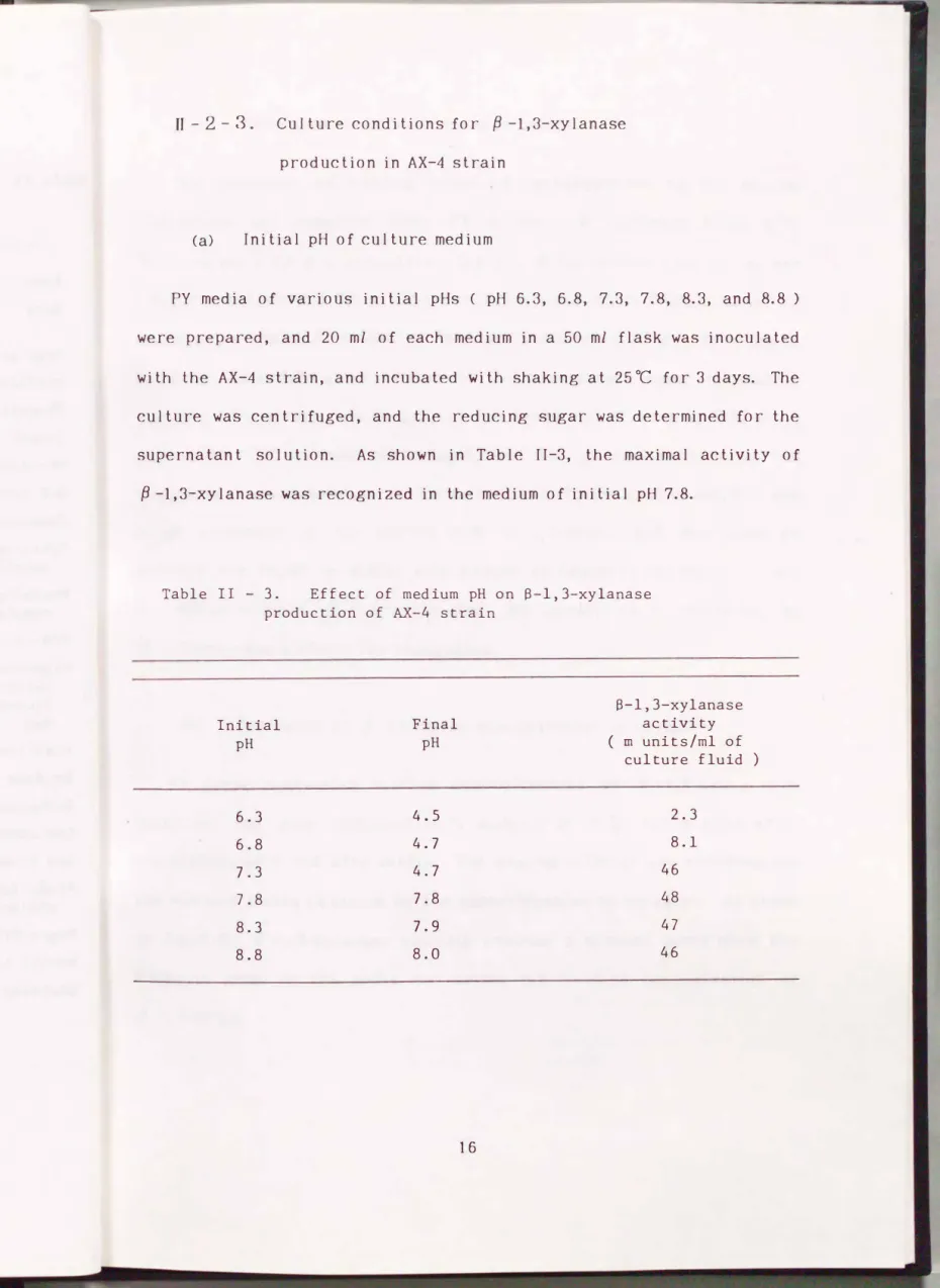
(a) Initial pH of culture medium
1
6
7
9
9 10
10 11 12 12
13 13
16
(c) Influence of
fJ
-1,3-xylan concentration medium(d) Composition of medium (e) Other examinations
ll-3. Discussion
ll-4. Summary
Chapter m Purification of
P
-1,3-Xylanase from Vibrio sp. AX-4ill-1. Materials and Methods ill -1-1. Materials
ill -1-2. Assay of enzymes ill-1-3. Analytical methods ill-2. Results
ill-2-1. Preparation of cui ture fluid ffi-2-2. Ammonium sulfate precipitation
ill-2-3. DEAE-Sepharose CL-6B chromatography ill-2-4. SP-Sephadex C-25 chromatography ill-2-5. Hydroxyapatite chromatography ill-2-6. Sephadex G-200 chromatography
ill-2-7.
fJ
-1,3-Xylan-affini ty chromatography ill-2-8. Homogeneity offJ
-1,3-xylanasein
17 19 19 19 22
23
24 24
24
25 25 26 27 27 27 29 31
ill- 3. Discussion ill-4. Summary
Chapter N Characterization of P-1,3-Xylanase from Vibrio sp. AX-4
N-1. Materials and Methods N-1-1. Substrates
IV-1-2. Thin layer chromatography of hydrolysis products IV-1-3. Assay of enzymes
IV-1-4. !so-electric focusing
IV-1-5. Molecular weight of P-1,3-xylanase IV-2. Results
IV-2-1. Action pattern of ,9-1,3-xylanase
(a) Action of ,9-1,3-xylanase on ,9-1,3-xylan
(b) Action of ,9-1,3-xylanase on xylooligosaccharides (c) Action of ,8-1,3-xylanase on PNP-glycosides
IV-2-2. Optimal pH of ,9-1,3-xylanase IV-2-3. Stability of ,9-1,3-xylanase N-2-4. Effect of temperature
IV-2-5. Inhibitors
IV-2-6. Isoelectric point
32 33
34
34 35 36 36 37
37 38 38 40 41 41 41 43
IV-2-7. Molecular weight of
{1-l
,3-xylanase IV-3. DiscussionIV-4. Summary
Chapter v I soIa tion of Porphyran-degrading Bacteria V-1. Materials and Methods
V-1-1. Po I ysaccharides
V-1-2. Sources of porphyran-degrading bacteria V-1-3. Media used for screening of bacteria v -1-4. Assay of porphyran-degrading enzyme
V -1-5. Porphyran-degrading enzyme productivity of isolated bacteria
V-1-6. Identification method for AP-2 strain
V -1-7. Examination for cui ture conditions of AP-2 strain
V -1-8. Preparation of porphyran-degrading enzyme from AP-2 strain
V-1-9. Thin layer chromatography of hydrolysis products
V-1-1
0.
Decomposing ratio of substrate V-2. ResultsV-2-1. Screening of porphyran-degrading hac teria V-2-2. Identification of AP-2 strain
44 45 45
47
49 50 50 51
52 52
53
53
54 54
55 56
V-2-3. Culture conditions for the production of porphyran
degrading enzyme in Vibrio sp. AP-2 (a) Influence of carbohydrates in medium (b) Initial pH of medium
(c) Culture temperature and period (d) Shaking of culture
V-2-4. Detection of hydrolysis products of porphyran
V-2-5. Decomposing ratio of substrate
V-2-6. Hydrolysis of various substrates by the enzyme from AP-2 strain
V-3. Discussion V-4. Summary
Chapter VI Purification of Agarase from Vibrio sp. AP-2 VI-1. Materials and Methods
VI-1-1. Materials
VI -1 -2. Assay of enzyme
VI -1-3. Po I yacrylamide disc ge I e lee tropho res is VI-2. Results
VI-2 -1. Preparation of cui ture fluid VI-2-2. Ammonium sulfate precipitation VI-2-3. CM-Sephadex C-50 chromatography
56 59 59 59 60
61
62 63 64
65
66 66 67
67 68 68
VI-2-4. Affinity chromatography
VI-2-5. To yo pearl HW-55 chromatography VI-2-6. DEAE-Toyopearl 650M chromatography VI-2-7. DNase I and RNase A t rea tmen t
VI-2-8. To yo pearl HW-55 chromatography VI-2-9. DEAE-Toyopearl 650M chromatography VI-2-1 0. Homogeneity of agarases
VI-3. Discussion VI-4. Summary
Chapter Vll Characterization of three Agarases from Vibrio sp. AP-2
VII-1. Materials and Methods VII- 1-1. Substrates
V1I -1-2. Molecular weight of three agarases
VII-2. Results
VII-2 - 1 . Action pat tern of agarases
(a) Action of agarases on agar and K -carrageenan
(b) Action of agarases on neoagarooligosaccharides
VII-2-2. Effect of pH on activity of agarases VII-2-3. Stability of agarases
VIT-2-4. Effect of temperature on activity of agarases
69
70
71
72
72
73
73
77
78
79
79
81
82
82
84
86
86
V1I-2-5. Molecular weight of agarases VJI-3. Discussion
VII-4. Summary
Chapter VIII Preparation and Regeneration of Protoplasts from Thalli of Seaweeds of Genus Porphyra Vlll-1. Materials and Methods
Wl-1-1. Vegetative thalli of seaweeds WI-1-2. Enzymes
VIII-1-3. Assay of enzymes
Wl-1-4. Preparation of pro top lasts Wl-1-5. Culture of protoplasts VIII-2. Results
Wl-2-1. Pre-treatment of seaweed thalli with papain
Vlll-2-2. Effects of temperature and pH on protoplast isolation
VIII-2-3. Effects of enzyme concentration and rea c t i o n time o n pro top I as t is o l a t i o n
VIII-2-4. Regeneration of protoplasts from P. yezoensis and P. tenera
vm- 3. Discussion
VIII-4. Summary
86 87 88
89
90 90 91 91 92
93
94
95
96 98 101
Chapter lX Summary Acknowledgements References
102 107 108
Chapter I Introduction
The cell fusion is one of the new biotechnologies, and has been developing markedly in recent years. By using the cell fusion, it is expected to produce new plant species in varieties having a good harvest yield, resistance to diseases, or to harsh climates. The most of works in the literature on the cell fusion, up to date, have been done with terrestrial plants1-4' but seaweeds.5-10'
It is necessary to release protoplasts from plant cell wall by enzymic degradation of the structural polysaccharides, for the study on the cell fusion. That is, the availability of the enzymes capable of degrading cell wall polysaccharides may be one of the most important factors to advance the study of cell fusion. The release of protoplasts from terrestrial plants is accomplished with cellulase and pectinase, because the main polysaccharides of the cell wall are cellulose and pectin. In seaweeds, however, the polysaccharides of the cell wall differ from those of terrestrial plants except chlorophyceae( cellulose), that is, those of rhodophyceae are {3 -1,3-xylan, {3 -1,4-mannan, and porphyran, and those of phaeophyceae are alginate and fucoidan.1u Lit tie work has been done on the seaweed polysaccharide-degrading enzymes, and in consequence the lack of knowledge on the enzymes may lead to a delay in the study of cell
fusion for seaweeds.
On the other hand, various o ligosaccharides with physio I ogical activity have been obtained from indigestible polysaccharides of terrestrial plants in recent years.12•13' Since most of polysaccharides
of seaweeds are indigestible, they are expected to be promising materials for the oligosaccharides with physiological activity. Recently, an oligosaccharide from alginate has been reported to be effective on the human body.14> Therefore, seaweed polysaccharides -degrading enzymes also may be beneficial to the preparation of the active o l igosaccharides.
The author noticed the enzymes capable of degrading cell wall polysaccharides of the seaweeds of the genus Porphyra, which were cui tivated abundantly and used as food stuff ( nori ) in Japan. Main polysaccharides of the cell wall in the seaweeds are {3 -1,3-xylan, porphyran, and {3-1,4-mannan.15•16> In this study, firstly, an attempt was made to obtain the three polysaccharide-degrading enzymes before the preparation of protoplasts and oligosaccharides.
{3-1,3-Xylan-degrading enzyme, {3-1,3-xylanase, has been reported to be produced by several strains of bacteria from marine environments ( e.g. sea water, bottom mud of coastal sea, and green, brown and red algae ).17> In the report of Fujisawa et al.18>, of the 64 strains of {3-1,3-xylan-degrading marine bacteria (unidentified) examined, at least 40 strains possessed {3-1,3-xylanase activity, and 36 strains were capable of hydrolyzing {3-1,4-xylan. {3 -1,3-Xylan-degrading enzymes also have been found in terrestrial fungi, Aspergillus batatae, Chaetomium globosum19>, and lrpex lacteus.20> These organisms produced the enzymes constitutively in addition to {3-1,4-xylanase when cultured on w he a t bran ( Asp. b a t a t a e and C h a. g l o b o sum ) , g l u co se ( C h a. g l o b o sum ) , and cellulose ( Irp. lacteus ).19> However, the {3 -1,3-xylanases from these organisms were not purified, and the properties were unknown yet.
Recently, Chen et al. purified six different {1-1,3-xylanases from a fungus Aspergillus terreus A-07, and characterized the enzymes. 2u All of the six enzymes were endo-type
f1
-1,3-xylanase ( EC 3.2.1.32 ).However, these enzymes are not expected to be used for the preparation of viable protoplasts from seaweeds because they hardly acted on the substrate in neutral condition ( the pH optima of these {1-1,3-xylanases were from 4.0 to 5.5 ). Exo-type {1-1,3-xylanase ( EC 3.2.1.72) was also reported by Fukui et al. in a fungus Chaetomium globosum19>, but the optimal pH of this enzyme was not reported ( the enzyme activty was determined at pH 3.5 in their study ). In 1990, another endo-type {1-1,3-xylanase was purified and characterized by Yamaura et al. from Pseudomonas sp. PT-5. 22> The purified enzyme had a pH optimum of 7.5, but the action pattern of the enzyme had not been fully investigated.
Porphyran-degrading enzyme ( {1-agarase, EC 3.2.1.81 ) has been found in several strains of bacteria isolated from marine environments, such as Cytophaga sp.23> and Pseudomonas atlantica.24"25> The enzymes from these bacteria hydrolyzed agar and porphyran to give neoagarotetraose as the predominant product. However, the enzymes degraded only a defined part of porphyran structure and exhibited a low hydrolyzing activity against porphyran as compared with agar. In the case of the {1-agarase from P. atlantica, decomposing ratio of porphyran was only 23% after exhaustive digestion.24> An extracellular {1-agarase ( {1- agarase I ) from P. atlantica was purified by Morrice et al., and the enzymatic property was investigated. 24"25> The purified {1-agarase I had a pH optimum of 7.0, and hydrolyzed agar to give neoagarotetraose as
the predominant product. The existence of another type of
intracellular agarase ( {:3-neoagarotetraose hydrolase26> or {:3-agarase ll, EC 3.2.1.- ) from P. atlantica was suggested by Morrice et a/.24•25>
The {:3 -agarase I1 was likely to be an endo-type enzyme the same as {:3 -agarase I , and hydrolyzed neoagarotetraose and agar to give neoagarobiose as the predominant product. However, the property of
{:3-agarase IJ has not been fully investigated.
As regards {:3 -1,4-mannanase, the isolation of bacteria with high productivity of the enzyme from natural habitats, and the purification and characterization of the enzymes from Vibrio sp. F-25 have been reported by Araki and Ki tamikado27> in the Laboratory of Fisheries Technology, Department of Fisheries, Faculty of Agriculture, Kyushu University. The author, therefore, prepared the endo- {:3 -1,4-mannanase from the bacterial strain in this study.
Little has been reported on the search for microorganisms which produce the potent {:3 -1,3-xylanase and agarase as mentioned above.
Therefore, the author focused the object of this study on the isolation of bacteria with high productivity of the two enzymes, and on the examination of properties of the enzymes.
This dissertation describes the above states divided into seven chapters. Chapter ll describes the isolation of {:3-1,3-xylanase-producing bacteria from natural habitats, and the identification of the bacterium with high productivity of the enzyme < AX-4 strain ). Chapters ill and N describe the purification of {:3 -1,3-xylanase from the AX-4 strain, and the characterization of the purified enzyme. Chapter V describes the
isolation of porphyran-degrading bacteria, and the identification of the bacterium with high productivity of porphyran-degrading enzyme ( AP-2
strain ). Chapters VI and VJI describe the puri fica tio n of
{3
-agarasesfrom the AP-2 strain, and the characterization of the enzymes. Chapter WI describes the use of
{3
-1,3-xylanase and agarases for the preparation of protoplasts from the thalli of Porphyra yezoensis var. narawaensis and green type of P. tenera var. tamatsuensis.Chapter ll Isolation of fJ -1,3-Xylanase-producing Bacteria
The main polysaccharides in the cell wall of seaweeds of the genus Porphyra are fJ -1,3-xylan, fJ -1,4-mannan, and porphyran.12•13) In these polysaccharides, fJ -1,3-xylan is a homo-polysaccharide which consists of D-xylopyranoses united to each other by fJ -1,3-linkage like a chain ( Fig. II-1 ). fJ -1,3-Xylan found in seaweeds differs from fJ -1,4-xylan which is mainly found in terrestrial plants. fJ -1,3-Xylan is also found in the cell wall from other groups of seaweeds, such as genera Caulerpa28) and Halimeda11) of chlorophyceae.
The fJ -1,3-xylan-hydrolyzing enzyme, fJ -1,3-xylanase, has been reported to be produced by several strains of bacteria from marine environmen ts17\ but no further biochemical studies on the purified bacterial enzyme were made until 1988. The enzyme has also been found from fungi.19"20) There have been some reports on the properties of f3 -1,3-xylanases from fungi, but the pH optima of these xylanases were from 4.0 to 5.5.21) They are not adequate to prepare protoplasts from seaweeds, because these enzymes hardly hydrolyzed fJ -1,3-xylan in a neutral condition. Therefore, the author searched for bacteria with a high productivity of fJ -1,3-xylanase which can degrade the substrate in a nearly neutral condition.
This chapter describes the isolation, culture conditions, and identification of a bacterium which is able to produce a large amount of
f3 -1,3-xylanase.
II- 1. Materials and Methods
ll- 1 - 1. Po I ysaccharides
{3
-1,3-Xylan was prepared by the method of Iriki et al.29) from a green alga Caulerpa racemosa collected from the coastal sea in Kagoshima Prefecture. Thalli of the seaweed were dried, and powdered with a millmesh 80 ). The powder was suspended in deionized water, and sufficiently washed with water. The suspension was filtered through a cloth sack. The residue obtained was treated with 1% HCl at room temperature for 45 min, and then washed as above mentioned. The residue
was treated successively with 1.25% NaOH and 1.25% H2S04 at 100 ac under N2 gas for 1 h. The residue was filtered through a cloth sack, suspended
again in water, and then bleached with 1% sodium chlorite. The bleached residue was thoroughly washed with water. The crude fibre obtained was extracted with 10% NaCl at room temperature. To the extract was added 2 volumes of ethanol, and the mixture was centrifuged at 6,000 r.p.m. for 20 min, and then the precipitate was washed with ethanol containing 1% HCl followed by ethanol, and ether, and dried in vacuo. Finally, 8.8 g of the white powder of
{3
-1,3-xylan was obtained from 100 g of the air-dried powder of the seaweed.Structural saccharide of the
{3
-1,3-xylan preparation was examined.The white powder , 0.1 g , was put in to a test tube, and 2 ml of trifluoroacetic acid was added. The tube was sealed by heating, and the
powder was hydrolyzed at 100 oc for 3 h. After cooling, the hydrolyzed solution was dried in vacuo. The hydrolyzates were analyzed by thin layer chromatography ( see IV-1-2 ). As the result, D-xylose was recognized on the chromatogram together with a trace amount of D-glucose, showing the high quality of the {3 -1,3-xylan preparation.
{3 -1,4-Mannan was prepared by the method of Love and Percival30>
from a green alga Codium fragile collected from the coastal sea in Fukuoka Prefecture. Porphyran was prepared by the method of Su and Hassid15> from the dry powder of Porphyra yezoensis (see V-1-1 ).
( 1
s 1,3 s 1,3
xylose xylose xylose
l J
n�0 � 0 £[1 °�0�H,OH
0 � l OH
OHH
nOh-L-( OH
IFig.II - 1. Structure of S-1,3-xylan.
II -1-2. Sources of
{3
-1,3-xylanase-producing bacteriaSeventy-eight samples, collected during 1985 in Fukuoka Prefecture, were examined. Those of marine origin included: 4 from coastal sea water, 21 from the bottom mud of the coastal sea, 22 from algae (green, brown, and red ), 5 from gills of marine fish, and 6 from intestinal contents of marine fish. Those from land and freshwater origins included: 10 from the soil, 4 from river water, and 6 from pond water.
ll -1-3. Method of screening for
{3
-1,3-xylanaseproducing bacteria
The medium for bacteria of marine origin was PY medium composed of 0.5% peptone trade name, polypeptone, Daigo Eiyokagaku Co. ), 0.1%
yeast extract ( Difco Lab., U.S.A. ), and 3% NaCl, pH 7.6 - 7.8. The author used a nutrient broth ( Difco Lab., U.S.A. ), pH 7.0 - 7.2, for those of freshwater and land soil origins. These media were used after adding suitable amounts of
{3
-1,3-xylan and solidifying with 1.5% agar, where required. Agar medium containing 1.0%{3
-1,3-xylan was whitish turbid because of the insolubility of{3
-1,3-xylan in water.Before screening, each solid sample was blended with a small volume of either sterilized 3% NaCl solution ( for samples of marine origin ) or 0.9% NaCl solution ( for those of land and freshwater origins). A small
quantity of the sample was streaked on the surface of agar medium containing 1.0% f3 -1,3-xylan in a petri dish, and the seeded medium was incubated aerobically at 25 oc for 4 - 7 days. Formation of the clear zone around the colony indicated the colony to be a
{3
-1,3-xylanase-producer. The f3 -1,3-xylanase-producing bacteria were then purified by repeated single-colony isolations. Finally, isolated
{3
-1,3-xylanase-producing bacteria were inoculated on agar slant media containing 0.3% f3 -1,3-xylan, stored in a refrigerator, and transferred monthly in homologous agar media to maintain viability.II-1 -4. Assay of f3 -1,3-xylanase
Enzyme solution, 0.5 ml, was added to 2.0 ml of 50 mM sodium acetate buffer, pH 6.0, containing 0.5% f3 -1,3-xylan and 2 mM CaCl2. After incubation at 37oC for an appropriate period, the reaction was halted by heating the mixture in a boiling water bath for 5 min. The reducing sugar produced was then determined by Somogyi-Nelson's method31' and expressed as D-xylose. One unit of the enzyme was defined as the activity which liberated 1 �mol of D-xylose per min, under the above condition.
II-1-5. f3 -1 ,3-Xylanase produc ti vi ty of isola ted hac teria
Cells taken from agar slant culture of each isolate were transferred
to 20 ml of sterile medium in a 100 ml flask. The preparation was then incubated with shaking by a reciprocal shaker ( 8 em amplitude, 90 strokes/min ) at 25 oc for 4 days. The medium used was composed of 0.1%
peptone, 0.1% yeast extract, 3% NaCl, 0.2% K2HP04, 0.05% KH2P04, 0.05%
MgS04·'iH20, and 0.3% f3 -1,3-xylan, pH 7.8 ( see II-2-3-(d) ). Each preparation was centrifuged, and the supernatant obtained was assayed for
{3
-1,3-xylanase activity.IJ- 1 - 6. Identification methods for AX-4 strain
Identification of the AX-4 strain was made according to the criteria given in Bergey's Manual of Systematic Bacteriology, Vol.l.32) The medium used was PY medium, and the incubation temperature was 25 oc except for special cases. Motility, morphology, and Gram-staining characteristics were determined by light microscopy. The Gram-staining was carried out by the modification of Hucker's method.33) Oxidase test was made by the method of Kovacs. 34) 0-F test was by the Hugh-Leifson' s method35), and the formation of acid from D-glucose was determined by color-transition of BTB, and the formation of gas was observed using Durham tube. The arginine test was performed by the method of Thorn ley. 36) The mol% G + C of the DNA range was measured by the method of Marmur37"38\ with the DNA from Escherichia coli K12 used as a standard.
Other identification methods were used, where required.39-41)
IT- 1- 7. Examination for cui ture conditions of AX-4 strain
The following conditions were examined: (a) initial pH of culture
medium; (b) addition of
fJ
-1, 3-xylan,fJ
-1, 4-xylan, porphyran,fJ
-1, 4-mannan, and cellulose etc. into medium as the inducer offJ -
1, 3-xylanase; (c) concentration of
fJ
-1, 3-xylan in medium; (d) concentration of peptone in medium; (e) addition of inorganic salts, cui ture temperature and period, and shaking by a reciprocal shaker.IT -1-8. Preparation of
fJ
-1, 3-xylanase from AX-4 strainThe strain was inoculated into 20 ml of liquid medium described in the paragraph on
fJ
-1, 3-xylanase productivity of isolated bacteria ( see II-1-5 ), and incubated with shaking ( 8 em amplitude, 52 strokes/min ) at 25 OC for 3 days. The cui ture was transferred to 1,000 ml of the same liquid medium in a 3 liter flask and incubated with shaking at the same temperature for 4 days. The culture obtained was centrifuged, and the supernatant fluid was subjected to the following precipitation Procedure. A II the f o II owing opera t i o n s were co n d u c ted a t 4 oc . So I i d ammonium sulfate was added to the supernatant fluid up to a final concentration of 75% saturation for ammonium sulfate precipitation. The mixture was kept overnight and centrifuged. The precipitate formed was dissolved in a small volume of 20 mM sodium acetate buffer, pH 6.0,containing 2 mM CaCl2, and the solution was poured into a cellulose tube and dialyzed overnight against several changes of the same buffer. The liquid inside the tube was used as
{3
-1,3-xylanase solution ( crude enzyme ).IT- 2. Results
II- 2- 1. Screening of
{3
-1,3-xylanase-producing bacteriaFour colonies formed a clear zone on the plate media, thereby showing the ability to decompose
{3
-1,3-xylan; two from the bottom mud of the coastal sea, and one each from a seaweed and from the intestinal contents of a marine fish ( Table II-1 ). A bacteriumoriginated from the bot tom mud exhibited the highest
( AX-4 strain
) {3
-1, 3-xylanase productivity, exhibiting 48 m units per ml after 4-day incubation. The enzyme productivities from all the other isolates were within the range of 7.9 to 32 m units per ml. Therefore, the AX-4 strain was used for the following experiments.IT- 2- 2. Identification of AX-4 strain
The morphological and biochemical characteristics of the AX-4 strain
Table II - 1. Sources of a-1,3-xylanase-producing bacteria
Places Samples Number of Number of
samples isolates
Sea water 4 0
Sea bottom mud 21 2
Coastal Sea weed 22 1
sea
Marine fish
gills 5 0
intestine 6 1
( Total 58 4
River water 4 0
River and
Pond water 6 0
pond
( Total 10 0
Land soil 10 0
Land
( Total 10 0
are shown in Table II-2. The bacterium was a Gram-negative and facultatively anaerobic rod with a single polar flagellum, it did not form endo-spores, formed acid but no gas from glucose, and was sensitive to 2,4-diamino-6,7-diisopropyl pteridine. Therefore, it was assigned to the genus Vibrio, according to the criteria given in Bergey's Manual of Systematic Bacteriology. All species of the genus listed in the Manual, however, differed from the AX-4 strain in some characteristics. The species of the AX-4 strain was not determined.
Table II - 2. MorpholoBical, cultural, and biochemical characteristics of AX-4 strain
Form Size
Gram stain Motility Flagellation Growth in air Unaerobic growth 0-F test
Catalase
Vibriostatic agent sensitivity Swarming on solid
complex media PHB-accumulation Pigmentation
Yellow-orange Blue-black
Red
Arginine dihydrolase Oxidase
Reduction of N03 to N02 Luminescence
Gas from D-glucose Production of acetoin
and/or diacetyl
rod 0 . 7-0 . 8 ).lffi X
1. 2-3. 2 .urn
Negative +
Single polar +
+
Fermentation +
+
+ + +
Voges-Proskauer test(24 h)
Methyl red test +
Thornley arginine +
Na + required for or-
ganic growth factors Growth at:
4°C 30°C 35°C 40°C
Production of:
Amylase Gelatinase Lipase Alginase Chitinase
Utilization of:
D-Xylose L-Arabinose D-Mannose D-Galactose Sucrose Trehalose Cellobiose Lactose Salicin Citrate D-Mannitol D-Sorbitol meso-Inositol Ethanol
L-a-Alanine
�-Alanine L-Serine L-Leucine L-Histidine L-Proline L-Tyrosine Mol% G + C of DNA
+
+ +
+ + + +
+ + + + + + +
+ + +
± + +
+ + + 46
IT- 2- 3. Cui ture conditions for
{3
-1,3-xylanase production in AX-4 strain(a) Initial pH of culture medium
PY media of various initial pHs ( pH 6.3, 6.8, 7.3, 7.8, 8.3, and 8.8) were prepared, and 20 ml of each medium in a 50 ml flask was inoculated with the AX-4 strain, and incubated with shaking at 25oC for 3 days. The cui ture was centrifuged, and the reducing sugar was determined for the supernatant solution. As shown in Table II-3, the maximal activity of
{3
-1,3-xylanase was recognized in the medium of initial pH 7.8.Table II - 3. Effect of medium pH on S-1,3-xylanase production of AX-4 strain
S-1,3-xylanase
Initial Final activity
pH pH ( m units/ml of
culture fluid
6.3 4.5 2.3
6.8 4.7 8.1
7.3 4.7 46
7.8 7.8 48
8.3 7.9 47
8.8 8.0 46
)
(b) Influence of carbohydrates in medium
The influence of various kinds of carbohydrates on the enzyme production was measured using PY medium.
f1
-1,3-Xylan ( 0.1 g ),{1
-1,4-xylan ( 0.1 g), porphyran ( 0.1 g),{1
-1,4-mannan ( 0.1 g), xylose ( 0.1 g), cellulose ( 0.1 g), powder( 0.2 g) and crude fibre C 0.2 g) of Caulerpa racemosa, or powder of Porphyra yezoensis ( 0.2 g) was added to 20 ml of the medium as an inducer off1
-1,3-xylanase. After inoculation with the AX-4 strain, the medium was in cuba ted with shaking at 25 oc for 4 days. The culture was centrifuged, and the enzyme activity of the supernatant was measured. As shown in Fig. II-2, the enzyme activity was found evidently in the medium withf1
-1,3-xylan, and the trace of activity was found in medium with powder of Caul erpa racemosa. In all the media withoutf1
-1,3-xylan and the powder of C. racemosa, no{1
-1,3-xylanase activity was recognized.(c) Influence of
{1
-1,3-xylan concentration in mediumPY media containing various concentrations of
f1
-1,3-xylan were prepared, and then incubated with shaking at 25 oc for 4 days after inoculation with the AX-4 strain. The enzyme activity was measured for the cui ture fluids obtained by the centrifugation of cultures. As shown in Fig.II-3,f1
-1,3-xylanase activity reached a maximal level when the organism grew in the media containing 0.2 to 0.5% concentration offJ
-1,3-xylan.Carbohydrate 0 None
Crude fibre from
]
C.racemosa
B-1,3-xylan Powder of C.racemosa
Powder of f.yezoensis
Xylose (3-1,4-xylan
B-1,3-xylanase activity ( m units/ ml of culture fluid )
15 30
I I
I I
Fig.II - 2. Induction of B-1,3-xylanase in AX-4 strain by different carbohydrates.
PY medium containing 0.5% of different carbohydrate was used as culture medium.
'"d :>-,:;:::) ·M +J...--1
·M �
>
·M aJ +-J H (.) :;:::) C'd +-J ....--1 Q) :;:::) Ul (.) C'd ��
C'd 0 ....--1
:>-,....-1 X 8
�'-..
.... Ul ...--I.U I ·M co.c :;:::) s
10
1.0 Concentration of B-1,3-xylan ( % ) Fig.II - 3. Influence of B-1,3-xylan concentration on
B-1,3-xylanase production of AX-4 strain.
(d) Composition of medium
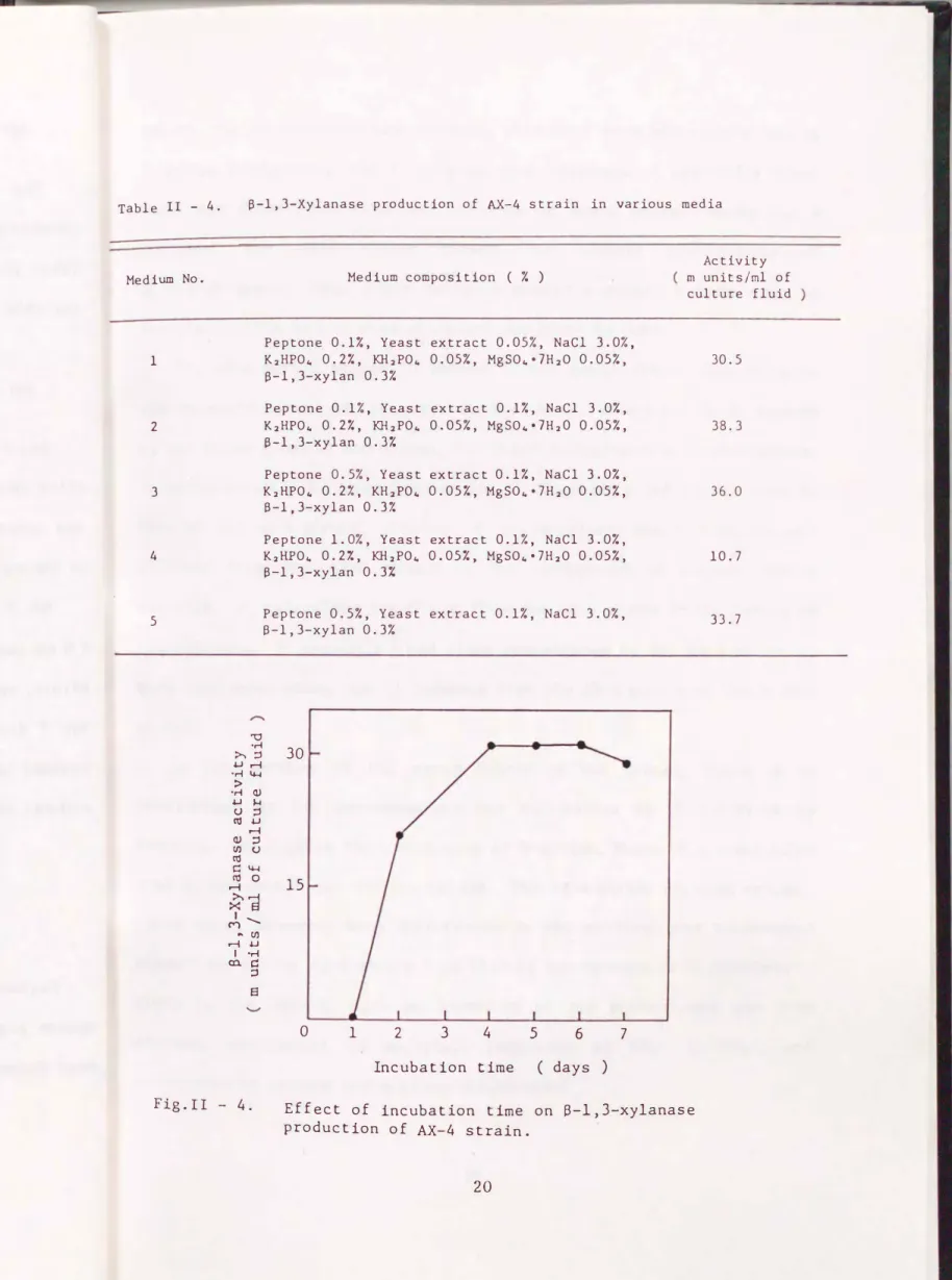
The enzyme activity of the culture fluids was measured after incubation with shaking at 25 oc for 3 days by using the media as shown in Table II-4. As the result of this experiment, media No. 2 and No. 3 were suitable for the {3 -1,3-xylanase production of the AX-4 strain.
(e) Other examinations
No. 2 media were incubated with shaking at 15, 20, and 25 oc for 3 days after inoculation with the AX-4 strain, respectively. When the organism was incubated at 25 oc, {3 -1,3-xylanase activity reached a maximal level in the cui ture fluid.
No. 2 medium, 1,000 ml, in a 3 liter flask was incubated with shaking ( 8 em amp I i tude, 52 strokes/min ) at 25 oc after inoculation with the AX-4
strain, and the enzyme activity of the culture fluid was measured daily for 7 days. As shown in Fig. II-4, {3 -1,3-xylanase activity reached a maximal level after 4 - 6 days. When the organism was incubated at 25 oc without shaking, no enzyme activity was found in the culture fluid.
ll- 3. Discussion
Fujisawa and Murakami17"18> reported the wide distribution and the sparse population of {3 -1,3-xylan-decomposing bacteria in the sea area near Shimonoseki. In this study, such bacteria were searched for in sea
Table II - 4.
Medium No.
1
2
3
4
5
S-1,3-Xylanase production of AX-4 strain in various media
""0
Medium composition ( % )
Peptone 0.1%, Yeast extract 0.05%, NaCl 3.0%, K2HP04 0.27., KH2P04 0.05%, MgS04•lH20 0.05%, S-1,3-xylan 0.3%
Peptone 0.1%, Yeast extract 0.1%, NaCl 3.0%, K2HP04 0.27., KH2P04 0.05%, MgS04•lH20 0.05%, B-1,3-xylan 0.37.
Peptone 0.5%, Yeast extract 0.1%, NaCl 3.0%, K2HP04 0.27., KH2P04 0.057., MgS04•7H20 0.057.,
13-1,3-xylan 0.37.
Peptone 1.0%, Yeast extract 0.1%, NaCl 3.0%, K2HP04 0.27., KH2P04 0.057., MgS04•7H20 0.05%, B-1,3-xylan 0.3:4
Peptone 0.5%, Yeast extract 0.1%, NaCl 3.0%, 13-1,3-xylan 0.3%
>,
·�
30·r1 IH Uri
:>
·r1 <l)
U H
(.) ;::j
ell u
<l) rl =' (J) (.) ell CIH
�
0 15�-- ��
... (J)rlW 1 -M co.t::
;::j El
0 1 2 3 4 5 6 7
Incubation time ( days )
Activity rn units/rnl of culture fluid
30.5
38.3
36.0
10.7
33.7
Fig.II - 4. Effect of incubation time on S-1,3-xylanase production of AX-4 strain.
water, sea bottom mud, and seaweeds collected from the coastal sea in Fukuoka Prefecture, and 4 isolates were obtained. A distinctly clear zone was detected around the colonies on plate media. Among the 4
isolates, the AX-4 strain showed the highest productivity of
{3
-1,3-xylanase. Some other bacteria showed a doubtful clear zone on the plate media, but no more attention was given to them.The AX-4 strain seemed to belong to the genus Vibrio, according to the Bergey's Manual of Systematic Bacteriology. Among all the 20 species of the genus given in the Manual, the characteristics of V. proteolyticus, V. angui ll arum I , V. spl endidus I , and V. flu via lis I had resemblance to that of the AX-4 strain. However, V. proteolyticus and V. anguillarum I differed from the AX-4 strain in the production of acetoin and/or diacetyl. V. splendidus differed from the AX-4 strain in the having of luminescence. V. fluvialis I had close resemblance to the AX-4 strain on many characteristics, but it differed from the AX-4 strain in the growth at 40°C.
I n the section of the genus Vibrio in the Manual, there is no description on the decomposition and utilization of
{3
-1,3-xylan by bacteria. As regards the utilization of D-xylose, there is a description that V. gazogenes can utilize xylose. The AX-4 strain utilized xylose.There were, however, many differences in the cultural and biochemical properties of the AX-4 strain from that of the species of V. gazogenes given in the Manual, such as formation of red pigment and gas from glucose, utilization of sorbitol, reduction of N03- to N02-, and production of oxidase and arginine dihydrolase.
ll- 4. Summary
A screening was undertaken to acquire bacteria with a high rate of
{3
-1,3-xylanase productivity. Such a strain was obtained from the bottom mud of the coastal sea in Fukuoka Prefecture. The organism neededf1
-1,3-xylan as an inducer to producef1
-1,3-xylanase, and released a large amount of the enzyme into the culture fluid, when incubated with shaking a t 2 5 oc f o r 4 to 6 days i n the f o ll owing I i quid me d i u m: 0. 1% o r 0.5% peptone, 0.1% yeast extract, 3.0% NaCl, 0.2% K2HP04, 0.05% KH2P04, 0.05% MgS04·?H20, and 0.3%f1
-1,3-xylan, pH 7.8. The bacterium was assigned to the genus Vibrio, according to the criteria given in the Bergey's Manual of Systematic Bacteriology, Vol.l. Species name was not determined for the organism, because some differences were recognized between the characteristic of the AX-4 strain and that of all the 20 species given in the Manual.Chapter ill Purification of
/l
-1,3-Xylanase from Vibrio sp. AX-4The AX-4 strain was assigned to the genus Vibrio, in the preceding chapter. Therefore, this strain is named Vibrio sp. AX-4 in the following chapters.
There have been reported purified
f3
-1,3-xylanases from Aspergillus terreus A-07 by Chen et al.21> In their study, six{3
-1,3-xylanases were purified by ammonium sui fate precipitation, successive column chromatography and isoelectric focusing. The specific activities of final enzyme preparations were 6.4 - 15.2 times more to the cui ture fluid, and the total recovery was 10.7%. These enzymes were not adequate for the preparation of protoplasts from seaweed, because they hardly acted on the substrate in neutral condition.This chapter describes the purification of the enzyme from Vibrio sp.
AX-4 by ammonium sulfate precipitation and successive column chromatography.
The properties determined with the crude enzyme preparation ( see II-1-8) were as follows : the enzyme was likely to exhibit a high activity in the range of pH 6.0 to 7.5, and was stable in a pH region from 4.5 to 10.0, and had a molecular weight of ca. 50 kDa. The enzyme split
{3
-1,3-xylan but not
f3
-1,4-xylan,f3
-1,4-mannan, porphyran, and cellulose.These properties were considered in the purification of the enzyme.
ill-1. Materials and Methods
ill -1-1. Materials
{3 -1,3-Xylan and {3 -1,4-mannan were prepared from green algae Caulerpa racemosa and Codium fragile, respectively ( see II-1-1 ).
Porphyran was prepared from a red alga Porphyra yezoensis (see V-1-1 ).
DEAE-Sephadex CL-6B, SP-Sephadex C-25, and Sephadex G-200 were purchased from Pharmacia Fine Chemicals C Sweden). Polyethylene glycol was purchased from Wako Pure Chern. Ind., and hydroxyapatite was from Seikagaku Kogyo Co.
ill-1-2. Assay of enzymes
{3 -1,3-Xylanase was assayed by the determination of increased reducing sugar as described in II-1-4. Protease activity was determined according to the method of Hagiwara et al.42>
ill -1-3. Analytical methods
Protein was determined by the method of Lowry et a /.43> with bovine serum al humin as the standard. Protein was also measured with a spectrophotometer by the absorption at 280 nm. Polyacrylamide disc gel electrophoresis was performed by the Davis's method44> on a 7% gel in a Tris-glycine buffer, pH 8.3. Protein developed on the gel was stained
with Coomassie brilliant blue R-250.
m-2. Results
ill- 2-1. Preparation of cui ture fluid
Vibrio sp. AX-4 was grown for 4 days in liquid medium ( medium No.2;
see II-2-3 ), pH 7.8, composed of 0.1% peptone, 0.1% yeast extract, 3% NaCI, 0.2% K2HP04, 0.05% KH2P04, 0.05% MgS04 · 7H20, and 0.3%
{3
-1,3-xylan, with shaking at 25°C. The culture was centrifuged at 10,000 r.p.m. for 30 min.From 7,200 ml of the cui ture, 6,840 ml of clear cui ture fluid was obtained.
All the following procedures were carried out at 4°C unless otherwise indicated.
ill-2-2. Ammonium sulfate precipitation
The cui ture fluid was adjusted to 75% saturation with solid ammonium sulfate, then left to stand overnight. The precipitate formed was collected by centrifugation, and dissolved in 190 ml of 50 mM sodium acetate buffer, pH 6.0, containing 2 mM CaCI2. The enzyme solution was dialyzed against the same buffer. This enzyme preparation showed a little activity of protease ( 0.26 unit/ml ), but the enzyme preparations from the following purification procedures were free from protease.
Ill-2-3. DEAE-Sepharose CL- 6B chromatography
The dialyzed enzyme solution was applied to a column ( 2.0 X 15 em) of DEAE- Sepharose CL-6B equilibrated with 50 mM sodium acetate buffer, pH 6.0, containing 2 mM CaCl2. After being washed with 10 bed volumes of the same buffer, the column was eluted with a linear gradient of 0- 0.8 M NaCl in 50 mM sodium acetate buffer, pH 6.0, containing 2 mM CaCl2 < total vol. 600 ml ). The enzyme activity was detected in tubes No. 74- 80, as shown in Fig. III-1, and these fractions were pooled and concentrated to 40 ml with polyethylene glycol ( M.W. 20 kDa ).
>-.
·.-4 u
>
-�
0.5tJ C'll
<1)
E·
>-.
N c w
0 '',
I l
I I
I l
I I
I I
I I
I I
I I I
I I
I I
I I
I I
I I
I I
' I I I I I
\
\
I I I \
'---- ...
---
20 40
"
/
� /
Tube no.
I I
�
I
\
'\ I
II I \l
I \I
I y
'' I
I '�
I
,
I I I
I
I I
60
2.0
�
....-1 E
u c
C'll
z 0
cc
� N
cc
1.0 C)
tJ c C'll ..0 )...J
0 Ul
..0 <
80
Fig.III - 1. Chro matography of S-1,3-xy1anase on a DEAE-Sepharose CL-6B column. • , enzyme activity;---- , absorbance at 280nm;
� , concentration of NaCl.
tubes no. 1 to 40, 15 ml/tube: tubes no. 41 to 95, 6 ml/tube.
ill- 2-4. SP-Sephadex C-25 chromatography
The enzyme solution from DEAE-Sepharose CL-6B chromatography was dialyzed against 50 mM sodium acetate buffer, pH 4.5, and then applied to a column ( 2. 0 X 6. 0 em ) of SP-Sephadex C-25 eq u iIi bra ted with the same buffer. Elution was performed with the same buffer. The enzyme activity was detected in tubes No. 3 - 20 ( Fig. III-2 ), and these fractions were concentrated to 15 ml with polyethylene glycol.
Ill-2-5. Hydroxyapatite chromatography
The enzyme solution was dialyzed against 2 mM phosphate buffer, pH 6.8, and the preparation was applied to a column ( 2.0 X 20 em) of hydroxyapatite equilibrated with the same buffer. After being washed with 6 bed volumes of the same buffer, the column was eluted with 100 mM phosphate buffer, pH 6.8. The enzyme activity was detected in tubes No.
49- 62 (Fig. III-3 ), and these fractions were concentrated to 10 ml with a UM-10 membrane of Amicon ultrafiltration apparatus.
Ill- 2-6. Sephadex G-200 chromatography
The enzyme solution from the preceding procedure was applied to a column ( 2.5 X 80 em) of Sephadex G-200 equilibrated with 50 mM sodium acetate buffer, pH 6.0, containing 2 mM CaCl2. Elution was performed
--- 100mM
2.0
I \ pH6.0
I \
I \ SOmN
I I
rl E 2.0 I I pH4.5 -til
w E
·rl �
� ;:l 0
co N
>, 1.5
·.-1 w <lJ
> 1.0 u
·ri �
w C1l
u ..0
C1l H
<lJ 0 til
E 1.0 �
>, N � t.Ll
Tube no.
Fig.III - 2. Chromatography of B-1,3-xy1anase on a SP-Sephadex C-25 column.
, enzyme activity;---, absorbance at 280 nm; --- , concentration and pH of acetic acid buffer.
tubes no. 1 to 30, 3.5 ml/tube; tubes no. 31 to 50, 3.0 ml/tube.
rl 1. 2 2mM
-E
�
w til I
•.-1 It
c II
;:l I I
..._, 0. 8 II
>, I I
w I '
·.-1 > I I I
·.-1
w I I
u I I
C1l 0.4 I I
llJ I I
» E I I
:-.J I I
� \
t.Ll I '
I ' ...
0 20
Tube
40 no. ( 5 ml
100mH
\ I I I I I I
60 each
c s 0 co N
u llJ
� C1l ..0 H 0 til ..0 -<
Fig.lll - 3. Chromatography of B-1,3-xy1anase on a hydroxyapatite column.
____._, enzyme activity;---, absorbance at 280 nm;
concencentration of phosphate buffer.
with the same buffer. The enzyme activity was detected in tubes No. 63 - 70 ( Fig. III-4 ), and these fractions were concentrated to 5 ml by ul trafi l tra tion.
m-2-7.
{3
-1,3-Xylan-affini ty chromatographySince
{3
-1,3-xylan is insoluble in water, the author used it for the specific adsorbent of the affinity chromatography. The affinity column was prepared as follows: 3 g dry powder of{3
-1,3-xylan was suspended in 30 ml of 5 mM sodium acetate buffer, pH 5.0, and centrifuged at 750 r.p.m.for 5 min, and this procedure was repeated several times. The precipitate obtained was mixed with Celite ( 2: 3, v/v ), and then put into a column C 1.0 X 5.5 em ) which had been loaded with Celite ( ca. 0.2 ml ) at the
bottom. A small amount of
.B
-1,3-xylan C ca. 0.2 ml) was put at the top of the column, and to tal bed volume was ad jus ted to 4 ml.The enzyme solution from the Sephadex G-200 procedure ( III-2-6 ) was dialyzed against 5 mM sodium acetate buffer, pH 5.0, containing 2 mM CaCl2. The enzyme solution was applied to the
{3
-1,3-xylan-affinity column which had been equilibrated with the same buffer, and the column was washed with 5 bed volumes of the same buffer ( flow rate: 1 ml/h) in aroom at 4 oc. The column was then transferred to a room at 370C, and eluted rapidly ( flow rate: 1 ml/min ) with the same buffer after preincubation for 20 min ( Fig. III-5 ). Active fractions C tubes No. 36 - 42 ) were pooled as the purified
{3
-1,3-xylanase, and stored in a...
-e
Ul E
w 3.0
·rl c::
1.0 0 c::
:l co
N
;>-.
w Q)
·ri u
>
·rl w 1.5
c::
0.5 .0 co
u ,._.
co 0
Ul Q)
�
.0 <
N c::
w 0
' ' ... /', 0
20 40 60 80 100
Tube no. ( 5 ml each
Fig.III - 4. Chromatography of �-1,3-xylanase on a Sephadex G-200 column.
• , enzyme activity;---, absorbance at 280 nm.
Fig.III - 5. Chromatography of S-1,3-xylanase on a affinity column.
_.,__ , enzyme activity;--- , absorbance at 280 nm ; , temperature of a column.
refrigerator.
The purification and yield of the
{3
-1,3-xylanase are summarized in Table III-1.ill-2-8. Homogeneity of
{3
-1,3-xylanaseThe
{3
-1,3-xylanase from the last procedure of purification gave a single protein band on polyacrylamide ge 1 e lee tropho res is ( Fig. III-6 ).Table III - 1. Purification of 8-1,3-xylanase from the culture fluid ( 6,840 ml ) of AX-4 strain
Total Total Specific Purifi-
Procedure protein activity activity Yield cat ion
( mg ) ( units ) units/ mg ( % ) fold
Culture fluid 2,630 324 0.123 100 1
< N1L.)lso .. precipitation 415 250 0.602 77.2 4.88
DEAE-Sepharose Cl-6B 88.9 205 2.30 63.2 18.7
SP-Sephadex C-25 48.7 159 3.26 49.0 26.4
Hydroxyapatite 24.0 139 5.80 42.8 4 7. 0
Sephadex G-200 10.7 137 12.9 4 2. 4 ] 05
Affinity chromatography 6.1 83.4 13.6 25.7 111
( - )
S-1,3-xylanase
BPB
( +
)Fig.III - 6. Polyacrylamide gel electrophoresis of final preparation of S-1,3-xylanase. The purified enzyme was subjected to disc electro
phoresis in 7.0% polyacrylamide gel at pH 8.3. Apporoximately SO�g of the enzyme was applied per tube. A current of 3 rnA was supplied per tube at room temperature.
(
+ ) , anode; ( - ) , cathode.m-3. Discussion
{3
-1,3-Xylanase was purified from the culture fluid of a marine bacterium, Vibrio sp. AX-4, by ammonium sulfate precipitation, and successive chromatography. Each procedure of purification improved effectively the enzyme purity, and the specific activity of each procedure was higher than that of the preceding procedure. However,the specific activity of the affinity chromatography was not much higher than that of the preceding procedure. This reason was supposed to be
that the enzyme activity was decreased in case of the elution at high temperature. This procedure was effective to purify the enzyme, though there was this shortcoming of the affinity chromatography.
Recently, another endo-type
fJ
-1,3-xylanase from a marine bacterium ( Pseudomonas sp. ) was purified by Yamaura et al. 22), and the recovery of final purification step was 6.0% to the cui ture fluid, while that of the enzyme from Vibrio sp. AX-4 was 23%. The low recovery of the enzyme from Pseudomonas sp. resulted from the coexistence of otherfJ
-1,3- xylanase in the cui ture fluid of the bacterium.m-4.
Summary
fJ
-1,3-Xylanase was purified from the cui ture fluid of a marine bacterium, Vibrio sp. AX-4, by ammonium sulfate precipitation and successive use of DEAE-Sepharose CL-68 chromatography, SP-Sephadex C-25 chromatography, hydroxyapatite chromatography, Sephadex G-200 chromatography andfJ
-1,3-xylan-affinity chromatography. The specific activity of the final purification procedure was 111 times higher than that of the culture fluid, and the recovery was 23%. The final enzyme preparation was homogeneous on polyacrylamide gel electrophoresis.Chapter IV Characterization of fl-1,3-Xylanase from Vibrio sp. AX-4
There have been reported two types of {1-1,3-xylanases, one type of the enzymes hydrolyzes {1-1,3-xylan to D-xylose ( exo-type enzyme, exo
,8-1,3-xylanase, EC 3.2.1.72 )45>, and another type hydrolyzes to D-xylose and D-xylooligosaccharides ( endo-type enzyme, endo- {1-1,3-xylanase, EC 3.2.1.32 ). AI though {1-1,3-xylanase was found in marine bacteria17•18>, fungi19•20', and land snail46>, the property of these enzymes has not been fully investigated. The mode of action had not been studied until 1986 except the {1 -1,3-xylanase from Chaetomium globosum19', and the unpurified enzyme proved to be an exo-type {1-1,3-xylanase. In 1986, six {1-1,3-xylanases were isolated from Aspergillus terreus A-072u, and all of the enzymes were reported to be endo-type fl-1,3-xylanase. However, the property and mode of action of bacterial {l-1,3-xylanase have not been investigated. This chapter describes the characterization of the
{1-1,3-xylanase purified from the cui ture fluid of Vibrio sp. AX-4.
IV-1. Materials and Methods
IV-1-1. Substrates
{J-1,3-Xylan was prepared from a green alga Caulerpa racemosa (see II-1-1 ). {l-1,3-Xylooligosaccharides ( xylobiose to xylopentaose ) were
prepared as follows: the enzyme solution ( 10 units ) from the AX-4 strain
was added to 200 ml of 2 mM sodium acetate buffer, pH 6.0, containing 1.0%
(3
-1, 3-xylan and 2 mM CaCl2, and the mixture was in cuba ted at 37oC for 12 h. The mixture was heated at 100oC for 5 min, and was concentrated invacuo. The hydrolysis product obtained was applied to a column ( 3.5 X
25 em ) of charcoal equilibrated with water. After being washed with 2 bed volumes of water, the column was eluted with 4, 9, 11, 30, and 50%
ethanol solution successively. The xylooligosaccharides obtained were spotted on a TLC-plastic sheet of Silica Gel 60. The chromatography was carried out by an ascending method ( see IV-1-2 ). After developing, the part of silica gel containing each oligosaccharide was torn off from the sheet, gathered up, and put into water. The solution was centrifuged, and the supernatant solution was obtained, and concentrated. Each oligosaccharide preparation was examined for structural saccharide< see II-1-1 ) and for polymerization degree by the method of French and Wild. 47) As the result of these examinations, the preparations were
evident to be purified xylooligosaccharides ( xylobiose to xylopentaose ).
{3
-1,4-Xylan, cellulose, D-xylose, and various p-ni trophenyl( PNP) - glycosides were obtained from Sigma Chemical Co. ( U. S. A. ).IV-1-2. Thin layer chromatography of hydrolysis products
Thin layer chromatography ( TLC ) of saccharide was performed on a plastic sheet of Silica Gel 60 ( Merck & Co. , Inc. ) by an ascending method,
and lhe saccharide was developed with a solvent of n-butanol - acetic acid- water ( 10 : 5 : 1, v/v ), and visualized by spraying the plate with a diphenylamine-ani I ine-phospha te reagent.
IV-1- 3. Assay of enzymes
{3 -1,3-Xylanase was assayed by the determination of increased reducing sugar as described in II-1-4. Activities of various glycosidases were determined as follows: the reaction mixture of 500 f-L l consisted of 50 J-L l of 6 mM PNP-glycoside, 350 J-L l of 2 mM sodium acetate buffer, pH 6. 0, containing 2 mM CaCI2, and 100 f-L l of the enzyme solution (ca. 0. 3 unit ). After incubation at 37oC for 20 h, 3 ml of 200 mM glycine-NaCl-NaOH buffer, pH 10.5, was added to the reaction mixture. The quantity of p-ni tropheno I liberated in the mixture was measured with a spectrophotometer at 400 nm.48)
IV-1-4. !so-electric focusing
A 110 ml column and Ampholines ( LKB Producter, Sweden) of pH range from 3. 5 to 10 or from 2. 5 to 4.0 and the method of Vesterberg and Svensson49) were used for iso-electric focusing. The electrophoresis was carried o u t a t 2 oc f o r 4 8 h, and then the so l u t i o n i n the co l u m n was fractionized ( flow rate: 2 mZ/min ). The fraction obtained ( 1. 5 ml each ) was measured at 2oC for the pH and the enzyme activity.
�
- - -�� -IV-1-5. Molecular weight of ,8-1,3-xylanase
The purified enzyme solution C 2.0 ml ) was applied to a column ( 2.2 X 100 em ) of Sephadex G-100 equilibrated with 20 mM sodium acetate
buffer, pH 6.0, containing 0.2 M NaCl and 2 mM CaCl2. Standard proteins C Myoglobin, 17.8 kDa; Chymotrypsinogen A, 25 kDa; Ovalbumin, 45 kDa;
Bovine albumin, 67 kDa ) were also applied to the column in the same condition. Elution was performed with the same buffer. The absorption of each fraction was measured at 280 nm for the protein assay.
IV- 2. Results
IV-2-1. Action pattern of ,8-1,3-xylanase
(a) Action of ,8-1,3-xylanase on ,8-1,3-xylan
To 10 ml of 10 mM sodium acetate buffer, pH 6.0, containing 50 mg ,8-1,3-xylan, 2 ml of the ,8 -1,3-xylanase solution C 2 units ) was added, and the mixture was in cuba ted at 37 oc. A part of the mixture was withdrawn at various intervals, boiled to inactivate the enzyme, and spotted on a TLC plate. The enzyme degraded ,8 -1,3-xylan to form xylose, xylobiose, xylotriose, and larger oligosaccharides, as shown in
Fig. IV-1. The result showed that the enzyme from Vibrio sp. AX-4 was an endo-
{1
-1, 3-xylanase.(b) Action of
{1
-1,3-xylanase on xylooligosaccharidesTo 0.2 ml of 10 mM sodium acetate buffer, pH 6.0, containing 2.0% each oligosaccharide, 0.2 ml of the enzyme solution ( 0.2 unit ) was added, and the mixture was incubated at 37oC for 20 h. The reaction mixture was concentrated in vacuo followed by spotting on a TLC plate. The enzyme acted on xylotriose and larger xylooligosaccharides, but not on xylobiose C Fig. IV-2 ). The hydrolysis products from xylotriose were xylose and xylobiose. Xylotetraose was degraded to xylose, xylotriose, and a small amount of xylobiose. The hydrolysis products from xylopen taose were main I y xylose and xylo tetraose, in addition to small amounts of xylobiose and xylotriose.
(c) Action of
{1
-1, 3-xylanase on PNP-glycosidesVarious glycosidase activities of the enzyme preparation were examined by using PNP- a - and -
{1
-D-ga lac to sides, PNP-f1
-D-N-ace tylgalactosaminide, PNP- a - and -
{1
-D-glucosides, PNP-f1
-D-N-acetylgl ucosaminide, PNP- a -L-fucoside, PNP-
{1
-D-fucoside, PNP- a- and -{1
-Dmannosides, PNP-
{1
-D-xyloside, PNP-sulfate, and PNP-{3
-D-glucuronide.The purified enzyme preparation was free from these glycosidases.
Xy E 0 0.25 0.5 1.0 3.0 5.0 20 Xy
Incubation time ( h )
Fig.IV - 1. Thin layer chromatogram of hydrolysis products of S-1,3-xylan with S-1,3-xylanase.
xl X,
X.) x., X.)
Xy, xylose; E, enzyme.
s X, xl
+ E
x ..
+ E
X, + E
s xl X:l·
X.) x�.
X.)
Fig.IV - 2. Thin layer chromatogram of hydrolysis products of several xylooligosaccharides with S-1,3-xylanase.
S, standard; E, enzyme; X1, xylose; X2-X�, xylobiose to xylopentaose.
IV-2-2. Optimal pH of
{J
-1,3-xylanaseThe reaction mixture of 80 Jl.l of purified enzyme solution ( 5 units ), 1,420 fl. l of buffer [ M/10 sodium acetate, pH 3.2 - 6.0; M/10 2-(n- morpholino) ethane sulfonic acid CMES), pH 5.0-8.0; M/15 phosphate, pH 6.0 - 8.0; M/20 Tris-HCl, pH 7.2 -9.0 ], and 1.0 ml of 1.0%
{J
-1,3-xylan aqueous suspension containing 4 mM CaCl2 were incubated at 37°C for 10 min. The enzyme exhibited a high activity in the range of pH 6.0 to 7.5 as shown in Fig. IV-3.Fig.IV -
3. pH-activity curve of B-1,3-xylanase.
o ,