緒 言
海綿静脈洞部硬膜動静脈瘻(cavernous sinus dural arteriovenous fistula;CSdAVF)は,一般的には良性 の疾患である6).しかし,眼圧上昇に伴う視力障害があ る場合や,脳表静脈への逆流(retrograde leptomeningeal venous drainage;RLVD)が強く見られる時には,すみ
やかに根治的治療を行う必要があり,その際は根治率が 高い経静脈的塞栓術(transvenous embolization;TVE)
が選択される2,4,12).通常,下錐体静脈洞(inferior petrosal sinus;IPS)経由でcavernous sinus(CS)にマイクロ カテーテルを進め,静脈洞内全体を塞栓することでシャ ントを消失させる3).このTVE時に注意すべき問題点と して,塞栓術中の静脈流出路の変化がある.後方のIPS
海綿静脈洞部硬膜動静脈瘻に対するdouble microcatheter法による経静脈的塞栓術
内山尚之1) 濵田潤一郎1) 毛利正直1) 東 良1) 廣田雄一1) 見崎孝一1)
林 裕1) 高畠靖志2) 山崎法明2) 荒川泰明3) 江塚 勇3)
Double microcatheter technique for transvenous embolization of cavernous sinus dural arterio-venous fistulas
Naoyuki UCHIYAMA
1)Jun-ichiro HAMADA
1)Masanao MOHRI
1)Ryo HIGASHI
1)Yuichi HIROTA
1)Koichi MISAKI
1)Yutaka HAYASHI
1)Yasushi TAKABATAKE
2)Noriaki YAMAZAKI
2)Yasuaki ARAKAWA
3)Isamu EZUKA
3)1) Department of Neurosurgery, Division of Neuroscience, Graduate School of Medical Science, Kanazawa University 2) Department of Neurosurgery, Fukui Saiseikai Hospital
3) Department of Neurosurgery, Joetsu General Hospital
●Abstract●
Objective: We describe a double microcatheter technique for transvenous embolization (TVE) of cavernous sinus dural arterio-venous fistulas (CSdAVFs).
Method: Eleven patients with CSdAVF were treated by TVE. We tried to navigate two microcatheters through the inferior petrosal sinus to the cavernous sinus (CS) and catheterized to the veins which had shown retrograde leptomeningeal venous drainage (RLVD), and the superior ophthalmic vein (SOV). We evaluated success rate of navigation of double microcatheters to the CS, success rate of superselective catheterization to the RLVD and the SOV, and angiographical and clinical cure rates.
Result: We performed 13 sessions of TVE for 11 patients. Two microcatheters were successfully navigated to the CS in 11 of 13 sessions (85%).
The microcatheters were successfully catheterized to all of the RLVD and the SOV superselectively. In one case, we were able to transfer one of the two microcatheters to the new RLVD, which appeared during embolization of pre-existing RLVD, and were able to occlude dangerous drainage immediately. Both angiographical and clinical cure rates at the initial treatment were 82%, and final angiographic and clinical cure rates were 100%.
Conclusion: A double microcatheter technique for TVE of CSdAVF is a safe and useful procedure with good angiographical and clinical outcomes.
●Key Words●
cavernous sinus, double microcatheter technique, dural AVF, transvenous embolization
(Received August 28, 2009:Accepted January 27, 2010)
1)金沢大学大学院医学系研究科 脳・脊髄機能制御学教室 2)福井県済生会病院 脳神経外科
3)上越総合病院 脳神経外科
<連絡先:内山尚之 〒920-8034 金沢市宝町13-1 E-mail:[email protected]>
か ら CS に 入 り 前 方 流 出 路 の 上 眼 静 脈(superior ophthalmic vein;SOV)から塞栓を開始した場合,シャ ン ト 血 流 量 の 減 弱 が な く, 新 た に 浅 中 大 脳 静 脈
(superficial middle cerebral vein;SMCV)や後方の脳 幹への静脈流出路が出現すれば,すみやかに静脈洞全体 の塞栓を行うか,新たに出現した静脈流出路を選択的に 閉塞するかの,いずれかの対応が必要となる.この対応 をより安全かつ容易に行うことを目的として,我々は,
CSdAVFに対するTVE時に,CS内に2本のマイクロカ テーテルを留置する方針で臨んでいる.本法の実際の手 技と有用性について報告する.
対象と方法
2007年3月 よ り2008年10月 ま で の1年8ヵ 月 間 に
TVEを施行した連続11例(男:女 1:10,年齢56~
81歳 平均66.9歳)のCSdAVFを対象とした.TVEを 施行した理由は,視力障害および眼圧上昇が4例,眼球 突出や結膜充血に加えRLVDがあることが6例,美容上 の問題(眼球突出)が1例であった(Table 1).
全例プロポフォールによる静脈麻酔下に治療を行っ た.大腿動脈に5Frシースを留置し,5Fr診断用カテ ーテルを患側の外頚動脈に誘導した.術中は,5Fr診 断用カテーテルより適宜造影しシャント血流を確認し た.大腿静脈に6Frシースを留置し,6Frガイディン グカテーテル ENVOY(Cordis, Miami Lakes, FL, USA)
もしくはSlimguide (メディキット株式会社,東京)を,
IPSの開存の有無に関わらず,患側の内頚静脈孔近傍に 誘導した.ガイディングカテーテルにWY-connector Table 1 Characteristics of the patients who underwent transvenous embolization for dural arterio-venous shunts at cavernous
sinus Case
No.
Age
Sex Symptoms RLVD IPS double catheter
RLVD catheterization
changed RLVD selection
RLVD embolization
angiographical outcome
clinical
outcome complications
1 81 F ocu SMCV closed no yes yes no filling cured transient VI palsy
(2 mo)
2 56 F ocu SMCV open yes yes yes recurrence
(1 yr) -
2-re 57 F - SPS → LMV open yes yes yes yes weak residual filling
→ no filling (6 mo) cured -
3 57 F ocu - open yes not
necessary - no filling cured -
4 62 F ocu, VI small v →
LMV open yes not
performned * no filling cured
(3 mo) -
5 57 F ocu SMCV open yes yes yes recurrence
(2 mo) -
5-re 57 F - SMCV closed yes yes yes no filling cured -
6 72 F exo, chem, III SMCV closed yes yes yes no filling cured
(3 mo) -
7 72 F chem uncal v closed no yes yes no filling cured -
8 57 M exo, chem - open yes not
necessary - no filling cured -
9 79 F exo, chemo, VI SPS, SMCV closed yes yes yes no filling cured
(3 mo) -
10 64 F exo, chemo, VI SPS → LMV open yes yes yes no filling cured
(6 mo) -
11 78 F exo, chemo, VI small v →
LMV closed yes not
performned * no filling cured
(1 mo) -
chem:chemosis, exo:exophthalmnus, IPS:inferior petrosal sinus, LMV:lateral mesencephalic vein, ocu:visual acuity ↓ , or intraocular pressure ↑ , RLVD:retrograde leptomeningeal venous drainage,
SMCV:superficial middle cerebral vein, SPS:superior petrosal sinus, III:oculomotor nerve palsy, VI:abducens nerve palsy.
*:RLVD disappeared after embolization of cavernous sinus
(Merit Medical Systems, South Jordan, UT, USA) を つけ,ヘパリン加生食で灌流した.続いてヘパリンを 100単位/kgで投与し,activated clotting timeを250~300 秒に維持した.
マイクロカテーテルは原則Excelsior 1018(Boston Scientific, Natick, MA, USA)を,マイクガイドワイヤ ーはTerumo GT wire(0.016inch, 45°,テルモ株式会社,
東京)を使用した.斜位30~45度で,診断用カテーテル からの造影にてCSの位置を確認し,頚静脈孔からIPS にマイクロカテーテルを進めた.IPSが造影される症例 ではロードマップ機能を使用し,IPSが造影されない症 例ではCSに向けてマイクロガイドワイヤーを少し進め,
続いてマイクロカテーテルを追随させた.マイクロガイ ドワイヤーはあまり強く押しすぎず,無理なく進む方向 にそのまま進めるようにして先端が進まなくなったらそ の位置までマイクロカテーテルを追随させた.この操作 を数回繰り返し最終的にCSに到達させた.2本目のマ イクロカテーテルも原則Excelsior 1018を使用し,1本 目と同様にWY-connectorを経由して同じガイディング カテーテルからIPS経由でCSに到達させた.RLVDが 存在する症例では1本のマイクロカテーテルをその逆流 静脈へ誘導し,もう1本のカテーテルを逆流している SOVへ誘導した.
TVEはRLVDを来した静脈(洞)においたマイクロ カ テ ー テ ル か らGDC(10soft も し く はUS,Boston Scientific, Natick, MA, USA)の2~4mmのコイルで 開始し,徐々にCSへ詰め戻った.RLVDが消失した後 にSOVを塞栓した.SOVの塞栓は,SOVと下眼静脈
(inferior ophthalmic vein) の 合 流 部 よ りCS側 か ら,
GDC(10soft もしくはUS)もしくはTRUFILL DCS Orbit(Cordis Neurovascular, Miami, FL, USA) の3
~4mmのコイルを用いて開始しCSへ詰め戻った.最後 にCS全体を塞栓した.CSの大きさによりGDCおよび Orbitを適宜使用した.Bare metal coil以外は使用しな かった.TVE施行中に新たなRLVDが出現した場合は,
もう1本のマイクロカテーテルをRLVDを来した静脈に 移動させた.最終的にRLVDおよびSOVへの逆流が完 全に消失し,シャントが著しく減弱もしくは消失した時 点で終了した.
今回の11例13回のTVEについて,double microcatheter technique(DMT)の有用性を検証するために以下の5 項目について検討を行った.
1)CSに2本のマイクロカテーテルが誘導可能か.
2) RLVDを来した静脈(洞)とSOVに選択的にマイ クロカテーテルが誘導可能か.
3) TVE中に新たに生じたRLVDにマイクロカテーテ ルを移動できるか.
4)解剖学的治癒率 5)臨床的治癒率
結 果
1.CSに2本のマイクロカテーテルが誘導可能か 2本のマイクロカテーテルをCS内に誘導できたのは 11/13回(85%)であった.血管撮影上IPSが開存して いた7例では,全例2本のマイクロカテーテルを誘導可 能であった.閉塞していたIPSであっても,6例中4例 で2本のマイクロカテーテルを誘導することができた
(Table 1, double catheter).
2. RLVDを来した静脈(洞)とSOVに選択的にマイク ロカテーテルが誘導可能か
治療開始時にRLVDが確認できたのは11/13例であっ た.そのうちの2例はCS後方の細いbridging veinから の流出であったので,選択的なマイクロカテーテルの挿 入は危険と判断し,誘導しなかった.それ以外の9例の RLVDを来した静脈(洞)の内訳は,SMCVが5例,上 錐体静脈洞(superior petrosal sinus;SPS)が2例,
uncal veinが1例,SPSとSMCVの共通幹が1例であっ た.これらの9本のRLVDを来した静脈(洞)に対して 全 例 で 選 択 的 に マ イ ク ロ カ テ ー テ ル を 誘 導 で き た
(Table 1, RLVD catheterization).
3. TVE中に新たに生じたRLVDにマイクロカテーテル を移動できるか
術中に新たなRLVDが出現した症例は1例であった.
その例では,SPSのTVEを開始したところ途中で新た にuncal veinへの逆流が出現した.その際,CS内に留置 していたもう1本のマイクロカテーテルをuncal veinに 移動させて塞栓することで,すみやかに逆流を止めるこ とが可能であった(Case 2)(Table 1, changed RLVD selection).
4.解剖学的治癒率
初回治療でシャントの完全消失に至ったのは5例で,
残りの6例はわずかなシャントが残存したが,うち4例 は3ヵ月後に完全消失した(初回治療解剖学的治癒率:
82%).残りの2例は新たなRLVDを生じたため再治療 を行った.再治療により1例は完全消失となり,1例は IPSへのわずかなシャントが残存したが,半年後に完全
閉塞を確認した(最終解剖学的治癒率:100%)(Table 1, angiographical outcome).
5.臨床的治癒率
視力障害,眼圧上昇を主訴とした4例,および美容上 の問題を主訴とした1例は,全例治療終了後に症状はす みやかに消失した.残りの6例の治療理由はRLVDの存 在であり,術前3例に外転神経麻痺,1例に動眼神経麻 痺が認められたが,全例3~6ヵ月後に症状が消失した
(初回治療臨床的治癒率:82%).また,1例で一過性の 外転神経麻痺が術後に生じたが,3ヵ月後には消失した
(最終臨床的治癒率:100%)(Table 1, clinical outcome
& complications).
代表症例
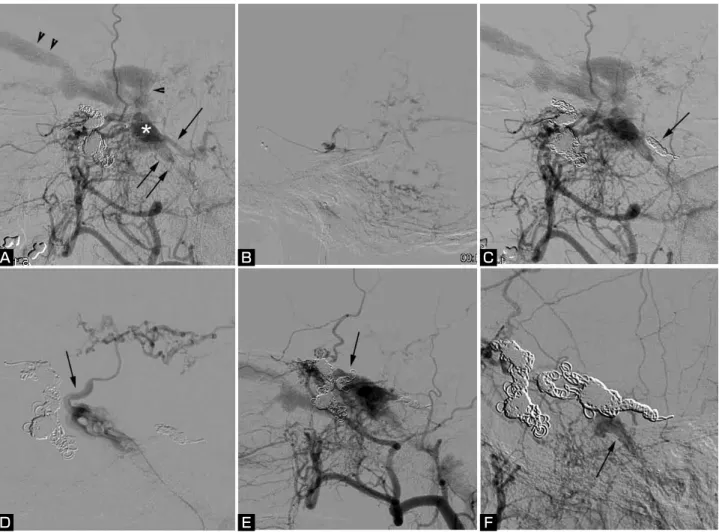
【Case 2】56歳 女性.耳鳴りと視力低下を主訴に来院 した.右CSにシャントを有するCSdAVFで右SMCVに 主たるドレナージがみられ,初回治療ではSMCVと SOVを塞栓して終了した.症状はいったん改善したが,
1年後に再発したため2回目の治療となった.シャント の 主 座 は 右CSで あ り, ド レ ナ ー ジ は, 右SPS, 右 IPS,intercavernous sinus経由で左CSから左SOVへ抜 ける3方向であった(Fig. 1A).まず右IPSからExcelsior 1018をCS経由で右SPSに誘導し(Fig. 1B),続いても う1本のExcelsior1018を同じくIPS経由で右CS内に置 いた.まず右SPSを,GDC(10soft 2DSR 3×6,3本;
US 2.5×4,2本;US 2×3,2本)で塞栓した(Fig.
1C).右CS内のExcelsior 1018からの撮影で,新たに uncal veinへの流出が確認された(Fig. 1D)ので,右 CS内のExcelsior1018をuncal veinへ移動しGDC(10soft 2DSR 3×6,2本;US 2.5×6,1本;US 2×4,4 本;US 2×3,1本)で塞栓した(Fig. 1E).最後に intercavernous sinusから右CS内までをGDC(18 2D 7
×30;18soft 5×8,2本;Vortex 4×6,4本;10 soft 2DSR 3×4,1本;US 2.5×4,1本;US 2×
4,1本)およびOrbit(4×7,1本;7×21,1本;
6×15,1本;5×10,3本)で塞栓し,右IPSへのシ ャントがわずかにみられるのみとなったので終了した
(Fig. 1F).
考 察
CSdAVFの治療方法には,①用手的頚部動静脈の圧 迫9),②TVE7),③経動脈的栄養動脈塞栓術5),④放射線
治療8,14),などがある.一般的には予後良好な疾患なので,
用手的に頚部動静脈の圧迫を行いながら経過を観察する 場合もあるが,眼圧上昇による視力障害や,脳表静脈へ の強い逆流がある場合には,より治癒率が高いTVEが 選択される11).
TVEは,通常1本のマイクロカテーテルをIPS経由で CSに誘導する.この場合,一般的にはもっともIPSか ら遠いSOVから塞栓を開始する.シャントはCSの後下 方に存在することが多いので,SOVの塞栓中にシャン ト量は減らず,逆に流出路が少なくなることで新たに脳 表静脈や脳幹静脈への逆流が生じる場合がある.また,
すでに存在するRLVDへの血流が急激に増加する場合も あり,時に出血や静脈性梗塞を起こす可能性がある11). このようなTVE中の静脈流出路の変化に対しては,マ イクロカテーテルを逆流静脈に移動してその静脈を塞栓 するか,すみやかにCS全体を塞栓する必要がある.マ イクロカテーテルが1本のみであると決して容易な手技 ではないが,2本あれば対応は比較的容易である.すな わち,当初塞栓を開始したマイクロカテーテル以外のも う1本を,新たなRLVDが生じた静脈にすみやかに移動 させることが可能であり,またCSそのものの塞栓を直 ちに行うこともできる.我々は, 2本のマイクロカテー テルをCSに誘導し,まずRLVDを確実に消失させ,続 いてSOVを塞栓し,最後にシャント部位を含めたCSを 塞栓してシャントを消失させるという方針で治療を行っ た.85%の症例で2本のマイクロカテーテルをCSに誘 導することができ,すべてのRLVDおよびSOVに選択 的にマイクロカテーテルを誘導できた.危険な静脈流出 路であるRLVDと眼症状の原因であるSOVへの逆流の 両者を,比較的短時間で確実に止めることができる点で 優れた方法と考えている.また,TVEの途中で新たな RLVDが出現したのは1例のみであったが,1本のマイ クロカテーテルを直ちにRLVDに移動させて塞栓を行う ことができ2本のカテーテルによるTVEの有用性を実 感した.
今回13回のTVE中6例でIPSが脳血管撮影上閉塞して いたが,全例でIPS経由でのCSへの到達が可能であり,
うち4例では2本のマイクロカテーテルを誘導できた.
閉塞したIPSを通る時の合併症として,静脈洞壁を穿孔 し硬膜下血腫やくも膜下出血の形成が報告されている.
静脈洞壁を損傷しないためには,抵抗が強い場合に無理 にガイドワイヤーおよびマイクロカテーテルを押さない ことが肝要である.0.035inchのガイドワイヤーでIPSの 探索を行う方法が推奨されているが3),我々の経験では
マイクロガイドワイヤーでも十分対応可能であり,静脈 洞壁の損傷もなかったことを考えると全例に0.035inch のガイドワイヤーの使用は必ずしも必要ではない.
CSdAVFに対するTVEは,従来のsingle microcatheter techniqueでも解剖学的治癒率71~89%,臨床的治癒率 77~96%と報告されている10).我々のDMTによる初回 治療後の解剖学的および臨床的治癒率はそれぞれ82%で あり,従来の治療成績を凌駕するものではない.しかし ながらCSに2本のマイクロカテーテルを誘導すること は特に困難でなく,RLVDを来した静脈(洞)とSOV
への選択的な留置や,術中の流出路変化への対応も速や かに行うことが可能であり,追加治療後の最終的な解剖 学的および臨床的治癒率はともに100%であった.今後,
targeted compartmental embolization1,13,15)の概念のもと でシャント部位のみの塞栓を試みる場合などには,
RLVDを確実に閉塞するための手段としてDMTは有用 と考えられた.
Fig. 1 Case 2 Angiograms (lateral views) of second session of transvenous embolization for dural arterio-venous (A-V) fistula at the cavernous sinus (CS).
A: A right external carotid angiogram, showing A-V shunt at the right CS (white asterisk), and triple venous drainage routes of the right superior petrosal sinus (SPS; arrow), the right inferior petrosal sinus (IPS; double arrows), and the left superior ophthalmic vein (double arrowheads) via the intercavernous sinus (arrowhead).
B:Venous drainage via the SPS.
C:A right external carotid angiogram, showing occlusion of the SPS (arrow).
D:Venography via a microcatheter in the CS, showing a new venous drainage through the uncal vein (arrow).
E:A right external carotid angiogram, showing occlusion of the uncal vein (arrow).
F: A final angiogram, showing occlusion of the triple venous drainage routes, and slight residual anterograde venous drainage via the IPS (arrow).
A B C
D E F
文 献
1) Agid R, Willinsky RA, Haw C, et al: Targeted compartmental embolization of cavernous sinus dural arteriovenous fistulae using transfemoral medial and lateral facial vein approaches. Neuroradiology 46:156-160, 2004.
2) Awad IA, Little JR, Akarawi WP, et al: Intracranial dural arteriovenous malformations: factors predisposing to an aggressive neurological course. J Neurosurg 72:839-850, 1990.
3) Benndorf G, Bender A, Lehmann R, et al: Transvenous occlusion of dural cavernous sinus fistulas through the thrombosed inferior petrosal sinus: report of four cases and review of the literature. Surg Neurol 54:42-54, 2000.
4) Brown RD Jr, Wiebers DO, Nichols DA: Intracranial dural arteriovenous fistulae: angiographic predictors of intracranial hemorrhage and clinical outcome in nonsurgical patients. J Neurosurg 81:531-538, 1994.
5) Elhammady MS, Wolfe SQ, Farhat H, et al: Onyx embolization of carotid-cavernous fistulas. J Neurosurg 112:589-594, 2010.
6) Halbach VV, Higashida RT, Hieshima GB, et al: Dural fistulas involving the cavernous sinus: results of treatment in 30 patients. Radiology 163:437-442, 1987.
7) Halbach VV, Higashida RT, Hieshima GB, et al:
Transvenous embolization of dural fistulas involving the cavernous sinus. AJNR 10:377-383, 1989.
8) Hirai T, Korogi Y, Baba Y, et al: Dural carotid cavernous fistulas: role of conventional radiation therapy-long-term results with irradiation, embolization, or both. Radiology
207:423-430, 1998.
9) Kai Y, Hamada J, Morioka M, et al: Treatment of cavernous sinus dural arteriovenous fistulae by external manual carotid compression. Neurosurgery 60:253-257, 2007.
10) Kim DJ, Kim DI, Suh SH, et al: Results of transvenous embolization of cavernous dural arteriovenous fistula: a single-center experience with emphasis on complications and management. AJNR 27:2078-2082, 2006.
11) Kiyosue H, Hori Y, Okahara M, et al: Treatment of intracranial dural arteriovenous fistulas: current strategies based on location and hemodynamics, and alternative techniques of transcatheter embolization. Radiographics 24:1637-1653, 2004.
12) Lasjaunias P, Chiu M, terBrugge K, et al: Neurological manifestations of intracranial dural arteriovenous malformations. J Neurosurg 64:724-730, 1986.
13) Nakamura M, Tamaki N, Kawaguchi T, et al: Selective transvenous embolization of dural carotid-cavernous sinus fistulas with preservation of sylvian venous outflow.
Report of three cases. J Neurosurg 89:825-829, 1998.
14) Onizuka M, Mori K, Takahashi N, et al: Gamma knife surgery for the treatment of spontaneous dural carotid- cavernous fistulas. Neurol Med Chir (Tokyo) 43:477-483, 2003.
15) Piske RL, Campos CM, Chaves JB, et al: Dural sinus compartment in dural arteriovenous shunts: a new angioarchitectural feature allowing superselective transvenous dural sinus occlusion treatment. AJNR 26:1715-1722, 2005.
JNET 4:3-8, 2010
要 旨
【目的】海綿静脈洞部硬膜動静脈瘻(CSdAVF)に対するdouble micocatheter technique(DMT)による経静脈的塞栓術(TVE)
の有用性について検討した.【対象と方法】CSdAVF 11例(男:女 1:10, 年齢56~81歳 平均66.9歳)に対して13回の
TVEを行った.2本のマイクロカテーテルをCS内に留置後,それぞれのカテーテルを逆流静脈(RLVD)および上眼静脈
(SOV)へ誘導し,RLVD,SOV,CSの順で塞栓した.【結果】13回のTVEのうち,11回(85%)で2本のマイクロカテー
テルをCS内に誘導できた.すべてのRLVDにマイクロカテーテルを選択的に誘導でき,塞栓術中に新たに生じたRLVDに 対しても,1本のマイクロカテーテルを移動させることですみやかに塞栓を行った.初回治療後の解剖学的および臨床的
治癒率は,ともに82%であった.【結語】CSdAVFに対してDMTによるTVEを行った.RLVDへの選択的なマイクロカテー
テルの誘導は困難ではなく,また術中の静脈流出路変化にも容易に対応できた.解剖学的および臨床的予後も良好であり,
CSdAVFのTVEの有用な方法である.