九州大学学術情報リポジトリ
Kyushu University Institutional Repository
展示空間に設置されるマルチメディアとのインタラ クションの認知的、行動的な理解
磯田, 和生
https://doi.org/10.15017/1866367
出版情報:Kyushu University, 2017, 博士(芸術工学), 課程博士 バージョン:
権利関係:
PhD thesis
Cognitive and Behavioral Understanding of Interaction with Multimedia in Exhibition Spaces
展示空間に設置される
マルチメディアとのインタラクションの認知的、行動的な理解
Kazuo Isoda
磯田 和生
Graduate School of Integrated Frontier Sciences Kyushu University
July 2017
Declaration
I hereby declare that this dissertation entitled “Cognitive and Behavioral Understanding of Interaction with Multimedia in Exhibition Space” is not substantially the same as any that I have submitted for a degree or diploma or other qualification at any other University.
Those parts of this thesis which have been published are as follows:
1. Chapter 2 is based on the paper “Effect of the Hand-Omitted Tool Motion on mu Rhythm Suppression. (Kazuo Isoda, Kana Sueyoshi, Yuki Ikeda, Yuki Nishimura, Ichiro Hisanaga, Stéphanie Orlic, Yeon-kyu Kim and Shigekazu Higuchi, Frontiers in Human Neuroscience 2016, Vol.10: 266), DOI:10.3389/fnhum.2016.00266”.
2. Chapter 3 is based on the paper “Tangible User Interface and Mu Rhythm Suppression: The Effect of User Interface on the Brain Activity in Its Operator and Observer. (Kazuo Isoda, Kana Sueyoshi, Ryo Miyamoto, Yuki Nishimura, Yuki Ikeda, Ichiro Hisanaga, Stéphanie Orlic, Yeon-kyu Kim and Shigekazu Higuchi, Applied Sciences 2017, Vol.7, No.4: 347), DOI:10.3390/app7040347”.
3. Chapter 4 is based on the paper “Effects of the Display Angle in Museums on User's Cognition, Behavior, and Subjective Responses. (Junko Ichino, Kazuo Isoda, Ayako Hanai and Tetsuya Ueda, Proceedings of the SIGCHI Conference on Human Factors in Computing Systems. 2013), DOI:10.1145/2470654.2481413”.
4. Chapter 5 is based on the paper “Effects of the Display Angle on Social Behaviors of the People around the Display: A Field Study at a Museum. (Junko Ichino, Kazuo Isoda, Tetsuya Ueda and Reimi Satoh, Proceedings of the 19th ACM Conference on Computer-Supported Cooperative Work & Social Computing. 2016), DOI:10.1145/2818048.2819938”.
Kazuo Isoda Kyushu University July 2017
Table of contents
Chapter 1 Background 1
1.1 Introduction ... 1
1.2 Research areas of interest ... 4
1.3 The purpose of this thesis ... 5
1.4 The structure of this thesis ... 6
Chapter 2 Effect of the Hand-omitted Tool Motion on mu Rhythm Suppression 8 2.1 Introduction ... 8
2.2 Methods ... 11
2.3 Results ... 14
2.4 Discussion ... 17
Chapter 3 Tangible User Interface and mu Rhythm Suppression: Effect of User Interfaces on Brain Activity in the Operator and Observer 20 3.1 Introduction ... 20
3.2 Methods ... 22
3.3 Results ... 27
3.4 Discussion ... 29
Chapter 4 Effects of the Display Angle in Museums on User’s Cognition, Behavior, and Subjective Responses 32 4.1 Introduction ... 32
4.2 Methods ... 35
4.3 Results ... 41
4.4 Discussion ... 49
Chapter 5 Effects of the Display Angle on Social Behaviors of the People around the Display: A Field Study at a Museum 54 5.1 Introduction ... 54
5.2 Methods ... 58
5.3 Results ... 66
5.4 Discussion ... 72
Chapter 6 Conclusion 77
6.1 Summary ... 77 6.2 Future work ... 78
Acknowledgment 80
Reference 81
Appendix 88
1
Chapter 1 Background
1.1 Introduction
· Advances in multimedia and the necessity of human understanding
In recent years, advances in information terminals known for displaying so-called multimedia are remarkable. For example, with respect to display performance, the resolution of a single unit has been increased from full HD (High Definition) to 4 K and 8 K, and the size has been miniaturized due to pixel integration, which is being in personal use. On the other hand, the size of the display is enlarging, not only as a single unit, but also as a combination of multiple units due to improvements in tiling technology.
Furthermore, due to diversification in devices, such as organic EL (Electroluminescence) and LED (Light Emitting Diode) projections in addition to conventional panels, the range of use has expanded from personal devices, such as smartphones, tablet PCs and HMDs (Head Mounted Display), to signage installed in public spaces (Nakamura & Ishido, 2009;
Digital Signage Consortium, 2016).
As a result, opportunities for coming into contact with multimedia in everyday life are expanding. Much of the information obtained from traditional media, such as printing media, has been digitized and is becoming available only via multimedia. Along with Internet connections and the development of various sensing technologies, opportunities will further expand, and not only the amount of information but also qualitative diversity will increase.
How will humans recognize, understand and act on the information obtained from multimedia? Understanding how humans interact with multimedia as information recipients is an indispensable factor in designing multimedia. As the information expands quantitatively and qualitatively, better ways of presenting information that understands human cognition and behavioral characteristics is required more than ever.
· Understanding the initiatives and layers of human computer interactions adopted up until now at the DNP Museum Lab
We have been searching for new forms of information communication using multimedia through such activities as the DNP Museum Lab (since 2006, http://www.museumlab.jp/).
This project focused on museums as places to utilize multimedia, such as interactive systems. We have been working from various approaches on how multimedia can contribute to the viewing experience in appreciation of works represented by art works.
2
We held exhibitions that added various kinds of multimedia equipment to the exhibitions of art works held by museums, such as the Louvre Museum and the French National Library, in the past. In the exhibitions, which have been held a total of eleven times in the past, each theme was set as a thematic approaches, and various matters which had been participants in past artwork exhibitions were solved by using new technology and application methods (Figure 1.1).
Figure 1.1 Development examples at the DNP Museum Lab.
3
The efforts that have been proposed and developed until now are classified into three layers as the interaction between human beings and the technical elements constituting the multimedia (Figure 1.2).
Figure 1.2 Factors related to equipment and humans.
The first layer is the technique of contents representation. We focused on how the information should be expressed so that it is easy for the viewers to understand it.This technology includes reproduction of artworks by digital representation, how to convey the background information of the work to help understand it, and so on.
The second layer is user interface technology. We focused on how to interact with the computer to acquire information. Initiatives to address issues that exist around the interface, including ease of operation, understanding operation methods, and so on.
The third layer is space design, including the design of hardware installed in the space.
Issues such as the shape and arrangement of the equipment to be viewed or operated, and the design of the viewing route. Initiatives to address issues in spatial design, which is not limited to appreciating alone, but also assuming use by multiple people.
Although these layers are not completely independent, it is possible to cover many problems by paying attention to each layer and considering issues related to multimedia in exhibition spaces.
4
· Introduce scientific approach to development site
Even in the past proposals so far, human cognitive and behavioral characteristics, which are known from human research findings, were taken into consideration.
In the content representation technology, studies in human visual characteristics were used for information arrangement, color expression, character size, and the like. In the user interface technology, research knowledge on human information perception has been applied to the size and arrangement of operating systems. Even in the space and hardware design, research findings, such as ergonomics, have been used as guidelines for the height and size of the screen (Sato, Katsuura, Sato, Tochihara, & Yokoyama, 1992; Itoh, Kuwano, & Komatsubara, 2003; Oshima & Okubo, 2005). Materials serving as guides for incorporating those findings were also published (Weinschenk, 2011).
However, there are methods that are proposed and adopted on the basis of experience, even if there is no research backing on the development site. Methods proposed from such site problems and optimized for actual exhibitions, etc., were fed back to the research and their effects were not often verified from human characteristics.
In this research, we focused on linking academic research with on-site tasks and proposals. We will apply challenges / proposals born from actual development sites to hypotheses in the research field and try to verify them from a multifaceted aspect. We believe that not only scientifically supporting empirical know-how, which is only known from examples, but also scrutinizing issues / proposals by scientific approaches will lead to discovering a part of human characteristics.
1.2 Research areas of interest
In this paper, we focused on three research areas that are closely related to each layer (shown in Figure 1.2), and applied them to solving the problem in each layer.
· Mirror neuron system
Brain activity that works when seeing other people's behavior is already known (Fadiga, Fogassi, Pavesi, & Rizzolatti, 1995; Rizzolatti, Fadiga, Matelli, et al., 1996). It is known that brain activities that are activated by merely observing behavior performed by other people without them being executing themselves are related to understanding and predicting the intention of others to act (Iacoboni et al., 1999). From the work of the mirror neuron system (MNS), we set a hypothesis that we can explain the expression method and the effect of the user interface and verify the possibilities.
· Tangible user interface
As one of the ideas for interaction with computers, an interface has been proposed in which information processed digitally is represented in the real world and the physical behavior of real objects are reflected in computer operations (Ishii & Ullmer, 1997). In terms of performance and subjectivity, the simplicity of this is well known (Shaer &
Hornecker, 2009), but we attempted to demonstrate it from the influence on brain activity.
5
· Social behavior around multimedia
Regarding behavioral characteristics to multimedia in public spaces, it is known that the honeypot effect (Brignull & Rogers, 2003) attracts the attention of users. In terms of communication, the space formed by a plurality of humans is defined as an instrumental F-formation (Kendon, 1990), and its social behavior is analyzed. Controlling the conditions for action in the actual exhibition environment and applying new approaches to social behavior analysis lead to acquiring new findings.
1.3 The purpose of this thesis
In the three layers related to exhibition technology, we focused on the empirical knowledge and tasks adopted for exhibitions as new experiments respectively. In order to explain this from the knowledge of human research and to discover new tasks, we will conduct a research approach and try to acquire knowledge newly supported by data by further examining new issues. Each related research area was set as follows, and approval verification approaches were made for each experiment.
· Effect of Hand-omitted Tool Motion on mu Rhythm Suppression (Chapter 2) In this chapter, we investigated the effect of the image of hands on mu rhythm suppression invoked by the observation of a series of tool-based actions in a goal-directed activity. As a source of visual stimuli to be used in the test, a video animation of the porcelain making process for museums was used. In order to elucidate the effect of the hand imagery, the image of hands was omitted from the original ("hand image included") version of the animation to prepare another ("hand image omitted") version. The present study has demonstrated that in individuals watching an instructive animation on the porcelain making process, the image of the porcelain maker's hands can activate the MNS. In observations of “tool included” clips, even the "hand image omitted" clip induced significant mu rhythm suppression in the right central area. These results suggest that visual observation of a tool-based action may be able to activate the MNS even in the absence of hand imagery.
· Tangible User Interface and mu Rhythm Suppression: Effect of User Interfaces on Brain Activity in the Operator and Observer (Chapter 3)
The intuitiveness of tangible user interfaces (TUI) is not only for the operator. It is quite possible that this type of user interface (UI) can also have an effect on the experience and learning of observers who are just watching the operator using it. To understand the possible effect of TUI, the present study focused on mu rhythm suppression in the sensorimotor area reflecting execution and observation of action, and investigated brain activity both in the operator and observer. In the observer experiment, the effect of TUI on observers was demonstrated through brain activity. Although the effect of the grasping action itself was uncertain, the unpredictability of the result of the action seemed to have some effect on the MNS-related brain activity. In the operator experiment, in spite of the same grasping action, brain activity was activated in the sensorimotor area when UI functions were included (TUI). Such activation of the brain activity was not found with a graphical user interface (GUI) that has UI functions without the grasping action. These
6
results suggest that the MNS-related brain activity is involved in the effect of TUI, indicating the possibility of UI evaluation based on brain activity.
· Effects of the Display Angle in Museums on User’s Cognition, Behavior, and Subjective Responses (Chapter 4)
In order to achieve the intended level of communication with visitors in museums where large displays are installed, it is essential to understand how various display factors affect visitors. We explored the effects of the display angle on individual users. In our experiment, we set up three types of flat displays -vertical, horizontal, and tilted- and comprehensively tested users’ cognitive, behavioral, and subjective aspects. The results showed that significant differences could be discerned with regard to cognitive and subjective aspects. Test results for the cognitive aspect showed that the display angle on which the displayed content was easy to understand and remember differed depending on age. Test results for the subjective aspect showed that irrespective of age, users rated tilted displays as being quicker to attract attention and easier to peruse, to understand and remember the content, and to interact with, and such displays were the most preferred.
· Effects of the Display Angle on the Social Behavior of People around the Display:
A Field Study at a Museum (Chapter 5)
In this chapter, we investigated through a field study how the angles (horizontal, tilted, and vertical angles) of displays deployed in a public space (at a museum) impact the social behavior of the people around the display. In the field study, we collected both quantitative and qualitative data of more than 700 museum visitors over a period of approximately three months. The findings of our study include the following: (1) the horizontal and vertical display angles have a higher honeypot effect, i.e., people interacting with a display attract other people, than the tilted display angle, (2) the vertical display angle, compared to the horizontal and tilted display angles, attracts several people to the display and encourages them to stay in the display space and share the space for a short period of time (88 seconds on average), and as a result, people frequently enter and leave the space with a display, and (3) display angles closer to the horizontal promotes the side-by-side arrangement, and display angles closer to the vertical promotes the L-shaped arrangement of an F-formation.
1.4 The structure of this thesis
In this paper, we focused on three layers related to exhibition technology (technology concerning content representation, interface technology, cabinet design and space design) in Chapter 2 to 5. Each research theme was set and tried by scientific examination using five experiments (Table 1.1).
In Chapter 2, we focused on the presence of human hand movement as one "technology on content expression" and measured human response to the movements of hands and tools expressed in the image. As a human response, we considered the influence on
"brain activity" from the MNS activity using "mu rhythm suppression".
7
In Chapter 3, we focused on the "tangible user interface" as one of the "interface technologies" and focused on the movement of hands that actually manipulate existing objects. This study consisted of two experiments, the influence on the observer and the influence on the operator, and we examined the influence on the brain activity from the same "mu rhythm suppression" activity as in Chapter 2.
Next, we focused on "angle of display that can interact with large sizes" as one of the factors of "space and display devices design". In Chapter 4, a simulated exhibition space was constructed in the laboratory, and individual responses were acquired and the influence on behavior, cognition and subjective response was considered. Based on the results of Chapter 4, in Chapter 5 we observed the response of multiple users with a field study in the actual exhibition space. Then, we considered the influence on 'social behavior around multimedia' in a more natural environment.
Finally, we summarized my conclusions in Chapter 6.
Table 1.1 Experimental conditions.
Exp. Layer Participants Location Survey items Chapter
1 Contents Observer
without
operation alone
Laboratory Experiment 1
・Cognition
<Brain Activity> Chapter 2
2 User Interface
Observer behaind Operator
without
operation alone
Laboratory Experiment 2
・Cognition
<Brain Activity> Chapter 3
3 User Interface Operator with
operation alone
Laboratory Experiment 3
・Cognition
<Brain Activity> Chapter 3
4
Hardware
and Space Operator with
operation alone
Laboratory Experiment 4
・Cognition
・Behavior
・Subjective
Responses Chapter 4
5
Hardware and Space
Operator Observer Passenger
with/without operation
mixed multiple Field Study
・Behavior
・Subjective
Responses Chapter 5
8
Chapter 2
Effect of the Hand-omitted Tool Motion on mu Rhythm Suppression
2.1 Introduction
・Mirror neuron system
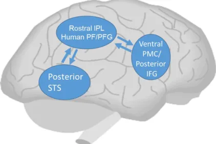
"Mirror neurons" that discharge during both action done and the same action observed were first identified in the F5 area (ventral premotor cortex) in macaque monkeys (Gallese, Fadiga, Fogassi, & Rizzolatti, 1996; Rizzolatti, Fadiga, Gallese, & Fogassi, 1996), and the neurons were subsequently found in the intraparietal sulcus, too (Fogassi et al., 2005). A number of experiments have suggested that this parieto-frontal cortical circuit in the observer of actions performed by other individuals encodes the goals and intentions of these actions (Rizzolatti & Craighero, 2004; Rizzolatti & Sinigaglia, 2010).
In humans, fMRI (functional magnetic resonance imaging) studies have revealed the presence of mirror-like brain regions similar to those in monkeys. It has been proposed that the MNS in humans (Figure 2.1) is involved not only in the recognition of the goals and intentions of actions (Iacoboni, 2005), but also in imitation (Iacoboni & Dapretto, 2006; Molenberghs, Cunnington, & Mattingley, 2009), empathy, facial expression recognition, and other social cognition functions (Preston & de Waal, 2002; Carr, Iacoboni, Dubeau, Mazziotta, & Lenzi, 2003; Jabbi, Swart, & Keysers, 2007; Schraa-Tam et al., 2012). In the study using fMRI, disassociation of visual processing between ventral and dorsal pathways was revealed during object and action recognition (Shmuelof &
Zohary, 2005).
・Mu rhythm suppression
In addition to fMRI, electroencephalography (EEG) has been used for the measurement of the activity of the MNS in humans (Pineda, 2005). Specific alpha ranges of EEG have long been known which are present over the central area, corresponding to the primary motor and other areas, in an individual physically at rest and suppressible by not only her/his performing an action but also just observing the same action performed by another individual (Gastaut & Bert, 1954; Chatrian, Petersen, & Lazarte, 1959) (Figure 2.2).
Those EEG which have mirror-like characteristics and occur in the central area on the scalp of an individual physically at rest are also called "mu rhythm". Since the first discovery of mirror neurons in 1996, a number of studies have revealed that mu rhythm are related to the MNS (Pineda, 2005).
9
According to some of these studies, mu rhythm can be suppressed to a varying degree depending on the goal of an action to be observed (Muthukumaraswamy, Johnson, &
McNair, 2004). Comparative EEG studies have also been reported using the test stimuli that are conventionally employed in fMRI studies (Perry & Bentin, 2009). Based on the study using both fMRI and EEG, mu rhythm are related to the activity of the primary motor cortex and the inferior parietal lobule that reflects the firing of the MNS (Arnstein, Cui, Keysers, Maurits, & Gazzola, 2011). These findings strongly suggest that, in humans, the level of mu rhythm suppression can be an index of the activity of the MNS.
Figure 2.1 Human mirror neuron system: Neural circuitry for imitation (Modified from Iacoboni and Dapretto, 2006, Page 943, Figure 1).
Figure 2.2 Example of mu rhythm suppression (Chatrian et al., 1959, Page 503, Figure 7).
10
・Tool-based action
The evolution of the brain has endowed humans with the ability of tool-based actions.
Although non-human animals can also perform tool-based actions, they cannot handle tools in as complex a way as humans. In humans and monkeys, visuomotor neural mechanisms that are involved in the visual observation and the handling of tools has been extensively investigated (Jeannerod, Arbib, Rizzolatti, & Sakata, 1995; Jarvelainen, Schurmann, & Hari, 2004; Maravita & Iriki, 2004; Peeters et al., 2009; Costantini, Ambrosini, Sinigaglia, & Gallese, 2011). However, the relation between the visual observation of the handling of tools and the activation of the MNS has been much less studied.
A magnetoencephalography (MEG) study has reported that the primary motor area in an individual can be activated by just watching the motion of hands of another individual in tool-based actions (Jarvelainen et al., 2004). fMRI studies in humans and monkeys also investigated the effect of watching a video featuring a tool-based action on the MNS (Peeters et al., 2009). As a result, a human-specific activity of the left inferior parietal lobule was identified, suggesting that this brain area is important in the tool-based actions in humans.
Similar mirror neurons are involved in brain activity induced by the visual observation of tools, which was first identified in monkeys (Murata et al., 1997). With the participant just focusing on the tool, these are neurons that respond, relying on the shape of the hand when it grabs the object and the pattern of movement. Rizzolatti et al. call them canonical neurons (Grezes, Armony, Rowe, & Passingham, 2003; Rizzolatti, Cattaneo, Fabbri- Destro, & Rozzi, 2014). According to a previous report in humans, the ventral premotor cortex, the posterior parietal lobe, and the precentral sulcus can be activated by the handling as well as the visual observation of a tool (Chao & Martin, 2000; Mecklinger, Gruenewald, Besson, Magnie, & Von Cramon, 2002; Grezes et al., 2003). These activities are regarded as important functions in the handling of tools. Furthermore, in tool action observations, mu rhythm is suppressed more according to participant's experience in the action (Cannon et al., 2014).
These previous reports are extremely intriguing as to the brain's response to tool-based actions, but there has been hardly any research that distinguishes between tool and the presence of hands. By controlling the task, Shmuelof et al. (2005) succeeded in distinguishing reaction to an object and reaction to an action; yet, this is purely the observation of a reaction to an object as a target matter. We will pay attention to the reaction to a tool that has been prepared as a medium to transmit the intended action to an object that is the participant. Research focusing on motion shows that mu rhythms are suppressed by biological motion but they will not be suppressed by random motion (Ulloa
& Pineda, 2007). Indeed, there are reports that, even though mu rhythm suppression occurs when a person watches an image of a ball being thrown, just watching the ball in flight will not suppress the mu rhythm (Oberman et al., 2005). It is also unclear whether the MNS can be activated by watching the image of tools, hands, or both, because the test visual stimuli employed in previous studies included not only the image of a tool but also the image of hands that was handling the tool. If the MNS is activated by transmitting the
11
intention of an action, even if the hand is omitted, it is probable that mu rhythm suppression will occur with just the movement of the tool.
In the present study, we investigated the effect of the image of hands on mu rhythm suppression invoked by the observation of a series of tool-based actions in a goal-directed activity.
2.2 Methods
[Experiment 1]
・Participants
The participants were 13 healthy, right-handed university students (7 females and 6 males, 22.2±1.3 years old) who normally do not engage in clay modeling work. The participants gave written informed consent to the present study only after they were provided with information on the test protocol. The study was approved by the Ethical Committee of Kyushu University (approval No. 84).
・Experimental conditions
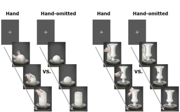
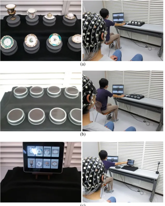
As a source of visual stimuli to be used in the test, a video animation was chosen which a museum used to instruct its visitors on the porcelain making process (DNP Museum Lab). This imagery consisted of processes such as clay kneading and wheel rotation (processes where tools are not used) as well as clay modeling using a kidney shaped profile and decoration (processes where tools are used), all performed by the hands of a porcelain maker.
In this study, first we observed the MNS activity under circumstances not related to tools in order to confirm whether or not MNS activity via EEG can be observed in the animation used for trials. Next, in accordance with the focus of this study, we looked to confirm the effect of only tool motion by comparing the presence / non-presence of hands under circumstances mediated by a tool. Note that, to avoid the influence by the order of conditions appearances, we shuffled the order of these experiments.
In order to elucidate the effect of the image of these hands, the image of hands were omitted from the original ("hand image included") version of the animation to prepare another ("hand image omitted") version. From each of these two versions, chapters on tool-free actions (e.g. clay kneading and wheel rotation: Figure 2.3, left panel) and chapters on tool-based actions (e.g. clay shaping with a kidney: Figure 2.3, right panel) were separately extracted and edited to make two shorter clips ("tool-free" and "tool included"). Each of the four shorter clips was presented repeatedly to each participant (70 seconds / clip). The control stimulus employed in the test was a static frame with a cross- mark at its center.
12
Figure 2.3 Chapters on "tool-free" actions (e.g. clay kneading and wheel rotation: left panel) and chapters on "tool included" actions (e.g. clay shaping with a kidney: right panel). The
stimulus for baseline data was a still frame with a cross-hair at its center.
First, the participants were allowed to watch the whole movies (the "hand image included" version and the "hand image omitted" version). Then each of the four shorter clips was presented as a test stimulus. Before and after each shorter clip, the still frame control was presented (40 seconds). The "hand image included" stimulus and the "hand image omitted" stimulus were counterbalanced between the participants for both the
"tool-free" stimuli and the "tool included" stimuli. While watching the video, the participants were instructed to move their eyes as little as possible from the center of the screen.
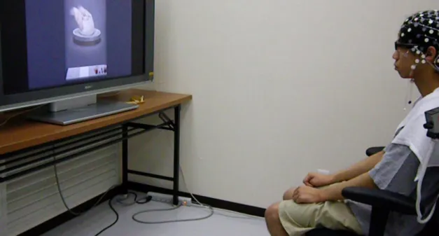
EEG were measured in an electromagnetically shielded room (illuminance = 200 lx, temperature = 25 degrees Celsius, moisture = 50 %). Each participant was seated on a chair and allowed to watch a series of the four clips and the still frame control on a liquid crystal display (19 in.), which was placed at 1.1 m from the chair. (Figure 2.4) EEG were detected using a 64-channel EEG cap (Hydrocel GSN 64 ver.1.0, Electrical Geodesics, Inc.), filtered with a low cut frequency of 0.3 Hz, a high cut frequency of 100 Hz, and a sampling rate of 250 Hz, which were A/D converted and recorded on a computer (PowerMac G5, Apple, Inc.) equipped with the Net Station 4.1.2 software (Electrical Geodesics, Inc.).
13
The obtained data, except for those from the initial 10 seconds, were subjected to a frequency analysis (Fast Fourier Transform; FFT) at 1 epoch (4.091 seconds long) per 2 seconds. An average power value in the 10-12 Hz range, which was considered to well reflect the activity of the motor cortex (Pfurtscheller, Neuper, & Krausz, 2000), was used as the mu power value. The calculated power values were normalized after logarithmic transformation, which were then analyzed using the EMSE Suite Data Editor 5.3 Release Candidate 3 software (Source Signal Imaging, Inc.).
For each channel, we calculated mu rhythm suppression, i.e. the difference in the mu power value between each test stimulus and the control stimulus before/after the test stimulus. We set the motor area of the cerebral cortex region as the region of interest (ROI) to provide us with an indicator for confirming whether or not the MNS activity was increased by the movement of the tool with intention. The data from two of the participants contained missing values and outliers, and thus were excluded from the analysis.
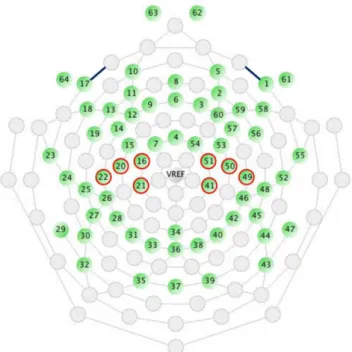
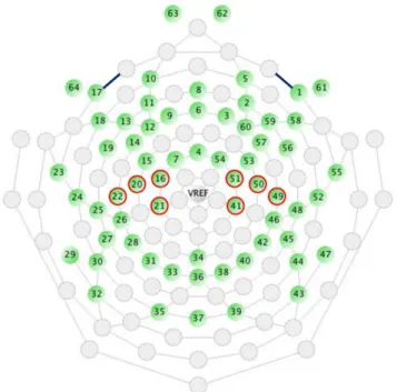
In order to enhance the reliability of the data, Two regions of interest (ROI) were defined and respective electrode sites were pooled: left central (LC: electrodes 16, 20, 21, 22) and right central (RC: electrodes 41, 49, 50, 51; Figure 2.5). A paired t-test was performed to determine the significant mu suppression from the baseline data of the still frame. Next, a two-ways (hand image included/omitted x Left/Right hemisphere) repeated measurements of analysis of variance (rm-ANOVA) were conducted to determine the significance. Additionally, a paired t-test was used by using each individual electrode site of 64 channels and a three-dimensional topographic map (t-map) was generated.
Figure 2.4 Settings of the Experiment.
14
Figure 2.5 Two regions of interest (ROIs):
ROIs were defined and respective electrode sites were pooled: left central (16, 20, 21, 22) and right central (41, 49, 50, 51).
2.3 Results
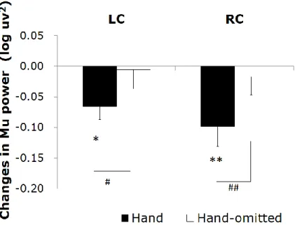
Analysis 1: Changes in the mu power value induced by watching the "tool-free" clips are shown in Figure 2.6. Compared to the control stimulus by the still frame, a significant decrease in the mu power value was induced by the "hand image included" clip in the left central area (t (10) = -3.13; p = 0.011) and the right central area (t (10) = -3.05; p = 0.010).
The "hand image omitted" clip did not induce any significant mu rhythm suppression.
We next compared the ability of the "hand image included" clip and that of the "hand image omitted" clip to induce mu rhythm suppression. As the results of two-way rm- ANOVA, although main effect of hand image was significant (F (1, 10) = 18.920; p = 0.001; ηp2 = 0.654), main effect of hemisphere and interaction were not significant. It turned out that, compared to the "hand image omitted" clip, the "hand image included"
clip induced a significant mu rhythm suppression in the right central area (t (10) = -4.01;
p = 0.002) and the left central area (t (10) = -2.57; p = 0.028). We made a similar comparison for each electrode site, and plotted the results in a three-dimensional t-map (Figure 2.7), demonstrating the difference between the "hand image included" and "hand image omitted" clips specifically in the right and left central. No significant differences were found in other areas.
15
Figure 2.6 Changes in the mu power value induced by watching the "tool-free" clips:
"hand image included" (■) and "hand image omitted" (□). Asterisks (*: p < 0.05, **: p < 0.01) mean the significant mu rhythm suppression from baseline to observation of video clips.
Sharps (#: p < 0.05, ##: p < 0.01) mean the significant differences between mu rhythm suppression by watching video clips "hand image included" and "hand image omitted".
Figure 2.7 Three-dimensional t-map:
Comparison for each electrode site, and plotted the results in a three-dimensional t- map, demonstrating the difference between the "hand image included" and "hand
image omitted" clips specifically in the right central.
16
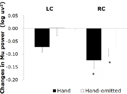
Analysis 2: Changes in the mu power value induced by watching the "tool included" clips are shown in Figure 2.8. Compared to the still image control, a significant decrease in the mu power value was induced by the "hand image included" clip in the right central area (t (10) = -2.26; p = 0.035). In addition, the "hand image omitted" clip also induced a significant decrease in the mu power value in the right central area (t (10) = -2.10; p = 0.050). As the results of two-way rm-ANOVA, although main effect of hemisphere was significant (F (1, 10) = 7.299; p = 0.022; ηp2 = 0.422), main effect of hand image and interaction were not significant. Mu suppression in right hemisphere was significant greater than that in left hemisphere. No significant differences were found for mu suppression between the "hand image included" and "hand image omitted" of the "tool included" clips. We made a comparison for each electrode site, and plotted the results by the corresponding three-dimensional t-map (Figure 2.9). In this analyze, we found also a significant difference from the left parietal region to the left temporal region (LP:
electrodes 27, 30; Figure 2.5). No significant difference was found in other regions between these results.
Figure 2.8 Changes in the mu power value induced by watching the "tool included" clips:
"hand image included" (■) and "hand image omitted"(□).
17
Figure 2.9 Three-dimensional t-map:
Comparison for each electrode site, and plotted the results in a three-dimensional t-map.
We found a significant difference from the left parietal region to the left temporal region.
2.4 Discussion
・"Tool-free" clips: comparison between "hand image included" and "hand image omitted"
In analysis 1, we confirmed mu rhythm suppression can be observed only when hands were present. As a result, we confirmed that MNS activity can be observed even with the animation video. In the observation of the "tool-free" clips by the participants, the "hand image included" clip induced a significant decrease in the mu power value in the central area (LC, RC), whereas the "hand image omitted" clip did not. These results are consistent with previous observation that the visual observation of the motion of hands induced mu rhythm suppression in the central area (Muthukumaraswamy et al., 2004; Oberman, McCleery, Ramachandran, & Pineda, 2007; Perry & Bentin, 2009), indicating that, in the animation of the porcelain making process used in the present study, the image of hands activated the MNS in the motor cortex. Moreover, activity was seen in both the LC and RC hemispheres. This is conjectured to be hand movement representation of a contralateral preference that appears in the dorsal stream (Shmuelof & Zohary, 2005) while, in this study, another factor is considered to be that both hands were active in the presented hand movement.
In the "tool-free" clips, the "hand image included" clip also induced a decrease in the power value in the 10-12 Hz range in some areas other than the central area (LC, RC).
This is thought to have happened because of the visual stimuli due to the clay images in the activity shared by presentations. The effect of the image of hands was investigated by comparing the effect of the "hand image included" clip and that of the "hand image
18
omitted" clip on the power value in the 10-12 Hz range. (Figure 2.7) A significant difference was only present in the right central area, where the "hand image included"
clip induced a significantly greater suppression of mu rhythm than the "hand image omitted" clip, which results in reaction to object (clay images) in the activity shared by presentations being offset, indicating a motor cortex-specific effect of the image of hands.
・"Tool included" clips: comparison between "hand image included" and "hand image omitted"
In analysis 2, regardless of whether or not the hand was present, we confirmed mu rhythm suppression in just the RC, and, depending on the tool movement, we confirmed that MNS activity could be seen. In the observation of the "tool included" clips, even the "hand image omitted" clip induced a significant mu rhythm suppression in the right central area, resulting in activity being seen just in RC, which is thought to stem from the fact that the movement was nearly all presented on the left side of the screen. This too matches the contralateral tendency as described by Shmuelof et al. (2005), indicating that the motion of a tool can induce mu rhythm suppression in its observer even in the absence of the image of hands handling the tool. This is possibly because the motion of the tools (e.g. a kidney) may have compensated for the omitted image of the hands. According to previous studies in monkeys, as a result of watching a tool-based action, neurons in the parietal lobe can merge the tool into the hands handling the tool, leading to a cortical magnification (Hihara et al., 2006). In addition, it has been reported that the primary motor area can be activated not only by watching the motion of hands but also by just imagining the same motion (Pfurtscheller & Neuper, 1997; Pineda, Allison, & Vankov, 2000). Mu rhythm suppression induced by the "hand image omitted" clip in the central area might be attributable to an ability of the motion of a tool to evoke the image of the hands handling the tool in the brain.
Another possibility is the involvement of a brain activity that is induced by the visual observation of tools. It is known that the areas involved in the handling of tools can also be activated by just watching the tools (Chao & Martin, 2000; Grezes et al., 2003). This characteristic activation of brain is seen in monkeys (Murata et al., 1997). Mecklinger et al. (2002) reported that the visual observation of a graspable object induces a stronger activity in the ventral premotor cortex than that of a non-graspable object. We suppose that such an activity induced by the observation of the motion of a tool may have induced mu rhythm suppression in the present study.
Similar to the "tool-free" stimuli, the "hand image included" clip in the "tool included"
stimuli also induced a decrease in the power value in the 10-12 Hz range in many areas over the scalp. Therefore we investigated the effect of the image of hands, by directly comparing the data from the "hand image included" and "hand image omitted" clips. As a result, the "hand image included" clip resulted in a significant mu rhythm suppression from the left parietal region to the left temporal region. From the two parameters compared, the only difference is the presence/non-presence of hands, so mu rhythm suppression in this case is considered to be dependent on caused by "hand movement".
As the presented "hand movement" is a right-hand one for manipulating a tool, a contralateral action that corresponds to the participant's own hand movement is appearing.
This matches the results showing a strong reaction to some of the images of a body shown
19
in Downing et al. (2001). Yet again, viewed from a different perspective, according to a previous report based on both fMRI and EEG, the activity of the inferior parietal lobule is strongly related to mu rhythm suppression (Arnstein et al., 2011). In addition, it has been known that the observation of a tool-based action can activate the left inferior parietal lobule (IPL) in the observer (Peeters et al., 2009). This activation is human- specific and not found in monkeys. In this study, the use of a tool is strongly related to the recognition of the difference in mu rhythm suppression between the "hand image included" and "hand image omitted" clips in the area corresponding to the inferior parietal lobule.
The present study has demonstrated that the visual observation of a tool-based action may be able to activate the MNS even in the absence of such an image of hands. This phenomenon may involve brain activities, which are known to fire in response to the visual observation of a tool. In the observation of the tool-based process, the image of hands induced mu rhythm suppression in the observer in the area corresponding to the inferior parietal lobule.
· Limitation
In this study, we adopted the commentary video animation of art works as a stimulus to evaluate museum information interfacing from the aspect of cerebral function. However, it is assumed that visitors to actual museums vary in characteristics, such as age, gender and profession. In previous research, it has been reported that mu rhythm suppression is influenced by experiences (Cannon et al., 2014). Therefore, we need to take into consideration the experiences of participants when undertaking research. Indeed, although we validated the presence/non-presence of hands when presenting tool movement, we did not validate the presence/non-presence of a tool. To further stringently isolate influences, we should probably also carry out validation that can compare the presence/non-presence of the tool concerned.
20
Chapter 3
Tangible User Interface and mu Rhythm Suppression: Effect of User Interfaces on Brain
Activity in the Operator and Observer
3.1 Introduction
・Tangible User Interface
Tangible User Interface (TUI) is a type of UI that allows a person to interact with digital information through the physical environment (Ishii & Ullmer, 1997). This type of UI involves the use of tangible objects that bridges the gap between digital information and physical space, and is characterized by its UI function based on the manipulation (e.g.
grasping and moving) of that objects (Fitzmaurice, 1996). Because TUI accepts specific user actions on the objects as inputs, the result of each action can be easily predicted, implying that it is more harmonious with intuition in comparison with other UIs. As compared with physical and digital representations, TUI provides more fun as well as intuitiveness to its users. Its applications in the design community include those focusing on user experience (e.g., Baskinger & Gross, 2010; Van Den Hoven et al., 2007) and learning at museums (Wakkary, Muise, Tanenbaum, Hatala, & Kornfeld, 2008).
・The intuitiveness of TUI
The intuitiveness of various TUIs provided at museums is likely to have an effect on the experience and learning of not only those who are operating them, but also of those who are just watching it. TUI can make observers understand the purpose of each user action more easily, because the user action on the tangible object is visible.
Such characteristics have led to its applications to education (e.g., Stanton et al., 2001;
Antle, 2007; O'Malley & Fraser, 2004), providing richer environment for education than conventional GUI (Shaer & Hornecker, 2009). For example, as proposed by Hornecker et al. (2006), TUI allows a variety of interaction styles and also has known social aspects.
Its application to collaboration was proposed early on (e.g., Arias, Eden, & Fischer, 1997;
Suzuki & Kato, 1995). TUI is also known to have an effect on its observers. When a person wants to use a UI for the first time, he/she often starts by watching someone actually operating it. Through such an observation to learn the operation, he/she can more easily find an interest in the operation and will become more motivated to operate the UI by himself/herself. Therefore, it is important to evaluate the intuitiveness of a UI for the observer.
21
There are a lot of UI evaluation methods, including those based on subjective, behavioral, and psychological approaches (e.g., Fitzmaurice & Buxton, 1997; Patten & Ishii, 2000;
Zuckerman & Gal-Oz, 2013). However, most studies to date have focused on the effect on the user. The effect on observers has been studied about their behaviors or experiences, but their physiological effects are rarely investigated. (Reeves, Benford, O'Malley, &
Fraser, 2005; Peltonen et al., 2008).
・Mirror neurons system
In the present study, we take a neuroscientific approach based on a brain function that is activated by observing the action of others. There are a set of neurons that fire both by performing and observing the same action. These are referred to as mirror neurons, first identified in specific areas in the brain of macaque monkey (Gallese et al., 1996).
It has been reported that mirror neurons play an important role in the understanding of the intention of an action of others (Rizzolatti, Fogassi, & Gallese, 2001). In humans, fMRI studies have shown that an MNS-like function involves a plurality of brain areas.
MNS in human is far more complex than that in macaque monkey, and has been reported to be involved in imitation and empathy (Iacoboni, 2009) as well as understanding of the purpose of an action of others (Iacoboni, 2005).
・Mu rhythm suppression
EEG has been proposed as a convenient method to monitor the activity of MNS (Pineda, 2005). Performing and observing an action both suppress the alpha band rhythm in the central sulcus (Gastaut & Bert, 1954). The alpha band rhythm around the central sulcus is called mu rhythm. Mu rhythm suppression is enhanced by observing a goal-directed action or performing a social task (Muthukumaraswamy et al., 2004; Oberman, McCleery, et al., 2007). In particular, the band power in 10-12 Hz was previously reported as a relatively sensitive indicator of the motor cortex activity (Pfurtscheller et al., 2000;
Muthukumaraswamy et al., 2004).
・Effect on the observer around the operator of UI
One characteristic of TUI is that it involves physical actions of the user to grasp a tangible object and the like, which are readily visible to its observers (e.g., Ishii, 2008; Shaer &
Hornecker, 2009). On the other hand, it is known that MNS is activated by the observation of the action of others. In Chapter 2, we suggested that MNS also responds to the movement of hands and tools shown in an introductory video for museum. In particular, it shows a pronounced response to the action of grasping an object (Rizzolatti & Matelli, 2003). With regard to the action of the operator of UI, such a grasping action makes TUI different from GUI, which is a more common type of UI (Fitzmaurice, 1996). What kind of effect on MNS is caused by watching others operating TUI, in comparison with GUI?
By monitoring the brain activity related to MNS during the operation of different UIs (i.e.
different information acquisition processes), it will be possible to evaluate the effect of each UI in terms of understanding of intentions, and the like.
・Approach from both sides of observer and operator
Because UI is primarily designed for its users, there is a non-negligible effect on a person actually operating it, besides its observer. Therefore, in the present study, we conducted two types of experiment to understand the effect of TUI on both its operator and observer
22
in terms of the MNS-related brain activity. First, we examined the effect of the observation of UI on the MNS-related brain activity in the observer (Experiment 2). Next, we investigated the same brain activity in the operator (Experiment 3). Based on the results of both experiments, the effect that was characteristic of TUI on the brain activity could be addressed.
3.2 Methods
[Experiment 2]
In Experiment 2, the brain activity in the observer watching the UI operation from behind was monitored to investigate the effect on the observer.
・Participants
The participants were 15 right-handed students (15 male, 21.9 ± 1.2 years old). All participants signed the informed consent form. The present study was approved by the Ethical Committee of Kyushu University (approval No. 84). No participant had a history of a psychiatric or neurological disorder.
・Experimental conditions
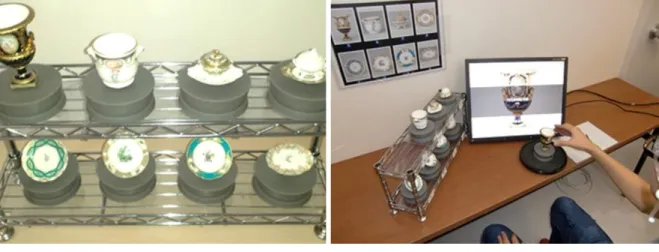
In the present study, each participant sat behind and watched an assistant (hereafter actor) operating a TUI. One of the art appreciation systems provided at the DNP Museum Lab was simplified into three different experimental TUIs composed of a screen and either tangible objects or a touch panel with object thumbnails. The effect of TUI was investigated in three conditions, i.e. using these different types of UI (two TUI and one GUI; Figure 3.1).
We adopted two TUI conditions to compare presence / absence of correspondence between the selected object and the displayed object. We also adopted third GUI condition to compare presence / absence of the grasping motion.
In the first condition, a set of small porcelain models (a total of eight different porcelains) was adopted as UI (TUI / OBJECT condition). In each task, the actor grasped one model and moved it onto a holder in front of the screen. As a result, the screen displayed a porcelain picture that corresponded to the model. Then the actor hid the porcelain picture from the screen by returning the model to the initial position. The actor conducted this task for all of the eight models (i.e. a total of eight tasks of displaying/hiding a porcelain picture).
In the second condition, eight identical can models were used as UI (TUI / CAN condition). The actor conducted a total of eight tasks of displaying / hiding a porcelain picture as in the first condition, except that the porcelain picture was not predicative from the appearance of the corresponding model.
In the third condition, thumbnails of eight different porcelains provided on the touch panel served as UI (GUI condition). In each task, the actor touched one thumbnail with a finger, as a result of which the screen displayed a porcelain picture that corresponded to the thumbnail. Then, the actor moved only a hand onto the holder in front of the screen. After that, the actor moved the hand and touched the same thumbnail to hide the porcelain
23
picture from the screen. Then the actor returned the hand to the holder. The actor conducted a total of eight tasks of displaying/hiding a porcelain picture as in the other conditions.
(a)
(b)
(c)
Figure 3.1 Settings of different conditions in Experiment 2:
(a) tangible user interface (TUI) / OBJECT condition; (b) TUI / CAN condition;
(c) graphical user interface (GUI) condition.
24
・Experimental procedure
Each participant put on an EEG electrode cap and sat 2 m behind the actor (Figure 3.2).
During the first 70 seconds, the participants stayed at rest looking at a fixation point displayed on the screen. Then, the actor started to operate the UI. The operation time for each picture was about 8.7 seconds, and a total of eight operation tasks were conducted which included all the eight different pictures in a random order. This set of tasks (about 70 seconds) was repeated once again. EEG was recorded while the participant was at rest and watching the actor's manipulation, and data from the resting state and the second set were used for the subsequent analysis. We carried out the above process for each of the three conditions, wherein the order of these conditions was counter-balanced between the participants. Each participant was instructed to sit still throughout the EEG measurement watching the hand of the actor during the manipulation and the screen during each picture displayed on the screen.
Figure 3.2 Two regions of interest (ROIs):
ROIs were defined and respective electrode sites were pooled: left central (16, 20, 21, 22) and right central (41, 49, 50, 51).
・EEG measurement and analysis
A 64-channel Ag-AgCl electrode cap, an EGI NetAmps 200 amplifier, and Netstation acquisition software (Electric Geodesics, Inc.) were used for the EEG measurement. The average of all channels was used as the reference, and sampling was done at 250 Hz, with a band-pass filter ranging from 0.3 to 100 Hz. FFT was applied to EEG segments (4.09- sec interval, 1024 points, Hanning window), which were overlapped by 50% (EMSE Data Editor 5.3, Source Signal Imaging, Inc.). Data from the first 10 seconds were excluded from the study for reason of transients in attention. The average power in 10-12 Hz was adopted as the mu rhythm power. Each power value obtained was converted into a logarithm to ensure the normality of the data. EMSE Suite Data Editor 5.3 Release
25
Candidate 3 (Source Signal Imaging, Inc.) was used for data analysis. Mu rhythm suppression in the observer watching the actor's action was adopted as the index of MNS.
Mu rhythm suppression in each condition was calculated using data from the resting state as reference. Because the sensorimotor area showed mu rhythm suppression, EEG was analyzed in two ROIs: left central (LC: 4 electrodes around C3) and right central (RC: 4 electrodes around C4).
The main effects of conditions (the TUI / OBJECT condition, the TUI / CAN condition, and the GUI condition) and brain areas (LC and RC), and their interactions were tested by two-way rm-ANOVA. For multiple comparisons, we used modified sequentially rejective bonferroni procedure. Before significance testing, outliers in each participant were identified by Smirnov-Grubbs test (p < 0.01). All the outlier channels were excluded from the subsequent statistical analyses.
[Experiment 3]
In Experiment 3, the brain activity in the operator actually operating UI was monitored to study the effect on the operator.
・Participants
The participants were 18 right-handed students (18 male, 22.1 ± 1.57 years old). Other details were the same as described above for Experiment 2.
・Experimental conditions
An ACTION condition, which involved the user action but no visual information as a results of each actions, was included in addition to the three conditions in Experiment 2 (i.e. four conditions in total). The ACTION condition was adopted to compare presence / absence of visual feedback of the consequence of the operation. The eight identical can models in the TUI / CAN condition were also used in this condition (Figure 3.3). The operator conducted a total of eight tasks as in the TUI / CAN condition, except that no porcelain picture was displayed on the screen.
26 (a)
(b)
(c)
Figure 3.3 Settings of different conditions in Experiment 3:
(a) TUI / OBJECT condition; (b) TUI / CAN condition and ACTION condition (in the ACTION condition, no porcelain picture was displayed on the screen); (c) GUI condition.
27
・Experimental procedure
Each participant put on an EEG electrode cap and sat in front of the screen. The operator stayed at rest for the first 60 seconds and then started to operate the UI. The operation time for each picture was about 13 seconds, and a total of eight operation tasks were conducted which included all the eight different pictures in a random order. This set of tasks (about 104 seconds) was repeated once again. EEG of the operator at rest and during the two sets of tasks was recorded, and data from the resting state and the second set were used for the subsequent analysis. We carried out the above process for each of the four conditions, wherein the order of these conditions was counter-balanced across the participants.
The participant was instructed to operate to a sound stimulus presented at a constant rhythm, for the sake of the consistency of the movement. The participant was also instructed to sit as still as possible during the EEG measurement and to keep looking at the manipulating hand during the manipulation and at the screen during a picture was displayed, so that the eye movement would be in line with the operation process. The participant went through enough explanation and exercise before the experiment.
・EEG measurement and analysis
For comparison with Experiment 2, the 10-12 Hz component of EEG was analyzed as in Experiment 3. Mu rhythm suppression in the operator was adopted as the index of brain activities. The main effects of conditions (the TUI / OBJECT condition, the TUI / CAN condition, the GUI condition, the ACTION condition) and brain areas (LC and RC), and their interactions were tested by two-way rm-ANOVA. The other analysis procedure was the same as Experiment 2.
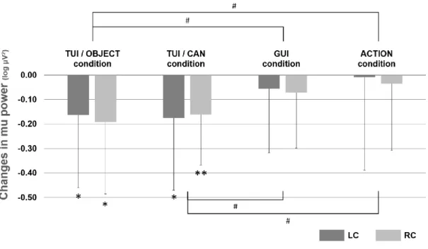
3.3 Results
[Experiment 2]
Mu rhythm suppression in each brain area due to UI was examined by comparing the power values in the mu band in the resting state and during the operation, by one sample t-test (Figure 3.4). As a result, RC in the TUI / CAN condition showed a significant difference (t (14) = -2.46; p = 0.028). In contrast, no brain area showed a significant difference in the TUI / OBJECT condition or the GUI condition. Based on the data from the resting state and the data from the operation, mu rhythm suppression was determined for LC and RC in different conditions (TUI / OBJECT condition, TUI / CAN condition, and GUI condition; Figure 3.4).
28
Figure 3.4 Changes in mu power:
Mu rhythm suppression (LC and RC) in different conditions in Experiment 2. Asterisk (*p <
0.05) means the significant mu rhythm suppression from baseline to observation of each actions. Daggers (+p < 0.10) mean the tendency in comparison with other conditions at RC.
Next, the main effect of conditions (TUI / OBJECT condition, TUI / CAN condition, and GUI condition) and brain areas (LC and RC), and their interactions were tested by two- way rm-ANOVA. The ANOVA results confirmed an interaction between condition and brain area (F (2, 28) = 4.245; p = 0.025; ηp2 = 0.233), and a main effect of condition in RC (F (2, 28) = 4.234; p = 0.025; ηp2 = 0.232). By t-test, there was a tendency that the TUI / CAN condition resulted in a higher suppression in comparison with other two conditions (t (14) = 2.50; p = 0.077 in TUI / CAN vs TUI / OBJECT, t (14) = 2.02; p = 0.077 in TUI / CAN vs GUI).
[Experiment 3]
As in Experiment 2, mu rhythm suppression in each brain area due to UI was examined by comparing the power values in the mu band in the resting state and during the operation, by one sample t-test (Figure 3.5). As a result, there are significant differences (t (17) = -3.30; p = 0.004 in TUI / CAN at RC, t (17) = -2.50; p = 0.023 in TUI / CAN at LC, t (17) = -2.32; p = 0.033 in TUI / OBJECT at LC, t (17) = -2.79; p = 0.013 in TUI/OBJECT at RC). In contrast, in the GUI condition and the ACTION condition, no brain area showed a significant difference. Based on the data from the resting state and the data from the operation, mu rhythm suppression was determined for LC and RC in different conditions (TUI condition, CAN condition, GUI condition, and ACTION condition; Figure 3.5).
29
Figure 3.5 Changes in mu power:
Mu rhythm suppression (LC and RC) in different conditions in Experiment 3. Asterisks (*p <
0.05, **p < 0.01) mean the significant mu rhythm suppression from baseline to observation of each actions. Sharps (#p < 0.05) mean the significant difference between conditions.
Then, the main effect between conditions (the TUI / OBJECT condition, the TUI / CAN condition, the GUI condition, and the ACTION condition) and between brain areas (LC and RC) were tested by two-way rm-ANOVA. In rm-ANOVA, the sphericity assumption was violated; to account for this violation, degrees of freedom were Greenhouse - Geisser adjusted. The ANOVA revealed a main effect of condition (F (2.3, 39.12) = 6.572; p = 0.002; ηp2 = 0.279). By t-test, it was shown that the TUI / OBJ condition and the TUI / CAN condition resulted in a significant suppression in comparison with the GUI condition and the ACTION condition (t (17) = 3.66; p = 0.012 in TUI / OBJECT vs GUI, t (17) = 3.65; p = 0.012 in TUI/OBJECT vs ACTION, t (17) = 3.12; p = 0.019 in TUI / CAN vs GUI, t (17) = 2.74; p = 0.042 in TUI / CAN vs ACTION).
3.4 Discussion
In Experiment 2, we found that the brain activity in the observer varied depending on the type of UI (Figure 3.4). In the TUI / CAN condition, watching the UI operation resulted in an elevated activity in RC in the observer in comparison with the resting state. This is consistent with the report that watching the action of others results in mu rhythm suppression in the somatosensory cortex, which roughly corresponds to RC (Pineda, 2005; Oberman, Pineda, & Ramachandran, 2007). Therefore, it is suggested that the brain activity was induced in this MNS-related brain area. Among brain areas, only RC showed the activity in this experiment. We suppose this was because the user action took place in the left hemifields of the observer (Shmuelof & Zohary, 2005) rather than the observer imagined imitating the right hand action of the operator as a result of watching it. This