Non-hypervascular hepatobiliary phase hypointense nodules on
Gd-EOB-DTPA-enhanced MRI as a risk factor for intrahepatic distant recurrence of
hepatocellular carcinoma after radiofrequency ablation
(肝細胞癌に対するラジオ波焼灼療法後の肝内他部位再発に寄与する因子とし てのGd-EOB-DTPA 造影 MRI の肝細胞相における低信号結節の検討) 千葉大学大学院医学薬学府 先端医学薬学専攻 (主任:露口利夫 講師) 井上将法
3
Abstract
Background and aims: Gadolinium-ethoxybenzyl-diethylenetriamine pentaacetic acid
(Gd-EOB-DTPA)-enhanced magnetic resonance imaging (MRI) performed before curative therapy for hepatocellular carcinoma (HCC) can distinguish between intrahepatic distant recurrence and hypervascularization. We aimed to retrospectively evaluate the presence of non-hypervascular hypointense nodules on hepatobiliary phase images (HBPI) from Gd-EOB-DTPA-enhanced MRI as a risk factor of the intrahepatic distant recurrence of early-stage HCC following radiofrequency ablation (RFA).
Methods: A total of 132 patients who underwent preprocedural
Gd-EOB-DTPA-enhanced MRI followed by initial RFA were retrospectively analyzed. Post-RFA intrahepatic distant recurrence, which excluded the hypervascularization of non-hypervascular hypointense nodules detected by preprocedural Gd-EOB-DTPA-enhanced MRI, was evaluated according to the presence of non-hypervascular hypointense nodules on preprocedural Gd-EOB-DTPA-enhanced MRI.
4
Results: Intrahepatic distant recurrence rates following RFA were higher in patients with
non-hypervascular hypointense nodules (1-year: 22.5, 2-year: 52.1%, 5-year: 89.1%) compared with in patients without non-hypervascular hypointense nodules (1-year: 7.0, 2-year: 28.8%, 5-year: 48.7%). The presence of non-hypervascular hypointense nodules was associated with markedly increased cumulative recurrence rates of both identical and different subsegment intrahepatic distant recurrence, being an independent risk factor for post-RFA identical and different subsegment intrahepatic distant recurrence (identical: HR = 2.365, P = 0.027. different: HR = 3.276, P <0.001).
Conclusion: The presence of non-hypervascular hypointense nodules on HBPI from
Gd-EOB-DTPA-enhanced MRI obtained prior to RFA is an important predictive factor of intrahepatic distant recurrence following RFA of HCC.
Keywords: hepatocellular carcinoma; Gd-EOB-DTPA-enhanced MRI;
non-hypervascular hypointense nodule; radiofrequency ablation; recurrence; hypervascularization
5
Introduction
Hepatocellular carcinoma (HCC) is the sixth most common cancer and third leading cause of cancer-related deaths worldwide, with about 667,000 new cases per annum [1]. Although surveillance systems of high-risk populations, radiological examination, and treatment methods have been developed and improved, most HCC patients suffer from the relapse even after curative therapy, resulting in a poor prognosis [2-4].
The newly introduced magnetic resonance imaging (MRI) contrast agent, gadolinium-ethoxybenzyl-diethylenetriamine pentaacetic acid (Gd-EOB-DTPA), has enabled the concurrent assessment of tumor vascularity and unique hepatocyte specific contrast during the acquisition of hepatobiliary phase images (HBPI) [5-9]. Gadoxetic acid is taken up by organic anion transporting polypeptide (OATP) B1/8 transporters into the hepatocytes [10-11]. OATP B1/8 expression gradually decreases during the process of hepatocarcinogenesis. Thus, borderline hepatocellular nodules that have potential to progress to overt HCC appear as non-hypervascular hypointense nodules on the HBPI (i.e., hypervascularization) [12, 13]. Hypervascularization is a new concept of recurrence
6
and proposed after the approval of Gd-EOB-DTPA-enhanced MRI.
Recent articles demonstrated the presence of non-hypervascular hypointense nodules on HBPI from Gd-EOB-DTPA-enhanced MRI observed prior to curative therapy conferred a high risk of post-curative therapy recurrence [14,15]. Moreover, these reports may indicate that the livers of patients with non-hypervascular hypointense nodules have a high potential of carcinogenesis. However, these reports did not identify the recurrence type by distinguishing between intrahepatic distant recurrence and hypervascularization. Because non-hypervascular hypointense nodules have a high probability of hypervascularization, the carcinogenetic potential of liver parenchyma cannot be estimated without classifying intrahepatic distant recurrence and hypervascularization. Furthermore, information on intrahepatic distant recurrence in patients with or without non-hypervascular hypointense nodules are required to consider treatment and follow-up strategies for patients with non-hypervascular hyponintense nodules along with hypervascular HCC. It remains controversial whether the presence of non-hypervascular hypointense nodules is a risk factor of intrahepatic distant recurrence after curative therapy. The aim of this study was to retrospectively evaluate the impact of the presence
7
of non-hypervascular hypointense nodules on HBPI of Gd-EOB-DTPA enhanced MRI on intrahepatic distant recurrence after radiofrequency ablation (RFA) of early-stage HCC.
8
Patients and Methods
Patients
The research ethics committee of our institution approved this study (No. 2,148), and the
requirement for the informed consent of treatment was waived. Medical records were
retrieved for patients with HCC treated at our institution.
Patients enrolled in this study were selected using the following inclusion criteria at
their initial RFA: (1) the presence of histologically confirmed or clinically diagnosed
HCC (fulfilling the criteria for lesions with typical imaging) [16], (2) presence of early-stage HCC (single hypervascular HCC lesion of ≤ 50 mm or ≤ 3 hypervascular HCC
lesions of ≤ 30 mm, without macrovascular invasion or extrahepatic metastasis), (3) Child–Pugh A or B, and (4) Gd-EOB-DTPA-enhanced MRI performed prior to initiation
of RFA.
The RFA procedure was performed percutaneously under conscious sedation. We
used real-time ultrasound guidance (Power Vision 8000, Aplio XV, Aplio XG, or Aplio
9
electrode (Cool-Tip; RF Ablation System, Covidien, Boulder, Colombia, CO) was
inserted into the tumor through the liver tissue, and then radiofrequency energy was
delivered for 6–15 min in each application. After one to two sessions of RFA, dynamic
computed tomography (CT) or Gd-EOB-DTPA-enhanced MRI was performed to
evaluate the efficacy of RFA. During treatment assessment, we compared imaging
findings for early, portal venous, and late phases prior to ablation and portal venous and
late phases following ablation. A lesion was judged to be completely ablated when the
non-enhanced area appearing in the portal venous or late phase of CT or MRI postablation
covered the entire lesion shown in all early, portal venous, and late phases of CT or MRI
preablation with a safety margin in the surrounding liver parenchyma [17]. To reduce
local recurrence after RFA, we consider a safety of at least 5 mm.
Gd-EOB-DTPA-enhanced MRI before intimal RFA
Gd-EOB-DTPA-enhanced MRI was performed prior to RFA for the diagnosis and
staging of HCC in all patients. MRI was performed using a 1.5-T scanner (Intera Achieva
10
coil, a 1.5-T scanner (Signa HDxt; GE Healthcare), or a 3-T scanner (Discovery MR 750;
GE Healthcare) with a twelve-channel Torso-array coil. We obtained T1-weighted dual
fast field echo images (in-phase and opposed phase) before injecting the contrast medium.
Dynamic fat-suppressed T1-weighted 3D turbo field-echo images were obtained before
and 23 s (arterial phase), 80 s (portal venous phase), 240 s (late dynamic phase), and 20
min (hepatobiliary phase) after the administration of the contrast agent. Breath-hold
multi-shot T2-weighted images and single shot spin-echo echo-planar diffusion-weighted
images (DWI) were obtained in the interval of the dynamic study. Patients received 0.025
mmol/kg body weight of gadoxetic acid (EOB Primovist; Bayer Health Yakuhin Co., Ltd.,
Osaka, Japan) at a rate of 2 mL/s through a cubital venous catheter, which was flushed
with 20 mL of saline using a power injector.
Imaging analysis
Pretreatment Gd-EOB-DTPA-enhanced MRI, including enhanced T1- and T2-weighted
images, DWI, dynamic imaging including arterial, portal venous, and late dynamic phases,
11
(To note, our hepatologists collectively have 13 years of experience with reading
Gd-EOB-DTPA-enhanced MRIs). First, the reviewers evaluated the HBPI and detected
hypointense nodules in the liver. For these nodules, the relative enhanced degree of the
nodules on the arterial phase images was determined with regard to whether or not
hyperintensity was present. Hemangiomas and hepatic cyst identified based on classical
enhancement pattern or nonenhancement on dynamic phase images and hyperintensity
on T2-weighted MR images, respectively, were excluded. When imaging assessment was
discordant between the two reviewers, consensus was obtained through discussion.
Follow-up and assessment of recurrence
During follow-up, tumor markers, including alfa fetoprotein (AFP) and des-γ-carboxy prothrombin, were measured every 1–2 months, and dynamic CT or Gd-EOB-DTPA-enhanced MRI was performed every 4–6 months. In case of suspected recurrence, at least two radiological imaging examinations, including dynamic CT, Gd-EOB-DTPA-enhanced MRI, contrast Gd-EOB-DTPA-enhanced ultrasound (US), and/or angiographic CT, were performed to confirm diagnosis.
12
In this study, the development of recurrence was further defined as local recurrence, intrahepatic distant recurrence, hypervascularization, or extrahepatic recurrence. Local recurrence was defined as the reappearance of enhancing tumor tissue adjacent to the ablation zone after the achievement of treatment success [18]. Intrahepatic distant recurrence was defined as the development of new hypervascular tumors, which were not classified as non-hypervascular hypointense nodules on HBPI from pretreatment Gd-EOB-DTPA-enhanced MRI and were not adjacent to the treated site. An emerging non-hypervascular hypointense nodule detected via Gd-EOB-DTPA-enhanced MRI at the follow-up assessment was not defined as intrahepatic distant recurrence until hypervascularization was confirmed. Hypervascularization of non-hypervascular hypointense nodules on HBPI of pre-RFA Gd-EOB-MRI-enhanced MRI was defined as nodules that showed hypervascularity in arterial phase images and hypovascularity in portal venous or late phase images on at least two radiological imaging examinations, including dynamic CT, Gd-EOB-DTPA-enhanced MRI, contrast-enhanced US, and/or angiographic CT [12, 13]. The appearance of extrahepatic metastasis was also assessed on follow-up imaging.
13
We also categorized intrahepatic distant recurrence as being subsegmentally either identical or different relative to the subsegment of the original nodules. In this study, subsegments 1–8 correspond to the Couinaud’s segments 1–8, respectively according to the General Rules for the Clinical and Pathological Study of Primary Liver Cancer, Third English Edition [19]. When a tumor was located in two or more subsegments, the subsegment involving the major part of the tumor was considered.
Statistical analysis
The cumulative incidence of each type of recurrence was estimated using the Kaplan– Meier method. Time to intrahepatic distant recurrence was defined as the period from initial RFA until the diagnosis of such recurrence. If extrahepatic metastasis was observed before intrahepatic distant recurrence, the censoring date was defined as the last date of radiological assessment not showing such evidence of recurrence. If local recurrence or hypervascularization was treated by resection or local ablation, we continued follow-up until the initial intrahepatic distant recurrence. If local recurrence or hypervascularization was treated by other therapies, such as Tranarterial chemoembolization, the censoring
14
date was defined as the last date of radiological assessment. Time to hypervascularization was defined as the period from initial RFA until the diagnosis of hypervascularization.
Univariate and multivariate Cox proportional-hazard regression models were used to estimate the hazard ratios for risk factors in relation to each type of recurrence. A p value of <0.05 was considered statistically significant. All statistical analyses were performed using SPSS statistical software (version 22; SPSS-IBM, Chicago, IL, USA). In multivariate analyses, we used the stepwise regression program. All variables indicated in the tables were included in the multivariate analysis. The results are demonstrated in the Tables.
15
Results
Patient characteristics at baseline
Between February 2008 and December 2012, 188 patients with HCC who received RFA for initial treatment of HCC were identified for potential inclusion in this study (Supplementary Fig. 1). Of the 132 patients who were eventually included in the study, the majority of patients were male (67%), Hepatitis C virus (HCV)-RNA-positive (67%), Child–Pugh class A (83%), and the median age was 72 years (range, 52–83) (Table 1). In 56 patients (42%), non-hypervascular hypointense nodules were detected on HBPI from pretreatment Gd-EOB-DTPA-enhanced MRI.
Intrahepatic distant recurrence after RFA according to non-hypovascular hypointense nodules
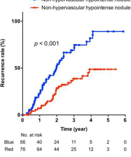
Sixty-eight patients (52%) developed intrahepatic distant recurrence during the follow-up period (median follow-follow-up, 24.6 months (range, 1.0–70.7 months). In the 56 patients with non-hypervascular hypointense nodules, the estimated 1-, 2-, 3-, and 5-year
16
cumulative recurrence rates were 22.5, 52.1, 69.0, and 89.1%, respectively, compared with 7.0, 28.8, 38.7, and 48.7%, respectively, in the 76 patients without non-hypervascular hypointense nodules (Fig. 1). We also analyzed data on the identical vs. different subsegmental intrahepatic distant recurrence (Fig. 2). The estimated 1-, 2-, 3-, and 5-year cumulative identical subsegmental intrahepatic distant recurrence rates were 7.4, 22.1, 33.5, and 56.6%, respectively, in patients with non-hypervascular hypointense nodules, compared with 1.5, 11.0, 12.8, and 23.1%, respectively, in patients without non-hypervascular hypointense nodules (Fig. 2A). On the other hand, the estimated 1-, 2-, 3-, and 5-year cumulative different subsegmental intrahepatic distant recurrence rates were 20.6, 48.3, 67.3, and 83.4%, respectively, in patients with non-hypervascular hypointense nodules, compared with 5.6, 19.5, 32.9, and 39.6%, respectively, in patients without non-hypervascular hypointense nodules (Fig. 2B). Multivariate analysis showed that the cumulative incidence of intrahepatic distant recurrence was significantly higher in patients with hypervascular hypointense nodules than in patients without non-hypervascular hypointense nodules (Table 2). Similarly, non-non-hypervascular hypointense nodules on HBPI from pretreatment Gd-EOB-DTPA-enhanced MRI was an independent
17
risk factor of both the identical and different subsegmental intrahepatic distant recurrence (Supplementary Tables 1 and 2).
Non-hypervascular hypointense nodules and hypervascularization
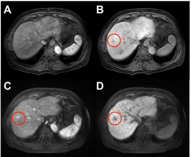
The agreement rate of detecting non-hypervascular hypointense nodules via pretreatment Gd-EOB-DTPA-enhanced MRI between the two reviewers was 76.5% (101 of 132 patients). The remaining 31 patients required consensus between the two reviewers. Consequently, we confirmed 56 patients with non-hypervascular hypointense nodules, and we determined a total of 127 non-hypervascular hypointense nodules with a median size of 8 mm (range: 3-17 mm). According to follow-up radiological assessment of these 56 patients, there was an 82.1% agreement rate between the two reviewers. In the follow-up period, 30 patients (46 nodules) developed the hypervascularization of non-hypervascular hypointense nodules as detected by pretreatment Gd-EOB-DTPA enhanced MRI (a typical case is shown in Figure 3). The 1-, 2-, 3-, and 5-year hypervascularization rates were 26.1, 51.9, 57.7, and 65.4%, respectively. The median time of progression to hypervascularization was 22.4 months (95% CI, 7.7–37.1 months; Supplementary Fig. 2).
18
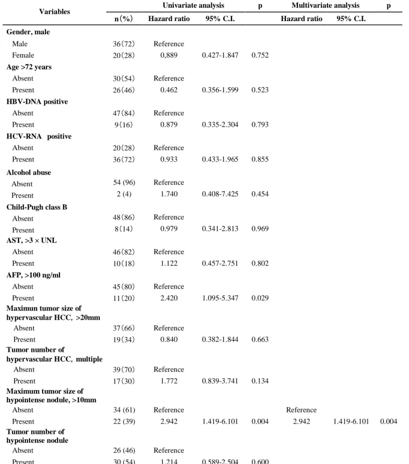
Table 3 shows the multivariate analysis of risk factors for hypervascularization in 56 patients with non-hypervascular hypointense nodules on HBPI from pretreatment Gd-EOB-DTPA-enhanced MRI. The maximum size of non-hypervascular hypointense nodules (>10 mm) was an independent predictive factor of hypervascularization.
19
Discussion
The present study showed that non-hypervascular hypointense nodules on HBPI from pretreatment Gd-EOB-DTPA enhanced MRI represent a risk factor of HCC intrahepatic distant recurrence. Our findings confirm observations from other investigations, extending these in several important directions. As established by the significant studies of Toyoda et al. and Lee et al., non-hypervascular hypointense nodules are at a higher risk of recurrence after curative therapy of HCC such as hepatectomy or RFA [14, 15]. Unlike previous studies, we classified post-RFA recurrence as intrahepatic distant recurrence occurring in the liver parenchyma and hypervascularization of non-hypervascular hypointense nodules on HBPI of pre-treatment Gd-EOB-DTPA-enhanced MRI. Our results indicate that livers with non-hypervascular hypointense nodules have a high potential of carcinogenesis.
Carcinogenesis and spread from the primary tumor are widely believed to represent the two major mechanisms underlying intrahepatic recurrence of HCC [20]. The diseased liver undergoes carcinogenesis (i.e., de novo recurrence) and dissemination of the
20
remnant tumor cells related to the primary tumor after curative therapy, leading to intrahepatic metastasis. Toyoda et al. indicated that the presence of non-hypervascular hypointense nodule was a risk factor for multicentric occurrence but not for intrahepatic metastasis, which was defined based on the radiological location and pathological examination of recurring nodules relative to non-hypervascular hypointense nodules [14]. The high risk of multicentric occurrence is attributed to the high potential of carcinogenesis. According to Toyoda’s study, hypervascularization of non-hypervascular hypointense nodules was classified as multicentric occurrence [14]. In contrast, we focused on intrahepatic distant recurrence, leaving out hypervascularization of non-hypervascular hypointense nodules. Intrahepatic distant recurrence in our approach was defined as recurrence in the liver parenchyma that lacked non-hypervascular hypointense nodules at pre-RFA. Specifically, intrahepatic distant recurrence included multicentric occurrence, except for the hypervascularization of non-hypervascular hypointense nodules at baseline and intrahepatic metastasis. As our study lacked pathological examination data, we could not classify multicentric occurrence and intrahepatic metastasis as well as the previous report did. This is a limitation of our study.
21
Conversely, hepatogenous spread of cancer cells may correlate with the local portal venous system, and notably, intrahepatic metastasis mainly occurs at the subsegment identical to that of the primary tumor. Based on this idea, anatomical resection involving the systemic removal of the hepatic spread contained in the tumor tissue is considered preferable [21, 22]. Thus, the risk of developing recurrence in the identical and different subsegments was separately analyzed in this study. Non-hypervascular hypointense nodules were found to be associated with a higher rate of intrahepatic distant recurrence at not only the different subsegment but also the identical subsegment. Taken together, differences in the incidence of intrahepatic distant recurrence between patients with or without non-hypervascular hypointense nodules was most likely not due to intrahepatic metastasis but due to potential carcinogenesis. Our results document the importance that patients with non-hypervascular hypointense nodules have high incidence of not only hypervascularizaiton but also intrahepatic distant recurrence. This information will be clinically useful during follow-up after RFA.
In this study, the incidence of non-hypervascular hypointense nodules on HBPI from Gd-EOB-DTPA-enhanced MRI (42%, 56/132) does not concur with that identified in
22
previous studies (Toyoda et al.: 23%, 18/77; Lee et al.: 79%, 110/139) [14, 15]. We consider that these differences might be caused by disease etiology and treatment procedures (resection/RFA) of the study patients. The etiology linked to HCC in the patients included in our study was HCV infection, whereas the etiology in the previous study by Lee et al. was HBV infection [15]. In general, liver function, tumor size, and tumor number are all taken into account when choosing between resection and RFA [16]. Selection bias might affect the rate of non-hypervascular hypointense nodules developing in the study population. On the other hand, the cumulative hypervascularization rate of non-hypervascular hypointense nodules in this study is similar to that found of previous studies [12, 13]. This result suggests that the method for the detection of non-hypervascular hypointense nodules in our study was appropriate.
Our study had several other limitations in addition to its inability to distinguish between multicentric occurrence and intrahepatic metastasis. First, this study was based on a retrospective analysis, and thus, there may be selection bias of patients and differences in the interval of follow-up radiological assessments. Additionally, the number of patients was small, and this study was limited to HCC patients treated by RFA.
23
To note, this study was not able to assess overall survival due to a small observation period. Large scale, multicenter, and prospective studies with longer observation periods to assess the dynamics of non-hypervascular hypointense nodules detected via Gd-EOB-DTPA-enhanced-MRI are warranted.
This study confirms that rigorous and careful follow-up after treatment are required to enable the identification of not only the hypervascularization of non-hypervascular hypointense nodules but also intrahepatic distant recurrence for patients presenting with non-hypervascular hypointense nodules on HBPI from pretreatment Gd-EOB-DTPA enhanced MRI. Furthermore, the finding that livers with non-hypervascular hypointense nodules have a high potential of intrahepatic distant recurrence supports the view that current post-curative therapies, including the early detection of hypervascularization of non-hypervascular hypointense nodules and intervention, are insufficient. Treatment strategies that deteriorate a potential of carcinogenesis (i.e., nucleos(t)ide analogues for HBV or interferon-free direct acting antivirals for HCV) should be considered for these patients [23-25].
24
from Gd-EOB-DTPA-enhanced MRI obtained prior to RFA is a significant predictive factor of intrahepatic distant recurrence following RFA. Observations from preprocedure Gd-EOB-DTPA-enhanced MRI are critical to developing post-RFA management strategies in patients with early-stage HCC.
25
References
1. Farazi PA, DePinho RA. Hepatocellular carcinoma pathogenesis: from genes to environment. Nat Rev Cancer. 2006;6:674-687.
2. Llovet JM, Schwartz M, Mazzaferro V. Resection and liver transplantation for hepatocellular carcinoma. Semin Liver Dis. 2005;25:181-200.
3. Tung-Ping Poon R, Fan ST, Wong J. Risk factors, prevention, and management of postoperative recurrence after resection of hepatocellular carcinoma. Ann Surg. 2000;232:10-24.
4. Shiina S, Tateishi R, Arano T, Uchino K, Enooku K, Nakagawa H, et al. Radiofrequency ablation for hepatocellular carcinoma: 10-year outcome and prognostic factors. Am J Gastroenterol. 2012;107:569-577.
5. Hamm B, Staks T, Mühler A, Bollow M, Taupitz M, Frenzel T, et al. Phase I clinical evaluation of Gd-EOB-DTPA as a hepatobiliary MR contrast agent: safety, pharmacokinetics, and MR imaging. Radiology. 1995;195:785-792.
26
al. Liver tumors: comparison of MR imaging with Gd-EOB-DTPA and Gd-DTPA. Radiology. 1996;200:59-67.
7. Kim SH, Kim SH, Lee J, Kim MJ, Jeon YH, Park Y, et al. Gadoxetic acid-enhanced MRI versus triple-phase MDCT for the preoperative detection of hepatocellular carcinoma. AJR Am J Roentgenol. 2009;192:1675-1681.
8. Ooka Y, Kanai F, Okabe S, Ueda T, Shimofusa R, Ogasawara S, et al. Gadoxetic acid-enhanced MRI compared with CT during angiography in the diagnosis of hepatocellular carcinoma. Magn Reson Imaging. 2013;31:748-754.
9. Van Beers BE, Pastor CM, Hussain HK. Primovist, Eovist: what to expect? J Hepatol. 2012;57:421-429.
10. Kitao A, Matsui O, Yoneda N, Kozaka K, Shinmura R, Koda W. The uptake
transporter OATP8 expression decreases during multistep hepatocarcinogenesis: correlation with gadoxetic acid enhanced MR imaging. Eur Radiol. 2011;21:2056-2066.
11. Kobayashi S, Matsui O, Gabata T, Koda W, Minami T, Ryu Y, et al. Intranodular
27
illustrate multi-step hepatocarcinogenesis within the nodule on Gd-EOB-DTPA-enhanced MRI. Eur J Radiol. 2012;81:3839-3845.
12. Inoue T, Hyodo T, Murakami T, Takayama Y, Nishie A, Higaki A, et al.
Hypovascular hepatic nodules showing hypointense on the hepatobiliary-phase
image of Gd-EOB-DTPA-enhanced MRI to develop a hypervascular hepatocellular
carcinoma: a nationwide retrospective study on their natural course and risk factors.
Dig Dis. 2013;31:472-479.
13. Hyodo T, Murakami T, Imai Y, Okada M, Hori M, Kagawa Y, et al. Hypovascular
nodules in patients with chronic liver disease: risk factors for development of
hypervascular hepatocellular carcinoma. Radiology. 2013;266:480-490.
14. Toyoda H, Kumada T, Tada T, Ninomi T, Ito T, Sono Y, et al. Non-hypervascular
hypointense nodules detected by Gd-EOB-DTPA-enhanced MRI are a risk factor for recurrence of HCC after heaptectomy. J Hepatol. 2013;58:1174-1180.
15. Lee DH, Lee JM, Lee JY, Kim SH, Kim JH, Yoon JH, et al. Non-hypervascular
heaptobilliary phase hypointense nodules on gadoxetic acid-enhaced MRI: Risk of HCC recurrence after radiofrequency adblation. J Hepatol. 2015;62:1122-1130.
28
16. Kudo M, Izumi N, Kokudo N, Matsui O, Sakamoto M, Nakashima O, et al.
Management of hepatocellular carcinoma in Japan: Consensus-Based Clinical Practice Guidelines proposed by the Japan Society of Hepatology (JSH) 2010 updated version. Dig Dis. 2011;29:339-364.
17. Motoyama T, Ogasawara S, Chiba T, Higashide T, Yokota H, Kanogawa N, et al.
Coronal reformatted CT images contribute to the precise evaluation of the radiofrequency ablative margin for hepatocellular carcinoma. Abdom Imaging. 2014;39:262-268.
18. Goldberg SN, Grassi CJ, Cardella JF, Charboneau JW, Dodd GD 3rd, Dupuy DE, et
al. Image-guided tumor ablation: standardization of terminology and reporting criteria. Radiology. 2005;235:728-739
19. Liver Cancer Study Group of Japan. General Rules for the Clinical and Pathological
Study for Primary Liver Cancer, Third English Edition. Tokyo; Kanehara 2010. 20. Sherman M. Recurrence of hepatocellular carcinoma. N Engl J Med.
2008;359:2045-2047
29 Surg Gynecol Obstet. 1985;161:346-350.
22. Hasegawa K, Kokudo N, Imamura H, Matsuyama Y, Aoki T, Minagawa M, et al.
Prognostic impact of anatomic resection for hepatocellular carcinoma. Ann Surg.
2005;242:252-259.
23. Papatheodoridis GV, Chan HL, Hansen BE, Janssen HL, Lampertico P. Risk of
hepatocellular carcinoma in chronic hepatitis B: assessment and modification with
current antiviral therapy. J Hepatol. 2015;62:956-967.
24. Shiratori Y, Kato N, Yokosuka O, Imazeki F, Hashimoto E, Hayashi N, et al.
Predictors of the efficacy of interferon therapy in chronic hepatitis C virus infection.
Tokyo-Chiba Hepatitis Research Group. Gastroenterology. 1997;113:558-566.
25. Kanogawa N, Ogasawara S, Chiba T, Saito T, Motoyama T, Suzuki E, et al.
Sustained virologic response achieved after curative treatment of hepatitis C
virus-related hepatocellular carcinoma as an independent prognostic factor. J
30
Table 1. Demographics and characteristics of study patients
Demographics/characteristics Value Gender [n(%)] Male 89 (67) Female 43 (33) Age, years [n(%)] ≤72 69 (52) >72 63 (48) Median (range) 72 (52-83) HBV-DNA positive [n(%)] Absent 117 (89) Present 15 (11) HCV-RNA positive [n(%)] Absent 44 (33) Present 88 (67) Alcohol abuse [n(%)] Absent 120 (91) Present 12 (9) Cirrhosis [n(%)] Absent 28 (21) Present 104 (79) Child–Pugh class [n(%)] A 109 (83) B 23 (17) AST [n(%)] ≤3.0×UNL 114 (86) >3.0×UNL 18 (14) AFP [n(%)] ≤100 112 (85) >100 20 (15)
Tumur size of hypervascular HCC, mm Maximum diameter,> 20 [n(%)]
Absent 85(64)
Present 47(36)
Median (range) 19 (6-40)
Tumor number of hypervascular HCC,multiple [n(%)]
Absent 95 (72)
Present 37 (28)
Non-hypervascular hypointense nodule on HBPI [n(%)]
Absent 76(58)
Present 56(42)
Abbreviations: HBV, hepatitis B virus; HCV, hepatitis C virus; AST, aspartate aminotransferase; AFP, alpha-fetoprotein; HCC, hepatocellular carcinoma; HBPI, hepatobiliary phase image
31
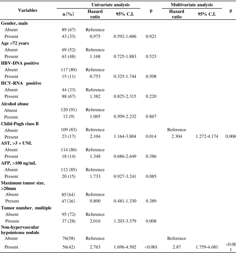
Table 2. Uni- and multivariate analyses of intrahepatic distant recurrence following radiofrequency ablation
Variables Univariate analysis p Multivariate analysis p n(%) Hazard ratio 95% C.I. Hazard ratio 95% C.I. Gender, male Absent 89 (67) Reference Present 43 (33) 0,975 0.592-1.606 0.921 Age >72 years Absent 69 (52) Reference Present 63 (48) 1.168 0.725-1.883 0.523 HBV-DNA positive Absent 117 (89) Reference Present 15 (11) 0.753 0.325-1.744 0.508 HCV-RNA positive Absent 44 (33) Reference Present 88 (67) 1.382 0.825-2.315 0.220 Alcohol abuse Absent 120 (91) Reference Present 12 (9) 1.065 0.509-2.232 0.867 Child-Pugh class B
Absent 109 (83) Reference Reference
Present 23 (17) 2.104 1.164-3.804 0.014 2.304 1.272-4.174 0.006 AST, >3 × UNL Absent 114 (86) Reference Present 18 (14) 1.348 0.686-2.649 0.386 AFP, >100 ng/mL Absent 112 (85) Reference Present 20 (15) 1.733 0.927-3.241 0.085 Maximum tumor size,
>20mm Absent 85(64) Reference Present 47(36) 0.800 0.481-1.330 0.389 Tumor number,multiple Absent 95 (72) Reference Present 37 (28) 2.016 1.203-3.379 0.008 Non-hypervascular hypointense nodule
Absent 76(58) Reference Reference
Present 56(42) 2.763 1.696-4.502 <0.001 2.87 1.759-4.681 <0.00 1 Abbreviations: HBV, hepatitis B virus; HCV, hepatitis C virus; AST, aspartate aminotransferase; AFP, alpha-fetoprotein
32
Table 3. Uni- and multivariate analyses of the hypervascularization of non-hypervascular hypointense
nodules
Variables Univariate analysis p Multivariate analysis p
n(%) Hazard ratio 95% C.I. Hazard ratio 95% C.I.
Gender, male Male 36(72) Reference Female 20(28) 0,889 0.427-1.847 0.752 Age >72 years Absent 30(54) Reference Present 26(46) 0.462 0.356-1.599 0.523 HBV-DNA positive Absent 47(84) Reference Present 9(16) 0.879 0.335-2.304 0.793 HCV-RNA positive Absent 20(28) Reference Present 36(72) 0.933 0.433-1.965 0.855 Alcohol abuse Absent 54 (96) Reference Present 2 (4) 1.740 0.408-7.425 0.454 Child-Pugh class B Absent 48(86) Reference Present 8(14) 0.979 0.341-2.813 0.969 AST, >3 × UNL Absent 46(82) Reference Present 10(18) 1.122 0.457-2.751 0.802 AFP, >100 ng/ml Absent 45(80) Reference Present 11(20) 2.420 1.095-5.347 0.029
Maximun tumor size of hypervascular HCC,>20mm Absent 37(66) Reference Present 19(34) 0.840 0.382-1.844 0.663 Tumor number of hypervascular HCC,multiple Absent 39(70) Reference Present 17(30) 1.772 0.839-3.741 0.134
Maximum tumor size of hypointense nodule, >10mm
Absent 34 (61) Reference Reference
Present 22 (39) 2.942 1.419-6.101 0.004 2.942 1.419-6.101 0.004 Tumor number of
hypointense nodule
Absent 26 (46) Reference
Present 30 (54) 1.214 0.589-2.504 0.600
Abbreviations: HBV, hepatitis B virus; HCV, hepatitis C virus; AST, aspartate aminotransferase; AFP, alpha-fetoprotein; HCC, hepatocellular carcinoma
33
Figure 1. Kaplan–Meier curve of cumulative intrahepatic distant recurrence (blue line: presece of hypervascular hypointense nodules, red line: absence of non-hypervascular hypointense nodules).
35
Figure 2. Kaplan–Meier curve of cumulative intrahepatic distant recurrence (blue line: presece of hypervascular hypointense nodules, red line: absence of non-hypervascular hypointense nodules) . A: Rates of intrahepatic distant recurrence at
the identical subsegment in patients with or without concurrent non-hypervascular hypointense nodules detected during the HBPI of pre-RFA Gd-EOB-DTPA-enhanced MRI. B: Rates of intrahepatic distant recurrence at different subsegments in patients with or without concurrent non-hypervascular hypointense nodules detected during the HBPI of pre-RFA Gd-EOB-DTPA-enhanced MRI.
36
Figure 3. A representative case of hypervascularization of non-hypervascular hypointense nodules in patients with hepatocellular carcinoma as detected by gadolinium-ethoxybenzyl-diethylenetriamine pentaacetic acid (Gd-EOB-DTPA) enhanced MRI. A 61-year-old male received radiofrequency ablation (RFA) as an
initial treatment for hepatocellular carcinoma (A: arterial image and B: hepatobiliary phase image (HBPI) of preprocedural Gd-EOB-DTPA-enhanced MRI).
Non-hypervascular hypointense nodules were observed in another site of the liver
parenchyma from which we performed RFA. Twenty-one months after RFA (C: arterial image and D: HBPI of preprocedural Gd-EOB-DTPA-enhanced MRI),
37
Supplementary Table 1. Uni- and multivariate analyses of identical subsegmental intrahepatic distant
recurrence following radiofrequency ablation
Variables Univariate analysis p Multivariate analysis p n(%) Hazard ratio 95% C.I. Hazard ratio 95% C.I. Gender, male Absent 89 (67) Reference Present 43 (33) 0.956 0.457-2.002 0.906 Age >72 years Absent 69 (52) Reference Present 63 (48) 1.012 0.497-2.060 0.975 HBV-DNA positive Absent 117 (89) Reference Present 15 (11) 1.417 0.542-3.705 0.477 HCV-RNA positive Absent 44 (33) Reference Present 88 (67) 0.888 0.431-1.832 0.749 Alcohol abuse Absent 120 (91) Reference Present 12 (9) 0.882 0.268-2.907 0.837 Child-Pugh class B
Absent 109 (83) Reference Reference
Present 23 (17) 3.190 1.464-6.954 0.004 3.474 1.570-7.688 0.002 AST, >3 × UNL
Absent 114 (86) Reference
Present 18 (14) 0.759 0.230-2.509 0.651 AFP, >100 ng/mL
Absent 112 (85) Reference Reference
Present 20 (15) 4.686 2.186-10.046 <0.001 5.609 2.473-12.722 <0.001 Maximum tumor size,
>20mm
Absent 85(64) Reference Reference
Present 47(36) 0.525 0.226-1.219 0.134 0.420 0.174-1.010 0.053 Tumor number,multiple Absent 95 (72) Reference Present 37 (28) 2.321 1.122-4.801 0.023 Non-hypervascular hypointense nodule
Absent 76(58) Reference Reference
Present 56(42) 2.980 1.423-6.239 0.004 2.365 1.100-5.082 0.027 Abbreviations: HBV, hepatitis B virus; HCV, hepatitis C virus; AST, aspartate aminotransferase; AFP, alpha-fetoprotein
38
Supplementary Table 2. Uni- and multivariate analyses of different subsegmental intrahepatic distant
recurrence following radiofrequency ablation
Variables Univariate analysis p Multivariate analysis p n(%) Hazard ratio 95% C.I. Hazard ratio 95% C.I. Gender, male Absent 89 (67) Reference Present 43 (33) 1.091 0.637-1.867 0.752 Age >72 years Absent 69 (52) Reference Present 63 (48) 1.124 0.679-1.860 0.649 HBV-DNA positive Absent 117 (89) Reference Present 15 (11) 0.612 0.245-1.531 0.294 HCV-RNA positive
Absent 44 (33) Reference Reference
Present 88 (67) 1.668 0.952-2.924 0.074 1.907 1.081-3.364 0.026 Alcohol abuse
Absent 120 (91) Reference
Present 12 (9) 1.029 0.468-2.265 0.943 Child-Pugh class B
Absent 109 (83) Reference Reference
Present 23 (17) 1.785 0.947-3.365 0.073 2.189 1.150-4.163 0.017 AST, >3 × UNL Absent 114 (86) Reference Present 18 (14) 1.626 0.821-3.220 0.163 AFP, >100 ng/mL Absent 112 (85) Reference Present 20 (15) 1.107 0.545-2.249 0.778 Maximum tumor size,
>20mm Absent 85(64) Reference Present 47(36) 0.797 0.463-1.370 0.411 Tumor number,multiple Absent 95 (72) Reference Present 37 (28) 1.329 0.758-2.331 0.321 Non-hypervascular hypointense nodule
Absent 76(58) Reference Reference
Present 56(42) 3.108 1.840-5.248 <0.001 3.276 1.937-5.540 <0.001 Abbreviations: HBV, hepatitis B virus; HCV, hepatitis C virus; AST, aspartate aminotransferase; AFP, alpha-fetoprotein
39
40
Supplementary Figure 2. Kaplan–Meier curve of the cumulative
hypervascularization rate of non-hypervascular hypointense nodules on pre-RFA
41