Invasive micropapillary carcinoma
成分を伴った S 状結腸癌の 1 例
田中 涼太
1)阿古 英次
1)亀谷 直樹
3)加藤 幸裕
1)河本 真大
1)金原 功
1)山本 篤
1)山田 靖哉
1)西村 重彦
1)妙中 直之
1)藤田 茂樹
2) 1) 一般財団法人住友病院外科 2) 一般財団法人住友病院病理部 3) 大阪市立大学付属病院腫瘍外科 症例は 64 歳の男性で,排便時の出血を主訴に近医受診,精査加療目的にて当科紹介受診となった.下部 消化管内視鏡検査で S 状結腸に表面不整の I 型腫瘍を認め,生検で高分化型腺癌と診断された.胸腹部造 影 CT では明らかなリンパ節転移や遠隔転移を認めず,腹腔鏡下 S 状結腸切除術,D2 郭清術を施行した. 病理組織学的検査所見では異型細胞が微小乳頭状構造を形成し,間質との間に空隙を伴う浸潤性微小乳頭 癌(invasive micropapillary carcinoma;以下,IMPC と略記)の像を呈した.最終診断は S 状結腸原発の IMPC,pT2(MP),N1,M0:fStage IIIa であった.術後経過は良好であり,現在は外来通院にて UFT+LV による術後補助化学療法施行中である.IMPC は最初に乳癌で報告されて以来,他臓器での報告も増加し てきているが大腸原発の IMPC の報告例は比較的少ない.また,発生臓器にかかわらず高率にリンパ管侵 襲やリンパ節転移を伴うことから予後不良であり,治療上注意が必要である.キーワード:浸潤性微小乳頭癌,IMPC,大腸癌
はじめに
浸潤性微小乳頭癌(invasive micropapillary carcinoma;以下,IMPC と略記)は 1993 年に Siriaunkgul ら1)
によって,高率にリンパ管侵襲やリンパ節転移を伴う生物学的悪性度の高い浸潤性乳管癌の一亜型として 提唱された概念である.最初に乳癌で報告されて以来,尿路系,肺,唾液腺など他臓器での報告例が増加 しているが,大腸原発の IMPC の報告例はまだ少ない.今回,我々は腹腔鏡下に切除しえた IMPC 成分を 伴った S 状結腸癌の 1 例を経験したので,若干の文献的考察を加え報告する.
症
例
患者:64 歳,男性 主訴:排便時出血 既往歴:高血圧 家族歴:特記事項なし. 現病歴:2014 年 7 月に排便時出血を主訴に近医を受診し,下部消化管内視鏡検査を施行したところ,S 状結腸に腫瘍性病変を認めたため,加療目的に当科紹介受診し,8 月に手術目的にて入院となった. 入院時現症:身長 169 cm,体重 54 kg.眼瞼結膜に貧血なく,眼球結膜に黄染なし.腹部に異常所見を 〈2015 年 10 月 27 日受理〉別刷請求先:田中 涼太 〒 530-0005 大阪市北区中之島 5 丁目 3 番 20 号 一般財団法人住友病院外科認めなかった.
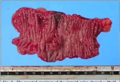
血液検査所見:Hb 12.7 g/dl,軽度の貧血を認めた.腫瘍マーカーは CEA 2.4 ng/ml,CA19-9 3 U/ml と上 昇を認めなかった. 下部消化管内視鏡検査所見:S 状結腸に表面不整の I 型腫瘍を認め,生検の結果,高分化型腺癌と診断 された(Fig. 1A). 胸腹部造影 CT 所見:S 状結腸の腫瘍は指摘できず,リンパ節転移や遠隔転移を疑う所見を認めなかっ た(Fig. 1B, C). 以上より,S 状結腸癌 cType I,cMP,cN0,cH0,cP0,cM0,cStage II の診断にて D2 リンパ節郭清を伴 う腹腔鏡下 S 状結腸切除術を施行した. 手術所見:肝転移巣や腹水,腹膜播種結節を認めず,腫瘍の漿膜外への浸潤も認めなかった. 切除標本肉眼所見:S 状結腸に 19×15 mm 大の I 型病変を認めた(Fig. 2). 病理組織学的検査所見:異型細胞が微小乳頭状構造を形成し,間質との間に空隙を伴う IMPC の像を呈 し,一部に高分化型腺癌を認めた(Fig. 3a, b).また,免疫組織学的検査では epithelial membrane antigen (以下,EMA と略記)染色では乳頭構造の表面が陽性となる inside-out growth pattern を示していた (Fig. 3c).また,リンパ管内皮細胞を染色する抗 D2-40 染色において,粘膜下層でのリンパ管侵襲を認め た(Fig. 3d).最終診断は一部に高分化型腺癌(5%を占める)を伴った S 状結腸原発の IMPC(95%を占め る)であり,S,type I,T2(MP),N1(1/9),int,INFb,ly1,v0,PN0,pPM0,pDM0,pRM0,fStage IIIaであった. 術後経過は良好であり,12 日目に軽快退院となった.現在は外来通院にて UFT+LV による術後補助化学 療法施行中であり,術後 7 か月の時点で明らかな再発は認めていない.
Fig. 1 A: Colonoscopic examination showed a type 1 tumor in the sigmoid colon. B, C: Abdominal contrast-enhanced CT images did not show the tumor in the sigmoid colon, and showed no swelling in the lymph nodes, or metastasis.
考
察
IMPCは 1993 年に Siriaunkgul ら1)によって,高率にリンパ管侵襲やリンパ節転移を伴う生物学的悪性度
の高い浸潤性乳管癌の一亜型として提唱された概念である.乳癌で報告されて以来,尿路系,肺,唾液腺
Fig. 2 The resected specimen of the sigmoid colon showed a 19×15-mm type 1 tumor.
Fig. 3 Microscopic findings of the tumor (a: HE ×12.5, b: HE ×200, c: EMA ×200, d: anti-D2-40 ×200). a: The tumor was mainly composed of micropapillary structures. The micropapillary component was about 95%, and the remaining 5% was well-differentiated adenocarcinoma. The tumor invaded the muscularis propria layer. b: The papillary cell clusters are surrounded by clear empty spaces (arrows). c: Immunohistochemical staining for epithelial membrane antigen (EMA) disclosed reverse polarity of the micropapillary carcinoma component, namely, an “inside-out pattern” (arrows). d: Immunohistochemical staining for anti-D2-40 revealed an extensive lymphatic invasion (arrow).
など他臓器での報告例が増加している.また,発生臓器にかかわらず高率にリンパ管侵襲やリンパ節転移
を伴うことから予後不良であるとされている2).
組織学的には,ホルマリン固定の影響により,腺癌成分と周囲間質との収縮率の差による人工的産物と して生じる裂隙に,一見脈管侵襲像様に浮遊するように微小乳頭状腫瘍細胞が存在するのが特徴である. また,免疫染色検査において腺細胞の管腔側表面に陽性を示す MUC1 染色や EMA 染色は,IMPC では微 小乳頭状癌胞巣の間質側表面に陽性となり,細胞における極性が反転した状態を呈することから inside-out
growth patternと呼ばれ,これが IMPC のもう一つの組織学的な特徴である3).
大腸における IMPC の本邦での報告例は,1977 年から 2014 年 3 月までの医学中央雑誌で「大腸」, 「IMPC」,「micropapillary carcinoma」をキーワードとして,1950 年から 2014 年 3 月までの PubMed で 「colon」,「micropapillary carcinoma」をキーワードとして検索すると,2014 年までに 36 例の報告例がみら れた4)~21).年齢は 26 歳から 89 歳まで男女比は 14:22 であった.記載のあった 32 例全ての症例(32/32, 100%)でリンパ管侵襲を認め,また 25 例(25/36,69.4%)でリンパ節転移を認めた.その中でも比較的 進行度が低いとされる T1・T2 の癌に対して切除された症例は自験例を含めて 10 症例であった(Table 1, 2). 内訳は SM が 8 症例4)14)~20),MP が 2 症例21)であり,年齢は 40 歳から 82 歳までで男女比は 7:3 であっ た.腫瘍の局在は上行結腸が 3 例,S 状結腸が 5 例,直腸が 2 例であった.腫瘍径は最大径が 8 mm から 65 mmであり,平均 29.4 mm であった.リンパ管侵襲,リンパ節転移に関してみると,記載のあった 9 例 全例(9/9,100%)でリンパ管侵襲を認め,リンパ節転移は 4 例(4/10,40%)とほぼ半数の症例で認め た.また,術後再発を認めたものは 3 例(3/10,30%)であった.1 例は Hisamori ら18)の報告で全身状態が 悪く SM 深部浸潤癌に対して内視鏡的治療が行われており,リンパ管侵襲は陽性であったが,明らかなリ ンパ節転移を認めていないにもかかわらず,術後 6 か月で多臓器に再発巣が出現し,術後 12 か月で原病死 Sex (male/female) 7/3 Tumor location Right-side colon 3 Transverse colon 0 Left-side colon 5 Rectum 2 Tumor size (mm) 29.4 Depth mucosa 0 submucosa 8 muscularis propria 2 Lymphatic invasion + 9 − 0 not described 1
Blood vessel invasion
+ 3 − 4 not described 3 Nodal metastasis + 4 − 6 Recurrence + 3 − 7
T
able 2 Resected cases of invasive micr
opapillary car
cinoma of the colon indicating submucosa or muscularis pr
opria No. Author Y ear Age/Sex Primary site T umor size (mm) Depth ly v N MPC% Stage Operative pr ocedur e Recurr ence Chemotherapy Outcome 1 Ueda 15 ) 2006 72/female R 27×26 SM 1 1 2 5% IIIb
superlow anterior resection
no done (ND) alive (8 months) 2 Kondo 4 ) 2008 70/male S 11 SM 3 ND 0 5% I sigmoidectomy no no
alive (close follow up)
3 Matsuzaki 16 ) 2009 60/male A 10×7 SM ND ND 0 ND I lap-ileocecal resection no no alive (8 months) 4 Sonoo 17 ) 2009 64/male S 30×25 SM 3 2 2 80% IIIB lap-sigmoidectomy no done (ND) alive (25 months) 5 Hisamori 18 ) 2009 71/female S 20×15 SM 2 0 0 (CT) 100% I (clinical) endoscopic mucosal resection
6 months (lung, liver)
no dead (12 months) 6 Nishijima 19 ) 2010 73/male R 65 SM 1 0 0 ND I
lap-lower anterior resection
19 months (lung)
no (after recurrence) lung lobectomy
, UFT+L V alive (39 months) 7 Takizawa 20 ) 2012 40/male A 8×8 SM 1 0 0 nearly 100% I lap-rt. hemicolectomy no no alive (24 months) 8 Koujima 21 ) 2012 78/female A 30×30 MP 2 1 0 75% I lap-rt. hemicolectomy 5 months (liver) no alive (5 months) 9 Mukai 14 ) 2012 82/male S 20 SM 3 ND 2 70% IIIb lap-sigmoidectomy no mFOLFOX6 alive (12 months) 10 Our case 64/male S 13 MP 1 0 1 95% IIIa lap-sigmoidectomy no UFT/L V alive (7 months)
後,肺転移巣に対して左上葉切除術を施行し UFT+LV の術後補助化学療法を施行され,その後は再発なく
初回手術後より 36 か月生存中である.最後に國府島ら21)の報告では,深達度は MP でありリンパ管侵襲は
陽性でリンパ節転移を認めなかったため,こちらも術後化学療法は施行されていなかったが,術 5 か月目 に肝臓に多発再発巣を認めた.
以上,術後再発を認めた 3 例18)19)21)は,いずれもリンパ管侵襲が陽性であったが,T1・T2 症例かつリン
パ節転移を認めない Stage I・II 症例であったため術後化学療法が施行されなかった.しかし,Table 1 で記
したように,リンパ節転移を認めた自験例を含む 4 例14)15)17)では,全て Stage IIIa 以上であり術後補助化学 療法が施行され,再発なく経過している. Kimら22)は 585 例の大腸癌症例の中から IMPC 成分を含む大腸癌 55 例と通常型大腸癌 119 例を比較し て,リンパ管侵襲,リンパ節転移,遠隔転移を来しやすく,stage も有意に高いことを報告している.Xu ら23)は大腸癌切除例 221 例中から 30 例の IMPC 成分を抽出し,深達度 T1・T2 症例において IMPC 成分は リンパ節転移のリスク因子となり,予後不良であると報告している. このことから,IMPC 成分を含む大腸癌では T1・T2 と比較的進行度が低い症例であっても高率にリンパ 管侵襲を伴っており,遠隔転移の確率も高く予後が悪いと考えられる.しかし,急速な転帰をたどり悪性 度の高さをうかがわせる報告もあるが,大腸癌に準ずる補助化学療法や遠隔転移に対する外科治療を施行 することによって,長期生存する症例も報告されていることから積極的な治療は生存期間の延長に寄与す ると考えられる.大腸癌診療ガイドライン24)では,リンパ節転移を伴わない T1・T2 症例にはリンパ管侵 襲陽性などの再発高リスクの Stage II 症例を除いては術後補助化学療法の施行は推奨されていないが,IMPC 成分を含む大腸癌の場合,たとえリンパ節転移を伴っていない T1・T2 であっても,積極的な術後補助化 学療法を検討すべきであると考える. また,Kim ら22)の報告では IMPC の腫瘍内の占有率と予後は無関係であったとしている.その一方で, Hauptら25)は IMPC 成分が全体の 10%以上である場合は有意ではないものの予後との関連がある可能性を 述べている.Table 1 の 10 症例では,IMPC 成分の腫瘍内の占有率については,記載のあった 8 例では 5~ 100%とさまざまであったが,その中で IMPC 成分の腫瘍内の占有率がほぼ 100%の症例でも長期生存して いる症例20)も認められ,生物学的悪性度との関係は不明であった. 大腸における IMPC の報告例が少ない理由として,第 8 版大腸癌取扱い規約26)でも分類されておらず, 臨床上,診断率に差異が生じ,十分な報告がなされていないことが考えられる.また,IMPC はその他の 組織型と混在していることが多く,IMPC 成分が腫瘍内のどの程度の割合を占めた場合に IMPC と診断す るか,まだ議論の余地がある. 前述したように,IMPC 成分を伴う場合は通常型大腸癌よりもリンパ管侵襲,リンパ節転移を高率に伴 うため,早期癌であっても十分なリンパ節郭清および積極的な術後補助化学療法が必要である.そのため, 組織学的な定義に加えて,術前診断に対するモダリティーが今後の課題と考えられる.自験例も含めて 37 の報告例のいずれもが術前診断に至っておらず,自験例では術前の生検組織では後方視的にみても術前診 断は不可能であった.今後は,IMPC 成分の腫瘍内の占有率にもよるが,占有率が高い場合においては新 たな pit pattern の確立の可能性もある20). 集積された大腸癌症例について,IMPC の病理組織学的な特徴や腫瘍内の占有率などを踏まえて統一化 された病理学的診断を行うことで,新たな術前診断のモダリティーの確立や,IMPC 成分の腫瘍内の占有 率と生物学的悪性度との関係や,通常型大腸癌との予後の比較などのさらなる解析が期待できる. 利益相反:なし
文献
1) Siriaunkgul S, Tavassoli FA. Invasive micropapillary carcinoma of the breast. Mod Pathol. 1993;6:660–2.
2) Nassar H. Carcinoma with micropapillary morphology: clinical significance and current concepts. Adv Anat Pathol. 2004;11:297–303.
3) 冨田 茂樹,山内 真悠子,市川 一仁,三富 弘之,藤盛 孝博.病理診断の最新動向.日本臨床.2014;72(1):63–70. 4) Kondo T. Colon invasive micropapillary carcinoma arising in tubulovillous adenoma. Pol J Pathol. 2008;59:183–5.
5) 高橋 祥,黒岩 厳志,平山 眞章,飛岡 弘敏.微小乳頭癌成分をともなった大腸原発 CK7 陽性,CK20 陰性,CDX2
陰性低分化腺癌の 1 例.日本消化器病学会雑誌.2011;108(12):2016–22.
6) Otsubo K, Kubo N, Nakashima N, Izumi M, Nakamori M, Koto H. A juvenile case of pulmonary lymphangitic carcinomatosis caused by sigmoid colon cancer with a component of micropapillary carcinoma. Intern Med. 2011;50:2361–5.
7) 竹内 正昭,山口 倫,笹冨 輝男,山口 圭三,内田 信治,村上 直孝,ほか.Invasive micropapillary carcinoma の部分 像を呈した上行結腸癌の 1 例.日本消化器外科学会雑誌.2012;45(6):680–7.
8) 岡野 美々,宮崎 正二郎,向後 正幸,杉木 孝章,大塚 亮,糟谷 忍.Micropapillary carcinoma 成分を伴った S 状結腸 癌の 1 例.日本臨床外科学会雑誌.2014;75(5):1370–5.
9) 山村 喜之,武藤 潤,鯉沼 潤吉,吉岡 達也,村川 力彦,大野 耕一.Invasive micropapillary carcinoma 成分を有した 大腸癌の 9 例.日本臨床外科学会雑誌.2014;75(3):621–6.
10) Kuroda N, Oonishi K, Ohara M, Hirouchi T, Mizuno K, Hayashi Y, et al. Invasive micropapillary carcinoma of the colon: an immunohistochemical study. Med Mol Morphol. 2007;40:226–30.
11) Kasashima S, Kawashima A, Zen Y. Invasive micropapillary carcinoma of the colon in ascitic fluid: a case report. Acta Cytol. 2010;54:803–6.
12) Doi H, Konishi K, Omori R, Yanagawa T, Katagiri A, Yamochi T, et al. Primary micropapillary carcinoma of the colon: a case report and literature review. Clin J Gastroenterol. 2011;4:99–103.
13) Shibuya H, Matsuda K, Shimada R, Horiuchi A, Iinuma H, Hayama T, et al. Invasive micropapillary carcinoma of the ascending colon—a report of a case. Int Surg. 2011;96:82–6.
14) Mukai S, Takakura Y, Egi H, Hinoi T, Saito Y, Tanimine N, et al. Submucosal invasive micropapillary carcinoma of the colon with massive lymph node metastases: a case report. Case Rep Oncol. 2012;5:608–15.
15) 上田 貴威,加島 健司,駄阿 勉,近藤 能行,穴井 秀明,森井 雄治,ほか.Micropapillary carcinoma 成分を伴う大腸 癌の 2 例.診断病理.2006;23(3):217–21.
16) 松崎 晶子,河内 由布子,新垣 淳也,西巻 正,吉見 直己.Micropapillary carcinoma 成分を伴う早期大腸癌の 1 例. 診断病理.2009;26(4):242–4.
17) Sonoo H, Kameyama M, Inatugi N, Nonomura A, Enomoto Y. Pedunculated polyp of early sigmoid colon cancer with invasive micropapillary carcinoma. Jpn J Clin Oncol. 2009;39(8):523–7.
18) Hisamori S, Nagayama S, Kita S, Kawamura J, Yoshizawa A, Sakai Y, et al. Rapid progression of submucosal invasive micropapillary carcinoma of the colon in progressive systemic sclerosis: report of a case. Jpn J Clin Oncol. 2009;39(6):399– 405. 19) 西島 弘二,宮下 知治,二上 文夫,西村 元一,藤田 秀人.術後 2 年目に肺転移再発をきたした腺腫内に発生した micropapillary carcionoma成分を有する直腸早期癌の 1 例.日本大腸肛門病学会雑誌.2010;63(4):201–5. 20) 瀧澤 初,曽 絵里子,中島 健,松田 尚久,斎藤 豊,九嶋 亮治.SMT 様の形態を呈した大腸浸潤性微小乳頭癌の 1 例.胃と腸.2012;47(8):1293–300. 21) 國府島 健,松本 祐介,渡邊 佑介,渡邊 貴紀,甲斐 恭平,佐藤 四三.Micropapillary carcinoma 成分を含んだ大腸癌 の 2 例.日本臨床外科学会雑誌.2012;73(12):3212–8.
22) Kim MJ, Hong SM, Jang SJ, Yu E, Kim JS, Kim KR, et al. Invasive colorectal micropapillary carcinoma: an aggressive variant of adenocarcinoma. Hum Pathol. 2006;37:809–15.
23) Xu F, Xu J, Lou Z, Di M, Wang F, Hu H, et al. Micropapillary component in colorectal carcinoma is associated with lymph node metastasis in T1 and T2 stages and decreased survival time in TNM stages I and II. Am J Surg Pathol. 2009;33:1287–92.
24) 大腸癌研究会編.大腸癌治療ガイドライン 医師用 2014 年版.東京:金原出版;2014.
25) Haupt B, Ro JY, Schwartz MR, Shen SS. Colorectal adenocarcinoma with micropapillary pattern and its association with lymph node metastasis. Mod Pathol. 2007;20:729–33.
Ryota Tanaka
1), Eiji Ako
1), Naoki Kametani
3), Yukihiro Kato
1),
Masahiro Komoto
1), Isao Kanehara
1), Atsushi Yamamoto
1), Nobuya Yamada
1),
Shigehiko Nishimura
1), Naoyuki Taenaka
1)and Shigeki Fujita
2)1) Department of Surgery, Sumitomo Hospital
2) Department of Diagnostic Pathology, Sumitomo Hospital
3) Department of Surgical Oncology, Osaka City University Hospital
A 64-year-old man with melena consulted our hospital, and was subsequently hospitalized. Colonoscopic examination showed a type 1 tumor in the sigmoid colon, and a biopsy indicated well-differentiated adenocarcinoma. Abdominal contrast-enhanced CT images did not show the tumor in the sigmoid colon, and showed no swelling in the lymph nodes, or metastasis. Laparoscopic sigmoidectomy with D2 dissection was performed. Pathological examination indicated pMP, pN1, M0: fStage IIIa, which was mainly composed of invasive micropapillary carcinoma (95%), as well as well-differentiated adenocarcinoma (5%). Postoperative adjuvant chemotherapy was performed. The patient is alive at 7 postoperative months with no recurrence. Invasive micropapillary carcinoma has a high incidence of invasive lymph node and lymphoid metastasis, and the prognosis is poor. Reports of invasive micropapillary carcinoma are very rare. This case requires careful follow-up.
Key Words: invasive micropapillary carcinoma, IMPC, colon cancer
[Jpn J Gastroenterol Surg. 2016;49(3):242-249] Reprint requests: Ryota Tanaka Department of Surgery, Sumitomo Hospital
5-3-20 Nakanoshima, Kita-ku, Osaka, 530-0005 JAPAN Accepted: October 27, 2015