はじめに 脳卒中後の肩手症候群(shoulder-hand syndrome; SHS)は 麻痺手の痛みをともなう浮腫と肩や手の可動域制限を特徴と する1)~3).単純な手の浮腫と SHS の間には肩に症状のない wrist-hand syndrome 4)5)も存在する.SHS の病態生理の解明 はその予防,早期発見,治療にとって重要である.SHS は reflex sympathetic dystrophy(RSD)の一つで CRPS I(complex regional pain syndrome type 1)に属する.CRPS I の初期には
交感神経活動の低下6)による皮膚温上昇や神経性炎症7)によ る痛覚過敏をきたす.Oaklander ら8)は皮膚の病理所見から CRPS Iは small-fiber neuropathy であるとしている.脳卒中後 の SHS において肩の軽微な外傷が誘発因子2)として重視され ているが,それに続く発症機序や病態生理はまだ明らかでな い.今回われわれは,脳卒中亜急性期の麻痺手の皮膚温を動 静脈吻合が豊富な apical skin であり交感神経活動を鋭敏に反 映する指尖で測定し,浮腫のみ例や浮腫に痛みをともなう例 における末梢での無髄神経障害の有無について検討した. 対象と方法 対象は当院にリハビリ目的で平成 18 年 4 月から平成 22 年 4月の間に入院した脳卒中患者 805 例から片麻痺をともなわ ない高次機能障害例や構音嚥下障害例,小脳病変の例を除外 し,本研究への同意がえられた初発の片麻痺患者 75 例,年齢 66.4 ± 12.4 歳,男性 43 例,女性 32 例,罹病期間 7.5 ± 3.1 週 (平均±標準偏差),上肢麻痺 Brunnstrom stage(以下 BRS) III: I~VI(中央値:レンジ),手指麻痺 BRSIII: I~VI である. なお,皮膚温測定は通常のリハビリ時間にリハビリ室にて担 当施療師がおこなったため,快適な室温 22.2~25.6°C または 暖かい室温 25.7~30°C 9)で皮膚温を測定できたものを今回の 検討対象とした. 方法としては,示指指尖10)11)で皮膚温を測定し(Fig. 1), SHSの特徴である手指の他動的屈曲時の痛覚過敏をともなう
原 著
脳卒中亜急性期の麻痺手の浮腫や痛みと皮膚温との関連
∼肩手症候群の病因∼
山中 弘子
1)*
山中 英賢
1) 要旨: 肩手症候群の病因は未だ不明である.われわれは無髄神経障害が肩甲下筋前面にあるとの仮説を立て, 脳卒中亜急性期片麻痺 75 例を手の浮腫や痛覚過敏の有無で 3 群に分け,示指指尖皮膚温を室温 22.2∼25.6°C か 25.7∼30°C で測定した.浮腫なし群は 23 例.浮腫・痛み群 20 例では,患側皮膚温が健側より高く,温暖条件で 差が小さく,75%で病変が内包・被殻・大脳白質と広い.内包,被殻病変の麻痺側上肢皮膚温は対側より低いと 報告があり,指の高値は末梢神経障害を示唆する.浮腫群 32 例に温度差はない.浮腫群は後神経束の無髄神経遮 断のみで軽症,浮腫・痛み群は後・外側神経束の無髄神経遮断と神経性炎症で重症の可能性がある. (臨床神経 2015;55:1-7) Key words: 肩手症候群,無髄神経線維,皮膚温,浮腫,痛み *Corresponding author: 厚地リハビリテーション病院〔〒 890-0068 鹿児島市東郡元町 11 番 6 号〕 1)厚地リハビリテーション病院 (受付日:2013 年 8 月 19 日)Fig. 1 Measurement positions on the index finger. Edema was calculated as the difference in the circumferences of the phalanx proximalis of the index fingers of the affected and unaffected hands. In addition, the circumferences of the affected forearms were smaller than those of the contralateral forearms, suggesting that hemi-local edema may serve as an obstacle of the radial nerve or median nerve or both.
浮腫1)3)例と痛覚過敏のない浮腫だけの例と浮腫のない例と を比較した.浮腫の判定は両示指の近位指節間皮線と手掌指 節皮線との中間点の周径を 4 mm 幅の紙帯を巻いて測定し (Fig. 1),その差 1 mm 以上を浮腫ありとした.静脈血栓やリ ンパ浮腫を除くため,浮腫のある例では両前腕周径も計測し た.痛覚過敏は他動的に優しく手指を屈曲させた際,痛みに よる逃避行動や強い苦痛の表情が生じたばあい,ありと判定 した.皮膚温は,リハビリ室で両側の示指尖端掌側に紙絆創 膏でテルモファイナー CTM303(本体精度± 0.1°C)の表面 型プローブ ME-PDK161(精度± 0.2°C)を固定し,数分後に 安定した値を測定し,同時に部屋の温度と湿度も記録した. また,両側の鼓膜温をテルモ耳式体温計(EM-30CP)で 1 回 ずつ測定し,平均値を鼓膜温とした.皮膚温や鼓膜温の測定 は施療担当の作業療法士が,手指の浮腫や前腕周径の計測と 痛覚過敏や手指の可動域制限の有無の判定は筆者が同一日に 独立しておこなった.互いの結果は評価判定でのバイアスを 避けるため検査の終了まで知らせなかった.また,共著者 (神経内科医)が retrospective に浮腫の有無と関係なく ID 順 に頭部 CT または MRI 画像を読影し,錐体路(大脳白質・ 内包)・被殻・視床・視床下部・脳幹の病変部位の有無を判定 した. 統計学的検定は,年齢,罹病期間,室温,湿度,鼓膜温, 示指皮膚温についてを,3 群間において 2 つの室温帯でそれ ぞれおこなった.正規分布で等分散ならば一元配置分散分析 を,正規分布でないか等分散でないならば Kruskal-Wallis 検 定をおこなった.浮腫の程度は 2 群で比較した.多重比較は 正規分布かつ等分散ならば Tukey-Kramer 法を,そうでない ものは Steel-Dwass 法をもちいた.麻痺の程度(BRS)につ いては,室温で分けずに全例を対象として 3 群間で Kruskal-Wallis検定・Steel-Dwass 法をおこなった.3 群間での性別, 疾患(梗塞・出血),麻痺側,中枢病変の有無の検定も室温で 分けずに全例において m × n 検定をおこなった.
Table 1 Subject demographics.
Characteristic No edema (n = 23) Edema (n = 32) Edema plus allodynia (n = 20)
Gender male/female 14/9 17/15 12/8
Hemorrhage/Infarction 12/11 14/18 7/13
Paralytic side right/left 7/16 17/15 12/8
Room temperature 22.2–25.6°C/25.7–30°C 11/12 16/16 10/10
Age (years; comfortable/warm) 66.2 ± 13.7/58.1 ± 12.9 66.2 ± 11.4/65.6 ± 12.1 73.4 ± 10.8/71.2 ± 6.7* Time from onset (weeks, comfortable/warm) 6.3 ± 1.4/6.7 ± 2.7 7.8 ± 4.0/8.1 ± 3.1 7.6 ± 3.0/7.9 ± 2.5
(days, range; comfortable/warm) (30–64)/(26–83) (22–137)/(28–112) (14–84)/(21–88) Data for age and time are means ± SD or range; all other data are number of patients. *P < 0.01, significant differences between no edema and edema plus allodynia groups in the warm condition.
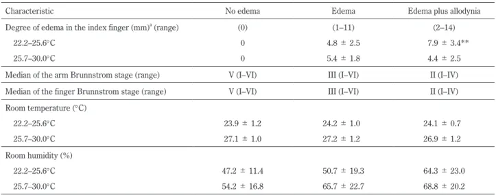
Table 2 Degree of paralysis and edema under various temperatures.
Characteristic No edema Edema Edema plus allodynia
Degree of edema in the index finger (mm)a (range) (0) (1–11) (2–14)
22.2–25.6°C 0 4.8 ± 2.5 7.9 ± 3.4**
25.7–30.0°C 0 5.4 ± 1.8 4.4 ± 2.5
Median of the arm Brunnstrom stage (range) V (I–VI) III (I–VI) II (I–IV) Median of the finger Brunnstrom stage (range) V (I–VI) III (I–VI) II (I–IV) Room temperature (°C) 22.2–25.6°C 23.9 ± 1.2 24.2 ± 1.0 24.1 ± 0.7 25.7–30.0°C 27.1 ± 1.0 27.2 ± 1.2 26.9 ± 1.2 Room humidity (%) 22.2–25.6°C 47.2 ± 11.4 50.7 ± 19.3 64.3 ± 23.0 25.7–30.0°C 54.2 ± 16.8 65.7 ± 22.7 68.8 ± 20.2
aEdema calculated as the difference in circumference of the phalanx proximalis of the index fingers of the two hands. **P < 0.01, significant difference between edema and edema plus allodynia groups (degree of edema).
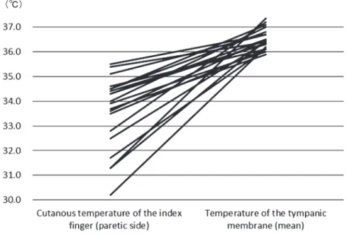
結 果 1)浮腫や痛覚過敏と患者特性 浮腫と痛みの有無で,Table 1 のように,室温 22.2~25.6°C では浮腫なし群 11 例,浮腫のみ群(以下浮腫群)16 例,浮 腫と痛み群(以下浮腫・痛み群)10 例,室温 25.7~30°C で は浮腫なし群 12 例,浮腫群 16 例,浮腫・痛み群 10 例に分 け,患者の特性を比較した.年齢は室温 25.7~30°C で浮腫・ 痛み群が浮腫なし群より有意に(P < 0.01)高かったが,室温 22.2~25.6°C では差がなかった.罹病期間,室温,湿度は 3 群間で差をみとめなかった.Table 2 のように,示指周径の左 右差でみた浮腫の程度は室温 22.2~25.6°C において浮腫・痛 み群が浮腫群より有意に強かった(P < 0.01).また,上肢と 手指の麻痺の程度(BRS 中央値)は浮腫なし群が V,浮腫群 が III,浮腫・痛み群が II であり,有意差(P < 0.05)をもっ て,浮腫群と浮腫・痛み群が浮腫なし群より,浮腫・痛み群 が浮腫群より麻痺が重かった.患側前腕周径が健側より大き いものはなかった.浮腫なし群に手指の屈曲制限はなかった. 浮腫群の中で 18 例(56.3%)は手指の屈曲制限をみとめた. 浮腫・痛み群は全例で手指の屈曲制限をみとめた. 2)浮腫や痛覚過敏の有無と示指の皮膚温との関連 Fig. 2に示すように,示指皮膚温の分散は室温 22.2~25.6°C が室温 25.7~30°C よりも大きかった.また,室温 22.2~25.6°C において浮腫・痛み群が浮腫なし群や浮腫群よりも有意に (P < 0.01,P < 0.01)麻痺側皮膚温引く健側皮膚温の値が大 きかった.室温 25.7~30°C においても浮腫・痛み群が浮腫な し群より有意に(P < 0.01)麻痺側皮膚温引く健側皮膚温の値 が大きかった. Fig. 3のように,浮腫・痛み群の患側示指皮膚温は常にそ れぞれの鼓膜温の平均よりも低かった. Table 3のように視床または視床下部病変の有無では 3 群間 に差をみとめなかった. Table 4のように,中枢病変の部位の数と偏在性は 3 群で有 意に差をみとめた(P < 0.01).浮腫・痛み群は大脳白質と内 包と被殻の 3 ヵ所におよぶ広範な病変が 75.0%と多いのがき わだっていた.浮腫群は同じ 3 ヵ所病変が 56.3%と多かった が,大脳白質と内包の 2 ヵ所の病変が 21.9%と多いのが特徴 であった.浮腫なし群も 3 ヵ所病変が 47.8%と多かったが, 大脳白質,内包,被殻のいずれか 1 ヵ所の病変が 26.1%とめ だっていた.脳幹の病変は浮腫なし群 8.7%,浮腫群 3.1%, 浮腫・痛み群 0%と多くはなかった. 考 察 今回われわれは,脳卒中後の SHS の特徴である手の浮腫や 痛みと示指皮膚温との関連について検討した.指先は皮膚血 管収縮線維により皮膚血流が調節され,温・冷刺激による血 流変化で皮膚温がいちじるしく変化する.SHS 急性期の患側 肢の皮膚温は健側より高い1)3).本報告でも浮腫・痛み群の患
Fig. 2 Temperatures of index fingers of stroke patients.
Temperatures of index fingers on the unaffected sides of patients with no edema (filled circles), edema (open squares), or edema with allodynia (crosses) plotted against those of index fingers on their affected sides. The line is y = x. a: Under comfortable condi-tions (22.2–25.6°C). b: Under warm condicondi-tions (25.7–30.0°C). Under the comfortable condition (sympathicotonia) skin tempera-tures of the affected index fingers in the edema and allodynia group did not decrease as they did in the other two groups. Under the warm condition, skin temperatures of the affected index fingers in the edema and allodynia group were slightly higher than those of the contralateral fingers.
側示指皮膚温は鼓膜温よりも低いが,健側示指皮膚温よりは 高い.同群の示指皮膚温の左右差は健側皮膚温が低いと大き い.これは健側皮膚血管が室温の低下に対応して収縮しても, 患側皮膚血管は収縮できずに皮膚温が高いままであることを 示唆する.Birklein ら12)は CRPS I において室温 24°C で 2 時 間馴化して皮膚温を計測したばあい,健側肢は急性期が慢性 期より低く,患側肢は急性期と慢性期で差がなかったと報告 している.今回の結果と Birklein らの報告は,CRPS I 急性期 には患肢の皮膚血管が拡張したまま熱を失う一方,健肢の皮 膚血管が収縮して熱の放出をおさえていることを示唆する. 脳卒中後の SHS における皮膚血管拡張が中枢性であるか 末梢性であるかはまだ明らかでない.今回,浮腫・痛み群の 中枢病変が広く内包・大脳白質・被殻におよぶ割合は他の 2 群より高く,視床・視床下部病変がある割合は他の 2 群と変 わらなかった.中里ら13)は 10 mm 以下の小梗塞病変と筋萎 縮のない麻痺側上下肢(手は除く)の皮膚温との関係をしら Fig. 3 Comparisons between cutaneous temperatures of the affected index fingers of patients in the edema plus
allodynia group and mean temperatures of their tympanic membranes.
In the edema plus allodynia group, temperatures of the affected index fingers were lower than the respective mean temperatures of their tympanic membranes, used as a measure of deep body temperature.
Table 3 Presence of infarction or hemorrhage in the thalamic or subthalamic reigions.
Patient group
Infarct or hemorrhage in thalamic and/or subthalamic region
+
-No edema (n = 23) 21.7 78.3
Edema (n = 32) 28.1 71.9
Edema plus allodynia (n = 20) 30 70 Values represent % of patients in the group.
Table 4 Presence of pyramidal lesions in various regions.
3 regions 2 regions A 2 regions B 2 regions C 1 region Brain stem
No edema n = 23 11 (47.8) 2 (8.7) 1 (4.35) 1 (4.35) 6 (26.1) 2 (8.7)
Edema n = 32 18 (56.3) 7 (21.9) 1 (3.1) 1 (3.1) 4 (12.5) 1 (3.1)
Edema plus allodynia n = 20 15 (75) 1 (5) 0 (0) 2 (10) 2 (10) 0 (0)
3 regions: Internal capsule plus cerebral white matter plus putamen. 2 regions A: Internal capsule plus cerebral white matter. 2 regions B: Internal capsule plus putamen. 2 regions C: Cerebral white matter plus putamen. 1 region: Internal capsule, or cerebral white matter, or putamen. (Values) represent % of patients in the group.
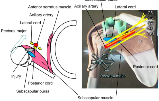
べ,内包・被殻病変では麻痺側上下肢の皮膚温が低下してい ることを示した.皮膚温度覚は Feedforward signal として視床 下部の体温中枢に働くが14),手の皮膚への冷刺激は数分単位 で上肢の皮膚血管を収縮させ,一時的に深部体温をも上昇さ せる15).おそらく麻痺肢では,筋の熱産生が減ることで温覚 からの feedforward 情報が減り,体温中枢が交感神経亢進を 介して麻痺側皮膚血管を収縮させ,結果として患側皮膚温が 低下しているものと思われる.ところが,もっとも交感神経 活動亢進を反映して皮膚温が低下するはずの指先でありなが ら,浮腫・痛み群の患側示指皮膚温は高い.この病態は手部 に末梢レベルでの交感神経遮断や局所の炎症があることを示 唆する. Wasnerらは,健常者の手へのヒスタミン塗布による皮膚温 上昇は体幹冷却で抑制できるが,CRPS I 患肢の皮膚温上昇は 体幹冷却で抑制できないことを示した6).これは,動静脈吻 合が開いて血流が皮下静脈叢にシャントされると皮膚血流が 減る16)ため,炎症で皮膚血管が拡張していても,交感神経亢 進によって皮膚温が低下することを示している. CRPS I患肢の皮膚温上昇が体幹冷却による中枢性の交感神 経活動亢進で抑制できなかったことは,末梢に交感神経伝導 遮断があることを示唆する.したがって,浮腫・痛み群の患 側示指皮膚温高値も末梢性の交感神経伝導遮断が原因と思わ れる. さらに,末梢の交感神経伝導遮断を示唆する所見として, 室温と浮腫との関係がある.SHS においては指の浮腫は富士 山様で,手の浮腫は中手骨上に横たわるようにあり,手関節 の浮腫は手根骨背側にあると報告されている3).つまり,指 先よりも指の近位で浮腫は強く,手の背側にめだつ.交感神 経活動亢進では,細動脈が細静脈より強く収縮するため平均 毛細血管圧が下がり,血管の外側から内側に細胞外液の移動 がおこって静脈還流が増え17),浮腫は軽減する方向に働く. 浮腫・痛み群において,健側示指は室温 22.2~25.6°C で Fig. 1 のように皮膚温が低下し,交感神経活動は亢進しており,交 感神経遮断のある患側示指とに明らかな周径差が出たと思わ れる.一方,室温 25.7~30°C のように暖かいと,健側の交感 神経活動は活発でなく,交感神経遮断のある患側との周径差 は出にくかったと思われる. 次に,交感神経遮断をきたした部位について考察する.浮 腫の測定部位である示指の基節骨部は背面が橈骨神経(後神 経束の末梢領域),掌側が正中神経(外側神経束の末梢領域) である.皮膚温の測定部位である示指尖端掌側は正中神経(外 側神経束の末梢領域)である.浮腫群は示指皮膚温の左右差 がなく浮腫はあるので,正中神経障害がなく橈骨神経障害は あると思われる.浮腫・痛み群は示指皮膚温の左右差があり 浮腫も重く,正中・橈骨両神経の障害があると思われる. 脳卒中後の SHS では他動的な肩の外旋外転で肩の痛みが 出現すること2)から肩甲下筋の損傷が示唆され,安静臥床中 におきる肩甲下筋と腕神経叢との癒着,その後の剝離が神経 周膜や神経栄養血管を傷つけている可能性がある.解剖学的 にみて,橈骨神経が先に正中神経が後に障害される場所とし ては腕神経叢の神経束後面がうたがわれる.神経束と肩甲下 筋は Fig. 4 のような位置関係にある.患者の安静臥位中に肩
Fig. 4 Locations of the cords of the brachial plexus against the subscapular muscle.
When the patient is lying on his back, the posterior cord of the brachial plexus is located under the axillary artery, such that the artery pushes the cord against the subscapular muscle. If there is bloody fluid around the brachial plexus from injury to the subscapular muscle or articular capsule, first the posterior cord and then the lateral cord may become adhered to the subscapular muscle.
甲下筋や滑液包が傷つき,腕神経叢周囲に血性浸出液が出現 すると,先に腋窩動脈の後方にある後神経束が癒着し,次に 外側神経束が癒着するだろう.腕神経叢神経束の交感神経節 後線維は無髄かきわめて薄い有髄からなる18)ため,もし出血 や虚血で無髄神経周囲のカリウム濃度が上昇したならば,膜 の過分極による伝導遮断をきたす19)と思われる.さらに,完 全な切断なく軸索まで損傷がおよぶならば,末梢に神経性炎 症をきたす20)可能性がある. 本研究における皮膚温の左右差や痛覚過敏の示指での有無 は,浮腫群と浮腫・痛み群が肩手症候群の軽症から重症の連 続した病態である可能性を示唆する.つまり,後神経束後面 に交感神経遮断をきたした軽症例が浮腫群に相当する.また, 後神経束後面だけでなく,外側神経束後面にも損傷がおよび, 交感神経遮断だけでなく軸索損傷で神経性炎症をもきたした 重症例が浮腫・痛み群に相当する.なお,浮腫群の一部は指 の可動域制限をきたしており,神経性炎症が治まった交感神 経遮断のみの後遺症状態である可能性がある.ただし,SHS の症状は肩と手にあって上腕・前腕にない.肩に症状のない wrist-hand syndromeも存在する.仮説が成り立つには,神経 束に segmentation があって,神経束後面により末梢側の神経 線維が位置している必要があり,今後の検討を要す. 謝辞:本研究に協力してくださった当院作業療法スタッフおよび 様々な助言をくださった査読の先生方,鹿児島大学大学院医歯学総合 研究科運動機能修復学講座リハビリテーション医学名誉教授川平 和美先生に深謝いたします. ※本論文に関連し,開示すべき COI 状態にある企業,組織,団体 はいずれも有りません. 文 献 1) 江藤文夫.肩手症候群の発生機序.総合リハ 1977;5:1037-1046. 2) Braus DF, Krauss JK, Strobel J. The shoulder-hand syndrome
after stroke: a prospective clinical trial. Ann Neurol 1994;36: 728-733.
3) Geurts AC, Visschers BA, van Limbeek J, et al. Systematic review of aetiology and treatment of post-stroke hand oedema and shoulder-hand syndrome. Scand J Rehabil Med 2000;32:4-10. 4) Falsetti P, Acciai C, Carpinteri F, et al. Bedside ultrasonography
of musculoskeletal complications in brain injured patients. J Ultrasound 2010;13:134-141.
5) Greyson ND, Tepperman PS. Three-phase bone studies in
hemiplegia with reflex sympathetic dystrophy and the effect of disuse. J Nucl Med 1984;25:423-429.
6) Wasner G, Heckmann K, Maier C, et al. Vascular abnormalities in acute reflex sympathetic dystrophy (CRPS I): complete inhibition of sympathetic nerve activity with recovery. Arch Neurol 1999;56:613-620.
7) Guo TZ, Wei T, Shi X, et al. Neuropeptide deficient mice have attenuated nociceptive, vascular, and inflammatory changes in a tibia fracture model of complex regional pain syndrome. Mol Pain 2012;8:85.
8) Oaklander AL, Rissmiller JG, Gelman LB, et al. Evidence of focal small-fiber axonal degeneration in complex regional pain syndrome-1(reflex sympathetic dystrophy). Pain 2006;120: 235-243. 9) 空気調和・衛生工学会.新版・快適な温熱環境のメカニズム 豊かな生活空間をめざして.改訂 2 版.東京:丸善;2006. p. 63, 76. 10) 入來正躬.体温生理学テキスト第 1 版.東京:文光社;2003. p. 80, 204-207. 11) 國本雅也.指尖部冷水負荷時の浸水部皮膚温の低下による皮 膚交感神経機能の評価.臨床神経 1989;29;1004-1008. 12) Birklein F, Riedl B, Claus D, et al. Pattern of autonomic
dysfunction in time course of complex regional pain syndrome. Clin Auton Res 1998;8:79-85.
13) 中里良彦,島津邦男,田村直俊ら.脳梗塞患者における全身 皮膚温異常の検討―とくに皮膚血管運動に対する中枢性支 配について―.臨床神経 1995;35:758-763.
14) Nakamura K. Central circuitries for body temperature regulation and fever. Am J Physiol Regul Integr Comp Physiol 2011;301: R1207-R1228. 15) 岩瀬 敏,澤崎直規,道上大策ら.ヒト皮膚交感神経中の血 管収縮神経活動の賦活と抑制が核心温変化に及ぼす影響.自 律神経 2001;38:7-10. 16) 大谷 修,堀尾嘉幸.人体の正常構造と機能 II 循環器.第 1 版.東京:日本医事新報社;2005. p. 81. 17) 本郷利憲,廣重力監修.標準生理学.第 5 版.東京:医学書 院;2000. p. 571. 18) 久野みゆき,秋田恵一.人体の正常構造と機能 IX 神経系(2). 第 1 版.東京:日本医事新報社;2005. p. 20.
19) Eng DL, Kocsis JD. Activity-dependent changes in extracellular potassium and excitability in turtle olfactory nerve. J Neurophysiol 1987;57:740-754.
20) Daemen MA, Kurvers HA, Kitslaar PJ, et al. Neurogenic inflammation in an animal model of neuropathic pain. Neurol Res 1998;20:41-45.
Abstract
Relationship between cutaneous temperature and hand edema and allodynia after stroke
—The etiology of shoulder-hand syndrome—
Hiroko Yamanaka, M.D., Ph.D.
1)and Hidekata Yamanaka, M.D.
1)1)Atsuchi Rihabilitation Hospital