要 旨: 79 歳,女性。左臀部,大 部痛のため受診し,CT で遺残坐骨動脈瘤と診断した。受診翌日に下肢痛 が出現し,坐骨神経圧迫症状に加え塞栓症が悪化したと判断した。塞栓症に血栓吸引・溶解療法を,遺残坐骨 動脈にステントグラフト(SG)留置術を施行した。下肢痛は消失したが,術後早期にSG は閉塞し軽度の間欠 性跛行が生じた。遺残坐骨動脈に対する SG 留置術の成績は確立しておらず,慎重に適応を考慮すべきである。 (J Jpn Coll Angiol 2021; 61: 39–43)
Key words: persistent sciatic artery aneurysm, distal embolization, endovascular treatment
受付:2020 年 5 月 22 日 受理:2021 年 4 月 2 日 公開:2021 年 6 月 10 日
序
言
遺残坐骨動脈(persistent sciatic artery; PSA)は胎生期 の臍動脈から分枝する坐骨動脈が遺残した,極めて稀 な疾患である。その組織学的,解剖学的特徴により PSA は瘤化,閉塞を来しやすい。また瘤内血栓による末梢動 脈塞栓症を生じることがある。今回,末梢動脈塞栓症 を合併した遺残坐骨動脈瘤(persistent sciatic artery aneu-rysm; PSAA)に対して血管内治療を行った症例を 1 例経 験した。
症
例
症 例:79 歳,女性 主 訴:繰り返す左下肢痛 既往歴・家族歴:脳梗塞,高血圧,高脂血症 現病歴:繰り返す左下肢痛で外来を受診した。初診時 に CT 検査で PSAA と診断した。左下 の動脈は下 中央 で閉塞しており,左下 に冷感を認めた。主に臀部,大 部の疼痛を訴えたため坐骨神経の圧迫による下肢疼痛 と判断した。待機的に血管内治療を行う方針としたが, 翌日に下肢痛が悪化し救急外来を受診し入院となった。 入院時現症:152 cm, 54 kg,血圧 122/82 mmHg,脈拍 84/分,整,左下 以下の色調は右に比して不良であり 安静時痛を認めた。臀部には軽度の圧痛を認めた。 入院時血液検査:血液検査ではLDHが320 U/Lと軽度 上昇を認めたのみで,CPKなど他は正常範囲内であった。 初 診 時 下 肢 CT 検 査(Figs. 1a∼c): 左 内 腸 骨 動 脈 か ら連続する PSA を認めた。大 骨頭レベルで最大短径 35 mm と瘤化し,開存しているものの多量の壁在血栓を 伴っていた。浅大 動脈は細く発達不良であったが,膝 窩動脈との連続性を認めた。左下 の動脈は下 中央で 閉塞していた。 診断・治療計画:入院時,PSAA からの末梢動脈塞栓 が悪化し下肢虚血が進行したと診断した。合併症を有 する高齢者であり,創感染,リンパ漏などの切開手術に よる合併症を回避できることから血管内治療のみで完遂 する方法を検討し,末梢塞栓症に対しては経カテーテ ル的血栓吸引・溶解療法,PSAA にはステントグラフト (SG)留置術を施行する方針とした。 血管内治療:局所麻酔下に右総大 動脈を逆行性穿 刺した。ヘパリン 3000 U を投与した。0.035 inch の Radi-focus Guidewire(Terumo Corp, Tokyo)と 4.2 F の Excellent EN Catheter(Cobra-Type)(Hanaco medical, Saitama) を 用 いて腹部大動脈分岐部を越えて左内腸骨動脈に進め, 7 F の Flexor Shuttle Guiding Sheath(Cook Medical Inc, IN, USA)の先端を PSAA を越える位置に留置した。造影す ると下 動脈の描出不良を認めた(Fig. 2a)。0.014 inch の Jupiter FC Peripheral Guidewire(Boston Scientific, MA, USA)を 4.2 F の Excellent EN Catheter(Cobra-Type)を用1 名古屋第一赤十字病院血管外科
2 独立行政法人地域医療推進機構中京病院血管外科
いて,腓骨動脈,前脛骨動脈,後脛骨動脈の順番に挿 入し,6 F の Thrombuster II(Kaneka Corp, Tokyo)で少量 の血栓を吸引した。血栓溶解を目的としてウロキナー ゼ 12 万単位を動注した。少量の赤色血栓が回収された ものの血流の再開はみられなかった。狭窄が関与して いる可能性を考慮し下 動脈に血管拡張術(PTA)を行 う方針とした。より広範囲に PTA を行うため,2.5/3.0× 210 mm RapidCross Balloon(Medtronic, CA, USA)でそれ ぞれの動脈に PTA を行った。PTA 後の造影でも下 動脈
の開通は確認できなかったが,0.014 inch の Command Pe-ripheral Guidewire(Abbott, CA, USA)を足底動脈まで進 めることができるようになったため,より末梢に狭窄が 存在している可能性を考慮し,2.0×220 mm Coyote Bal-loon(Boston Scientific, MA, USA)で足底動脈まで PTA を 行った。結果として明らかな狭窄は確認されなかった が,PTA 後に造影で後脛骨動脈の血流改善を確認するこ とができた(Fig. 2b)。引き続いて,ガイドワイヤーを 0.018 inch の V18 Guidewire(Boston Scientific, MA, USA) に交換し,PSAA をまたぎ,瘤の中枢(血管径 6.5 mm), 末梢(血管径 7.0 mm)に 20 mm 以上のランディングを確 保するように 8×150 mm Viabahn を留置した。Viabahn が 十分に拡張したことが確認できたため,圧着は行わずに 終了した(Figs. 3a, b)。
Figure 1 a, b) Computed tomography showed left complete persistent sciatic artery with aneurysmal change. c) 3D Computed tomography angiogram showed incomplete development of the su-perficial femoral artery and the occlusion of the crural arteries.
Figure 2 a) Left anterior tibial artery, posterior tibial artery and peroneal artery were occluded. b) After endovascular treatment, the posterior tibial artery was revascularized.
Figure 3 a) Intraoperative angiogram showed the persistent sciatic artery aneurysm. b) Viabahn was deployed to exclude the persistent sciatic artery aneurysm.
Figure 4 Postoperative CT revealed Viabahn was occluded and the crural arteries were patent.
考
察
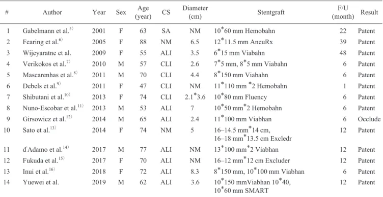
PSA は,0.01–0.06% の非常に稀な頻度でみられる。高 い確率で瘤化することが報告されており,PSA の閉塞, 末梢塞栓による末梢循環不全,伴走する坐骨神経の圧迫 による神経症状,瘤破裂が生じるとされている1)。本症 例は初診時には坐骨神経圧迫による疼痛が優位に出てい たものの,塞栓による虚血が進行したことで,下 の 虚血痛が前面に出てきたものと考えられた。Bower らは PSA と膝窩動脈の連続性から完全型と不完全型に分類し た。完全型は PSA が膝窩動脈に連続し,下 の主たる 血流源となっており全体の 80% を占める。不完全型は PSA と膝窩動脈の連続性はなく,通常のように大 動脈 例あるのみで,良好な成績が報告されている。長期成 績については 4 年が最長であり,より長期の開存率につ いては不明である17)。本症例では,術後 8 日目には閉塞 が判明し,坐位時の圧迫により閉塞が生じた可能性があ る。圧迫による閉塞を危惧し,術中に Viabahn 内にベア メタルステントを留置した報告もみられるが,効果,長 期成績は不明である18)。 不完全型に分類されるPSAは膝窩動脈への連続性が ない。そのため,治療対象になるのは坐骨神経の圧迫や PSAAの破裂など瘤化に伴う症状を呈するものに限られ る。下肢の血行再建を要さず,PSAの結紮術や塞栓術が 選択される。本症例はPSAが膝窩動脈に連続した完全型 であり,PSAの瘤化による神経圧迫症状と瘤内血栓の末Table 1 The published cases found in PubMed regarding endovascular stent graft repair for persistent sciatic artery aneurysm
# Author Year Sex (year)Age CS Diameter (cm) Stentgraft (month) ResultF/U
1 Gabelmann et al.5) 2001 F 63 SA NM 10*60 mm Hemobahn 22 Patent
2 Fearing et al.6) 2005 F 88 NM 6.5 12*11.5 mm AneuRx 39 Patent
3 Wijeyaratne et al. 2009 F 55 ALI 3.5 6*15 mm Viabahn 48 Patent
4 Verikokos et al.7) 2010 M 57 CLI 2.6 7*5 mm, 8*5 mm Viabahn 6 Patent
5 Mascarenhas et al.8) 2011 M 70 CLI 4.4 8*150 mm Viabahn 6 Patent
6 Debels et al.9) 2011 F 47 CLI NM 11*110 mm *2 Hemobahn 1 Patent
7 Shibutani et al.10) 2013 F 74 CLI 2.1*3.6 10*80 mm Fluency 6 Patent
8 Nuno-Escobar et al.11) 2013 M 53 ALI 7 10*50 mm*2 Hemobahn 6 Patent
9 Girsowicz et al.12) 2014 M 65 ALI 2.4 11*100 mm Viabhan 6 Occlude
10 Sato et al.13) 2014 F 74 NM 5 16–14.5 mm*14 cm,
16–18 mm*13.5 cm Excledr 12 Patent
11 d Adamo et al.14) 2017 M 77 ALI NM 13*100 mm*2 Viabhan 12 Patent
12 Fukuda et al.15) 2017 F 70 ALI NM 16–12 mm*12 cm Excluder 12 Patent
13 Inui et al.16) 2018 F 72 ALI 8.3 8*150 mm, 10*100 mm Viabhan 6 Patent
14 Yuewei et al. 2019 M 62 ALI 3.6 10*150 mmViabhan 10*40,
10*60 mm SMART 12 Patent
F: female, M: male, CS: clinical symptom, SA: subacute, NM: not mentioned, ALI: acute limb ischemia, CLI: chronic limb ischemia F/U: follow up
梢動脈塞栓により徐々に下肢虚血が進行し下肢の疼痛が 生じたと考えられた。下肢塞栓症に対しては血栓吸引・ 溶解療法を施行する方針とした。PSAAに対しては動脈 瘤神経圧迫症状の解除と末梢動脈塞栓の予防のために, PSAにSGを留置する方法,およびPSAの塞栓術と総大 動脈–膝窩動脈バイパスを行う方法が検討され,低 侵襲な前者が選択された。血管径を考慮し,SGとして Viabahnを選択した。術前にSGの早期閉塞のリスクにつ いて考慮したが,発達不良ながら膝窩動脈に連続する浅 大 動脈が存在し,SGが閉塞してもある程度は下 の 血流量が保たれると予想した。もし閉塞時に重症虚血の 症状が発生した場合でも総大 動脈–膝窩動脈バイパス により治療できると考えた。今回の症例では術後にVia-bahnの閉塞を認めたものの閉塞時には自覚的にも他覚的 にも気づくことなく,最終的に軽度の跛行症状を残すの みであった。本症例については,まずPSAに対するコイ ルもしくはプラグによる塞栓と末梢塞栓に対する血栓吸 引・溶解療法を行い,下肢の虚血症状に応じて二期的に バイパス手術を行う方法も有効であったと思われる。
結
語
PSA に対する Viabahn 留置は保険適用を受けていない。 そのうえ,本症例では SG 留置により塞栓術を施行した のと同じ結果となった。以上から現状では坐骨神経圧迫 症状と下肢動脈血流の保持を目的とした PSAA に対する SG 留置術は全身状態などから血管内治療のみで完遂す べき場合など,適応を限って施行すべきと考える。利益相反
著者全員が利益相反はない。文
献
1) Greebe J: Congenital anomalies of the iliofemoral artery. J Cardiovasc Surg 1977; 18: 317–323
2) Bower EB, Smullens SN, Parke WW: Clinical aspects of persistent sciatic artery: report of two cases and review of the literature. Surgery 1977; 81: 588–595
3) Ahn S, Min SK, Min SI, et al: Treatment strategy for persis-tent sciatic artery and novel classification reflecting anatomic status. Eur J Vasc Endovasc Surg 2016; 52: 360–369
4) van Hooft IM, Zeebregts CJ, van Sterkenburg SMM, et al: The persistent sciatic artery. Eur J Vasc Endovasc Surg 2009; 37: 585–591
5) Gabelmann A, Kramer SC, Wisianowski C, et al: Endovas-cular interventions on persistent sciatic arteries. J Endovasc Ther 2001; 8: 622–628
6) Fearing NM, Ammar DA, Hutchinson SA, et al: Endovascular stent graft repair of a persistent sciatic artery aneurysm. Ann Vasc Surg 2005; 19: 438–441
7) Verikokos C, Avgerinos ED, Chatziioannou A, et al: Endo-vascular repair of a persistent sciatic artery aneurysm. Vascu-lar 2010; 18: 162–165
8) Mascarenhas de Oliveira F, de Souza Mourao G: Endovascu-lar repair of symptomatic sciatic artery aneurysm. Vasc Endo-vascular Surg 2011; 45: 165–169
9) Debels H, De Gendt G: Persistent sciatic artery aneurysm: a case report. Acta Chir Belg 2011; 111: 256–259
10) Shibutani S, Hayashi E, Obara H, et al: Rapid development of aneurysmal formation after successful endovascular treatment of chronic total occlusion of a persistent sciatic artery. Ann Vasc Surg 2013; 27: 499.e5–499.e8
11) Nuño-Escobar C, Pérez-Durán MA, Ramos-López R, et al: Persistent sciatic artery aneurysm. Ann Vasc Surg 2013; 27: 1182.e13–1182.e16
12) Girsowicz E, Georg Y, Lejay A, et al: Midterm failure after endovascular treatment of a persistent sciatic artery aneurysm. Ann Vasc Surg 2014; 28: 1323.e7–1323.e12
13) Sato H, Nakai M, Sato M, et al: Retrograde popliteal endovas-cular stent-graft repair for a growing persistent sciatic artery aneurysm (type IIa): case report and review of the literature. J Vasc Interv Radiol 2014; 25: 1997–2000
14) d Adamo A, Sirignano P, Fanelli F, et al: Endovascular solu-tion of acute limb ischemia engendered by persistent sciatic artery pseudoaneurysm due to stent fracture. Ann Vasc Surg 2017; 43: 310.e9–310.e12
15) Fukuda H, Onitsuka S, Yoshida S, et al: Endovascular stent graft repair of a persistent sciatic artery aneurysm. Ann Vasc Dis 2017; 10: 246–249
16) Inui TS, Picel AC, Barleben A, et al: Endovascular manage-ment of a large persistent sciatic artery aneurysm. Ann Vasc Surg 2018; 52: 312.e13–312.e16
17) Wijeyaratne SM, Wijewardene N: Endovascular stenting of a persistent sciatic artery aneurysm via retrograde popliteal ap-proach: a durable option. Eur J Vasc Endovasc Surg 2009; 38: 91–92
18) Wang Y, Xin H, Tan H, et al: Endovascular stent graft repair of complete persistent sciatic artery aneurysm with lower limb ischemia: a case report and review of the literature. SAGE Open Med Case Rep 2019; 7: doi: 10.1177/2050313X19841462
(W.L. Gore, AZ, USA) was deployed to exclude the PSAA. Although the lower limb pain was alleviated,
the stent graft occluded in the early postoperative period and caused intermittent claudication. As the long
term results of stent graft placement for persistent sciatic artery is unknown, the indication should be
care-fully considered.
(J Jpn Coll Angiol 2021; 61: 39–43) Online publication June 10, 2021