Clin Case Rep. 2020;00:1–2. wileyonlinelibrary.com/journal/ccr3
|
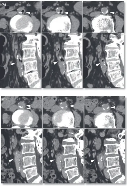
1A 70-year-old diabetic woman was admitted to our hospital with a complaint of high fever accompanying shaking chills for 3 days. The patient's body temperature was increased to 39°C, and blood analysis showed elevations of leukocytes (17 400/µL) and C-reactive protein (16.5 mg/dL). Computed tomography revealed air-density spots in the aortic wall (Figure 1A), and Salmonella enteritidis serotype O9 was de-tected by blood culture. She had no other notable symptoms including digestive symptoms and had no history of eating raw food. Under a tentative diagnosis of Salmonella-induced infective saccular aneurysm in the infrarenal abdominal aorta, the patient underwent antibiotic treatment with am-picillin/sulbactam. On day 11, the air had disappeared and the aortic wall showed a varicose deformity (Figure 1B). The patient recovered completely with 8-week antibiotic therapy without any operation.
Cases of infective aortic aneurysms account for only about 1%-3% of total cases of aortic aneurysm. However, the case fatality has been reported to be as high as 18% in 2 years.1 Salmonella species, as detected in this case,
are known as a common pathogen of crucial infection.2
Not only intra-aortic gas but also the presence of a pene-trating aortic ulcer suggests acute aortic syndrome, which requires urgent management.3 Without treatment,
my-cotic aneurysms are associated with high mortality from rupture or uncontrolled sepsis. Thus, clinicians should pay attention to these findings for early diagnosis of the disease.
ACKNOWLEDGMENTS
None to report.
CONFLICT OF INTEREST
We have no financial relationships to disclose.
AUTHOR CONTRIBUTIONS
DO and HN: wrote the first draft and managed all of the submission process. MO, YS, and RM: supervised clinical management of the patient. HH and FO: contributed to clin-ical management of the patient and revised the manuscript.
Received: 1 July 2020
|
Revised: 21 August 2020|
Accepted: 25 August 2020 DOI: 10.1002/ccr3.3367C L I N I C A L I M A G E
Intra-arterial gas, a clue for diagnosis of peri-aortic
inflammation due to infection
Daisuke Omura
1,2|
Masatoshi Ogata
2|
Yoshio Sakane
2|
Ryuichi Matsuo
2|
Harushige Nakatsukasa
2|
Hideharu Hagiya
1|
Fumio Otsuka
1This is an open access article under the terms of the Creative Commons Attribution License, which permits use, distribution and reproduction in any medium, provided the original work is properly cited.
© 2020 The Authors. Clinical Case Reports published by John Wiley & Sons Ltd.
1Department of General Medicine,
Okayama University Graduate School of Medicine, Dentistry and Pharmaceutical Sciences, Okayama, Japan
2Department of Internal Medicine,
Mizushima Central Hospital, Kurashiki, Japan
Correspondence
Daisuke Omura, Department of General Medicine, Okayama University Graduate School of Medicine, Dentistry and Pharmaceutical Sciences, 2-5-1 Shikata-cho, Kita-ku, Okayama 700-8558, Japan. Email: [email protected]
Abstract
We have presented a case of Salmonella-induced infective aortic aneurysm in which the presence of peri-aortic gas was a clue for diagnosis. The disease is clinically in-frequent but potentially has a high mortality rate. Clinicians should consider this fatal disease from any trivial findings.
K E Y W O R D S
2
|
OMURA etAl.ORCID
Daisuke Omura https://orcid.org/0000-0002-7878-3813
Hideharu Hagiya https://orcid. org/0000-0002-5086-1891
Fumio Otsuka https://orcid.org/0000-0001-7014-9095
REFERENCES
1. Kan CD, Lee HL, Yang YJ. Outcome after endovascular stent graft treatment for mycotic aortic aneurysm: a systematic review. J Vasc Surg. 2007;46(5):906.
2. Hsu RB, Chen RJ, Wang SS, Chu SH. Infected aortic aneurysms: clinical outcome and risk factor analysis. J Vasc Surg. 2004;40:30-35.
3. Bansal S, Singhania N, Yadav M, Singhania G. A Penetrating Aortic Ulcer. J Emerg Med. 2020;S0736-4679(20):30362.
How to cite this article: Omura D, Ogata M, Sakane
Y, et al. Intra-arterial gas, a clue for diagnosis of peri-aortic inflammation due to infection. Clin
Case Rep. 2020;00:1–2. https://doi.org/10.1002/ ccr3.3367
FIGURE 1 Abdominal computed tomography on admission (A) and on day 11 (B). Air-density spots were found in the aortic wall (A, arrowheads). They later disappeared, and outpouching of the arterial wall emerged at the corresponding site (B)
(A)