• 検索結果がありません。

Department of Neurosurgery, Graduate School of Medical Science, Kanazawa University, Kanazawa, Japan

N/A
N/A
Protected

Academic year: 2021

シェア "Department of Neurosurgery, Graduate School of Medical Science, Kanazawa University, Kanazawa, Japan "

Copied!
26
0
0

読み込み中.... (全文を見る)

全文

(1)

Chapter

D EVELOPMENT OF S YMPTOMS IN A RACHNOID C YSTS

Yasuhiko Hayashi

, MD, PhD

Department of Neurosurgery, Graduate School of Medical Science, Kanazawa University, Kanazawa, Japan

A BSTRACT

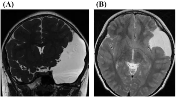
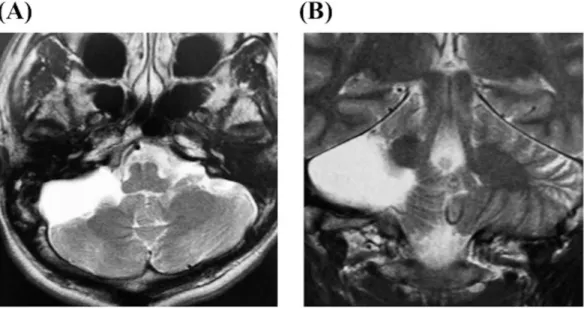
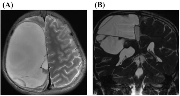
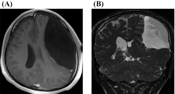
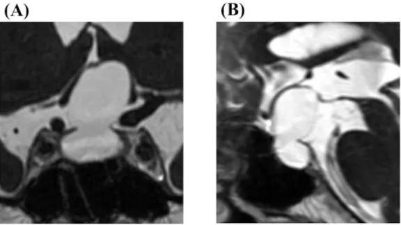
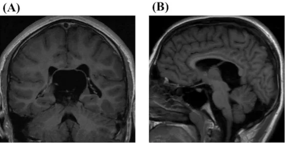
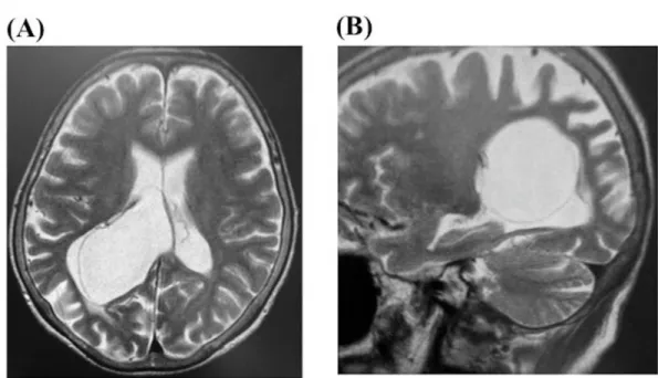
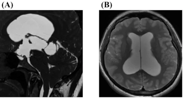
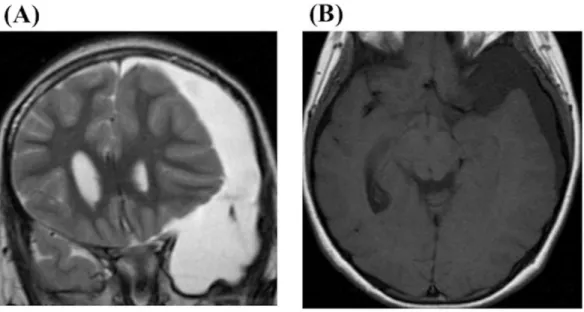
Arachnoid cysts are benign developmental anomalies of the central nervous system and arise in virtually all locations where arachnoid membrane exists. It is reported that they account for approximately 1% of all intracranial lesions and are caused by the splitting of the arachnoid membrane. Many authors have indicated that arachnoid cysts typically remain constant in size or grow very slowly. Therefore, most opportunities to detect arachnoid cysts are incidental. However, recent advent of more advanced imaging techniques has increased the frequencies to find them. The enlargement of arachnoid cysts are reported to occur most frequently in patients under 5 years of age, and the symptoms and signs derived from arachnoid cysts include elevated intracranial hypertension and focal signs due to the effects of compression to the surrounding cerebral parenchyma. Even in cases in which neurological symptoms are present, it is often difficult to properly correlate nonspecific signs and symptoms with the findings of arachnoid cysts. This is especially true in the cases common symptoms, such as headache, that arise frequently in the general population. In most of the cases, arachnoid cysts usually bring with a thin and bulging inner table of the skull. These bone deformities suggest a long process accompanying a gradual increase in intracystic pressure that probably begins in early infancy.

Three mechanisms of the cyst expansion in arachnoid cysts have been described by many authors as follows: First, osmotic gradient between the cyst and the surrounding subarachnoid space induced fluid influx into the cyst cavity; second, fluid is secreted from ependymal cells on the cyst wall; third, a one-way ball-valve mechanism develops on the cyst wall. Almost symptomatic arachnoid cysts belong in children, the progressive growth of the skull and plasticity of the brain in children can buffer the mass effect

Corresponding Author Address: Yasuhiko Hayashi, M.D., Ph.D. 13-1 Takara-machi, Kanazawa, Ishikawa, 920- 8641, Japan, Tel: +81-076-265-2384, Fax: +81-076-234-4262, E-mail; yahayashi@med.kanazawa-u.ac.jp.

(2)

caused by the cysts. Therefore, the arachnoid cysts in children and adolescence are usually asymptomatic or minimally symptomatic. The patient’s symptoms develop once the breakdown of compensatory mechanism in the brain surrounding the arachnoid cyst happens. Even in elderly patients with or without cyst expansion, the breakdown of compensatory mechanisms can lead to relatively rapid progression of symptoms.

However, the factors that induce the breakdown of compensatory capacity remain unknown.

In addition to those mechanisms, the symptomatic arachnoid cyst infrequently associates with intracranial hematoma, such as chronic subdural hematoma, acute subdural hematoma, acute epidural hematoma, and intra-cyst wall hematoma. Especially, it is well known that arachnoid cysts located in the middle cranial fossa can present with chronic subdural hematoma. The hematomas are frequently induced by minor trauma.

The symptoms in arachnoid cysts are presented according to the various locations arachnoid cysts arise, and the developmental mechanisms are also discussed with the aspects of accompanying hydrocephalus or intracranial hematomas, and patients’ ages.

Keywords: arachnoid cyst, development, congenital, symptom

L IST OF A BBREVIATIONS CPA cerebellopontine angle

CSDH chronic subdural hematoma CSF cerebrospinal fluid

EEG electroencephalogram

FIESTA fast imaging employing steady-state acquisition MRI magnetic resonance imaging

WI weighted image.

I NTRODUCTION

Arachnoid cysts are well known as common anomalies of the central nervous system.

They are mainly benign congenital origins, and the cysts consist of the wall lined by arachnoid cells and intra-arachnoid collections of cerebrospinal fluid (CSF) within the cyst formed by a splitting or duplication of the arachnoid membrane during the complex folding of the neural tube and formation of the subarachnoid cistern [1-6]. On the other hand, some arachnoid cysts have been documented as being acquired resulting from inflammation, hemorrhage, tumors, trauma, connective tissue disorders, or even from iatrogenic causes [7, 8]. Although, in most cases, arachnoid cysts are clinically asymptomatic and remain static in size, these cysts may cause various neurological findings depending on their locations, mainly because cysts occasionally and slowly expand, and displace the adjacent structures [2, 3, 9].

Arachnoid cysts can occur relatively rare in approximately 1% of all intracranial space-

occupying lesions [10-12], and recent reports about magnetic resonance imaging (MRI)-

screening examinations found intracranial arachnoid cysts in 1.1-1.7% of healthy and

asymptomatic volunteers [13, 14]. They can exist all along the craniospinal axes, but a large

majority of cases are located intracranially [11, 15]. Most commonly, arachnoid cysts are

located in or adjacent to the Sylvian fissure and in the cerebellopontine angle (CPA) [16].

(3)

Regardless of the benign histological nature of arachnoid cysts, the associated expansive effects may occasionally cause the compression of adjacent neural tissue and obstruction of CSF flow, ultimately resulting in a variety of symptoms such as developmental delay, seizures, macrocephaly, insufficiencies of motor and sensory functions, and headaches [17, 18]. Recent advances in neuroimaging techniques and its widespread availability have achieved an increased detection of arachnoid cyst in a neuroradiological screening of outpatient clinic, with the diagnoses made more frequently at earlier stages. Therefore, many authors have been reported that the natural history of arachnoid cysts turns to be quite diverse and unpredictable in each individual [19, 20]. As described above, the arachnoid cysts can occasionally increase in size and intracystic pressure, resulting in producing of symptoms due to mass effect or obstruction. However, the detail mechanism of enlargement of the cysts remains controversial.

S YMPTOMS AND T HEIR D EVELOPING M ECHANISMS IN E ACH L OCATION OF A RACHNOID C YST Middle Cranial Fossa Arachnoid Cyst

Arachnoid cysts can occur all along the craniospinal axes, but a large majority of cases are located intracranially [16]. Intracranial arachnoid cysts demonstrate deviated preponderance of location for the middle cranial fossa. This finding from literature suggests a possible genetic component in the development of some arachnoid cysts as well as sidedness and sex distribution [21, 22]. It is reported that those cysts located in the middle cranial fossa account for 34% to 50% of all arachnoid cysts [16, 23, 24].

Initially, arachnoid cysts in the middle cranial fossa were believed to arise secondary to primary temporal lobe hypogenesis [25]. Patients with those cysts may present various signs or symptoms, including headache, focal neurological deficits, macrocrania, epilepsy, developmental delay and hydrocephalus [23]. Headaches are the most common symptoms in older children (reported in as many as 70% of symptomatic cases) and usually presented as chronic recurrent attacks. It is speculated that headaches are caused mainly by increased intracranial pressure, which is frequently evidenced by the presence of papilloedema [23].

Meanwhile, macrocrania and a temporal bulge are the most common symptoms in infant [22,

26]. Expansion of arachnoid cysts induces the remodeling process of the bone structure,

resulting in deformity of the frontal part of the temporal bone. The remodeling process is that

bone tissue undergoes continuous renewal with osteoclasts, which resorbs the calcified matrix

and osteoblasts and synthesizes a new bone matrix [3]. It is still controversial whether there is

a relationship between epileptic seizures and intracranial arachnoid cysts without obvious

intracranial pressure signs. However, arachnoid cysts in the middle cranial fossa can present

epileptic seizure most frequently in all arachnoid cysts. One of the possible mechanisms is

mass effect derived of arachnoid cysts, such as compression and irritation of the surrounding

cortex, and disturbance of local CSF dynamics [27]. Some authors indicated positive results

concerning the surgical treatment of arachnoid cysts under the conditions of simultaneous

epileptic seizures [28, 29]. In our case, a young male patient with an arachnoid cyst in the

middle cranial fossa suffered with syncope attack accompanied with electroencephalogram

(4)

(EEG) abnormality on the ipsilateral to the arachnoid cyst. Endoscopic fenestration was successfully performed, and the syncope attack and EEG abnormality were immediately disappeared. However, some authors suggested that arachnoid cysts might not be related to a specific seizure and EEG focus [30, 31]. Although, in some cases, arachnoid cyst is considered as epileptic focus and its surgical treatment is considered to be effective, it remains not to be conclusive. However, seizures and headaches often persist despite adequate surgical treatment of the cyst is performed. Therefore, some have suggested a role for intracranial pressure monitoring or cerebral blood flow measurement to assist with surgical decision making [32, 33].

The mechanism of cyst expansion remains unclear and Williams et al. suggested to be due to CSF pumping through persisting communication pathways between the cyst and CSF space caused by venous and arterial pulsation, which induce not only bony remodeling but also direct bulbar compression [34]. As another possible mechanism of cyst expansion, a slit- valve mechanism was appeared at the point of communication [23, 35]. These two mechanisms, the “water pump mechanism” and the “valve mechanism” force a one-way CSF flow into the lumen of arachnoid cyst leading to cyst expansion [36]. Trauma is considered to be a possible developmental factor of arachnoid cyst, and causes splitting and duplication of the endomeninx in infancy, which is a period of incomplete cistern formation [37].

Representative case is presented as Figure 1.

Figure 1. A 9-year-old boy complained of headache presented with a huge arachnoid cyst located in the middle cranial fossa. MRI (A; coronal section on fast imaging employing steady-state acquisition (FIESTA), B; axial section on T2 weighted image (WI)) showed that the arachnoid cyst compressed the surrounding cerebral parenchyma and bulged onto the temporal convexity.

Although a light motor retardation had been recognized on preoperative neurological examination,

it improved after endoscopic fenestration between the cyst and the carotid cistern.

(5)

Cerebellopontine Angle Arachnoid Cyst

The CPA represents the second most common location and accounts for about 11% in all arachnoid cysts [16]. These cysts have been considered to become symptomatic in early childhood. Although there have been only a few small series reporting the CPA arachnoid cyst about the clinical presentation and the natural history, they have been reported to become symptomatic with cranial nerve dysfunctions, ataxia, headache, nystagmus and vertigo [38, 39]. It has been suggested long-term compression of the cranial nerves in the CPA may cause irreversible nerve damage [40]. The most affected cranial nerve is vestibular nerve followed by facial nerve. However, some authors indicated that decompression of vestibular nerve and facial nerve with making fenestration of CPA arachnoid cyst resulted in complete recovery from dysfunctions of both nerves [41, 42]. They addressed that the thick arachnoid wall of the CPA arachnoid cyst transmitted the pulsatile activity of a displaced CPA artery as a mechanism of development and recovery of symptoms [41]. The timing of surgery and the ages are thought to have some role to resolve the symptoms, and children are generally more resilient to neural tissue trauma and have more chances to recovery from the compression with arachnoid cyst compared to an adult [42]. In the cases associated with hydrocephalus, it has been postulated that there is a direct relationship between the development of an arachnoid cyst and abnormal CSF dynamics. However, the development and the expansion of other arachnoid cysts located in the CPA in early childhood remain unknown [43].

Representative case is presented as Figure 2.

Figure 2. A 35-year-old male suffered from mild headache presented with arachnoid cyst at the

cerebellopontine angle on MRI (A; axial section and B; coronal section on T2WI). His symptoms

did not include the insufficiencies of vestibular nerve and facial nerve, therefore, no operative

indication was considered. And headache disappeared after a little while.

(6)

Interhemispheric Arachnoid Cyst

Interhemispheric arachnoid cysts arise from arachnoid membrane of the interhemispheric fissure or parasagittal region, and they account for 5-6% of all intracranial arachnoid cysts [12]. They usually accompany agenesis or hypogenesis of corpus callosum [44], and exist both sides on the same extent manner. Neuroradiological evaluation showed remarkable compression onto the surrounding cerebral parenchyma, and the anterior horn of the lateral ventricle present characteristic configuration, which is called “bat wing appearance.” The interhemispheric arachnoid cysts frequently present multi-lobular, different from other types of intracranial arachnoid cysts [45].

They are made a diagnosis until 2 months old in half of the cases and present various symptoms, including intracranial hypertension syndrome (headache, vomiting, and papilloedema), macrocrania, seizure, psychomotor delay, motor insufficiencies, gait disturbances, urinary incontinence mainly due to compression onto the surrounding cereberum [27,46]. Therefore, if the cysts are so large that they are close to the ventricle or the cistern, it is necessary to build a stable communication between the cyst and the normal CSF pathway. The procedures of endoscope are reported to be effective with few complications [47]. Representative case is presented as Figure 3.

Figure 3. An 11-year-old boy manifested mild hemiparesis presented with interhemispheric arachnoid cyst with multiple cystic components on MRI (A; axial section on T2 WI and B; coronal section on FIESTA). His symptoms improved after endoscopic fenestration between multi-cyst components.

Cerebral Convexity Arachnoid Cyst

Cerebral convexity arachnoid cysts arise from arachnoid membrane of the cerebral

convexity, and they account for 4% of all intracranial arachnoid cysts. Their symptoms are

different depending on the patients’ group of ages. In childhood, they are progressing in size

(7)

and characteristically present an asymmetrical macrocrania. In adulthood, their symptoms are seizure, headache, and progressive hemiparesis due to compression of elevated intracystic pressure. Among the symptoms the cysts can manifest, seizure is the most popular and usually associated with headache. Their neuroradiological evaluation revealed convexity of the skull asymmetrically enlarged with bony thinness or erosion, and suture split [16, 48].

Representative case is presented as Figure 4.

Figure 4. A 22-year-old male complained of headache and unsteadiness presented with a huge convexity arachnoid cyst at the parietal region. MRI (A; axial section on T1WI, B; coronal section on FIESTA) showed that the arachnoid cyst revealed marked compression on the adjacent cerebral parenchyma and ipsilateral lateral ventricle. His symptoms were completely disappeared after endoscopic fenestration between the cyst and the surrounding subarachnoid space.

Suprasellar Arachnoid Cyst

Suprasellar arachnoid cysts arise from an anomaly of the diencephalic membrane of

Liliequist, either as a diverticulum or from splitting of the membrane and secretion of CSF

within the cyst, and account for 1-10% of all intracranial arachnoid cysts [49]. They can cause

obstructive hydrocephalus as a result of compression or direct occlusion of a cerebral

aqueduct or the foramen Monro in the majority of them, and progressively enlarges and

become symptomatic in early childhood, and 60-90% of the patients are reported to be

children [50]. Macrocrania and psychomotor retardation are the most common symptoms in

infants, whereas intracranial hypertension syndrome, psychomotor retardation, seizure, and

precocious puberty are the most common symptoms in older children [23, 51]. Stretching of

the optic nerve and chiasm over the cyst wall causes the visual function insufficiencies, such

as visual field defect, optic atrophy, and papilloedema. In addition, the compression to the

proximity of the hypothalamus and pituitary induces endocrinological abnormality, such as

amenorrhea, short stature, and obesity as well as precocious puberty [50].

(8)

Two types of suprasellar arachnoid cyst can be demonstrated with neuroradiological evaluation and neurosurgical observation. First, the most frequent type, a communicating cyst as a result of dilation of the interpeduncular cistern. It has been understood as the Liliequist membrane, which is probably congenitally imperforate, expands upward to form a communicating cyst with the prepontine cistern by CSF pulsations and elevated intracystic pressure below [52]. Second, less frequent type is a non-communicating cyst of the diencephalic membrane of the Liliequist membrane to form a suprasellar cyst [53]. Caemaert et al. addressed another possible mechanism of cyst expansion, which is a slit-valve mechanism at the point of communication [35]. He reported the first endoscopic observation of a slit-valve created when the basilar artery pierces the prepontine arachnoid membrane, and the valve action was found to be synchronous with arterial pulsation. Crimmis et al. described the intraoperative endoscopic finding about the slit-valve mechanism as “duck-beak” and recognized the same findings in 80% of their study [54]. El-Ghandour reported that this suspected valve mechanism was verified with endoscopic observation in 68% of patients of their series. These cysts are thought to be originated in the prepontine cistern, rather than in the suprasellar cistern, and can extend upward into the suprasellar cistern [23]. Bobble-head doll syndrome is a rare movement disorder associated with suprasellar arachnoid cyst, and characterized by a 2- or 3-Hz back and forth movement of head, and sometimes also of the neck, trunk, and upper extremities. The pathophysiological mechanism of this bobble-head is speculated as the rhythmic movement caused by the intracystic pulsation pressure on the surrounding structures, such as thalamus and on its connections to the basal ganglia and the motor cortex by the dilated third ventricle [55].

Intrasellar Arachnoid Cyst

A major difference between intrasellar arachnoid cysts and other arachnoid cysts is that these arachnoid cysts are not associated with normal CSF cistern because leptomeninges are considered to be absent in the sellar turcica [56]. On the other hand, cystic lesions within the sella turcica extending into the suprasellar cistern are frequently detected. These lesions include pituitary adenoma, Rathke cleft cyst, arachnoid cyst, pituitary cyst, and other rarer cysts, however, intrasellar arachnoid cysts are very rare [57, 58]. Although their pathogenesis remains unclear, Beneditti et al. presented an ectiopathogenetic correlation between primary empty sella syndrome and intrasellar arachnoid cysts. Initially, largely communicating subarachnoid spaces extending into the sella turcica, and the subarachnoid spaces are secondarily isolated by closure of the communication because of meningitis, hemorrhage, and inflammation. Subsequently, intrasellar arachnoid cysts are formed. Dubuisson et al.

suggested a developing mechanism of these arachnoid cysts as follows. There is no arachnoid membrane below the diaphragm sellae under the normal condition. A large diaphragmatic aperture and pulsatile CSF force would let the suprasellar cistern enter into the sella turcica.

The pituitary stalk and gland could work as a ball-valve mechanism to reocclude the dural defect after CSF entrance. Apposition of arachnoid membrane at some point could result in a communicating cyst in the sella turcica [57].

They become symptomatic mostly in adulthood, and this is quite different from

suprasellar arachnoid cysts, which become symptomatic mostly in childhood [59]. Visual

function disturbances and headache are common presenting symptoms associated with these

(9)

intrasellar arachnoid cysts (55-60%). Endocrine symptoms are less common, however, mostly suffered axis of the hypothalamus and pituitary is gonadotropin, followed by thyrotropin and adrenocorticotropin. Their symptoms are menstrual irregularities, infertility, decreased libido, or impotence [56, 58, 60]. Representative case is presented as Figure 5.

Figure 5. A 62-year-old female manifested with visual function impairment showed a large intrasellar arachnoid cyst extending into the suprasellar cistern. MRI (A; coronal section and B; sagittal section on FIESTA) revealed the arachnoid cyst compressed the optic chiasm remarkably. Her symptom completely diminished after endoscopic cyst removal with endonasal approach.

Quadrigeminal Cistern Arachnoid Cyst

In 1993, a quadrigeminal cistern arachnoid cyst was first classified as “quadrigeminal

plate cyst,” originating in the quadrigeminal cistern region by Di Rocco et al. It has been

reported that quadrigeminal arachnoid cysts are rare, and account for 5-10% of all intracranial

arachnoid cysts [16, 61]. These cysts may have different extension toward surrounding

regions, according to the presence of “loci minoris resistentiae,” such as the region of the

trigone cranially, the supracerebellar cistern caudally, the third ventricle anteriorly, and the

ambient cistern laterally. Because of their intimate relationship with dorsal midbrain, the

quadrigeminal cistern arachnoid cysts make the cerebral aqueduct distort or compress at an

early stage, resulting in symptomatic with obstructive hydrocephalus in almost all the cases

[62]. Symptoms are commonly caused by associated hydrocephalus, such as macrocrania,

intracranial hypertension syndrome (headache, vomiting, lethargy, and papilloedema), or

compression of the dorsal midbrain, such as visual impairment (convergence spasms,

abnormality of the pupils, upper lid retraction), and Parinaud syndrome. Seizure,

developmental delay, gait ataxia are other uncommon symptoms. The duration of symptoms

are reported as a range from 1 month to 1.3 years (mean 9.5 months). Because symptomatic

quadrigeminal cistern arachnoid cysts are almost inevitably associated with hydrocephalus, it

(10)

has been increasingly reported that endoscopic treatment for these cysts can be performed safely and provide a high rate of success [63]. Representative case is presented as Figure 6.

Figure 6. A 13-year-old boy associated with diplopia presented with a large quadrigeminal cistern arachnoid cyst on MRI (A; coronal section and B; sagittal section on T1WI). His symptom improved immediately after endoscopic fenestration between the lateral ventricle and the cyst.

Intraventricular Arachnoid Cyst

The origin of solely intraventricular arachnoid cyst is not fully clarified. There are isolated case reports in the literature discussing the presence of arachnoid cyst located in the lateral ventricle or the fourth ventricle [64]. They are frequently associated with obstructive hydrocephalus. Therefore, intraventricular arachnoid cysts can present with intracranial hypertension syndrome (headache, vomiting, lethargy, and papilloedema), macrocephaly and hydrocephalus in infants, and with position-related headaches in older patients [65, 66].

Ependymal, choroid plexus, epidermoid and parasitic cysts in the fourth ventricle were

considered to be differential diagnosis [67]. Other manifestations of the lateral ventricle

arachnoid cysts are seizure, psychomotor delay, visual function insufficiencies (disturbance at

the visual tracts), and those of a fourth ventricle arachnoid cyst are progressive vertigo and

ataxia [68]. Representative case is presented as Figure 7.

(11)

Figure 7. A 54-year-old female associated with headache and mild burred vision on her left showed a large arachnoid cyst in the right lateral ventricle on MRI (A; coronal section and B; sagittal section on T2WI). Her symptom improved after endoscopic fenestration between the lateral ventricle and the cyst.

Posterior Fossa Arachnoid Cyst

Posterior fossa arachnoid cysts are divided according to their location, the vermis-cisterna magna, the cerebellar hemisphere, the cerebellopontine angle, the quadrigeminal cistern, and the cerebellocervical junction. These cysts are characteristically associated with obstructive hydrocephalus by obstructing the openings of the fourth ventricle or by trapping of these orifices at the foramen magnum, and may evolve with cerebellar tonsillar descent (acquired Chiari malformation) and syringomyelia. There exists a pressure gradient across the cranial and spinal compartment, due either to the increased intracranial pressure. Oldfield et al.

proposed that the descended cerebellar tonsils could obstruct the CSF flow to and from the

spinal compartment at the foramen magnum, leading to the development of syringomyelia

from the findings of dynamic MRI study [69]. In addition to these findings, these cysts

frequently accompanied with other congenital anomalies at the skull base, including

achondroplasia and platybasia [65, 70]. Their symptoms are intracranial hypertension

syndrome (headache, vomiting, lethargy, blurred vision, 6

th

cranial nerve palsy, and

papilloedema), followed by macrocrania, split sutures, irritability, and developmental delay in

infants and young children, and neuralgia, ataxia, dizziness, and vertigo in older children [46,

70]. Among them, headache is a clinical relevant factor for these patients. In the series by

Helland and Wester [24], 11 out of 13 surgically treated posterior fossa arachnoid cysts

presented with headache, and only one of them had a poor clinical outcome after surgical

decompression.

(12)

We present with a rare case of quadrigeminal cistern arachnoid cyst [71]. A 28-year-old woman complained of chronic and progressively worsening headache following the delivery of her first child. MRI revealed marked triventriculomegaly, arachnoid cyst located in the quadrigeminal cistern, and cerebellar tonsillar descent (Figure 8). Papilloedema suggested a long-standing elevation of intracranial pressure. Endoscopic third ventriculostomy was performed successfully and resulted in complete recovery from her headaches and papilloedema. Postoperative MRI revealed resolution of ventriculomegaly and cerebellar tonsillar descent, suggesting that the forth ventricle outlet obstruction was associated with the development of the hydrocephalus.

Figure 8. A 28-year-old female complained of chronic and progressively worsening headache after delivery of her first child. MRI (A; coronal section and B; sagittal section on T1WI) revealed an arachnoid cyst located in the quadrigeminal cistern, and cerebellar tonsillar descent with marked ventricular dilatation. Endoscopic third ventriculostomy was performed successfully and resulted in complete relief from her headaches.

S YMPTOMS M ANIFESTATION A CCORDING TO D IFFERENT

C ONDITIONS OF A RACHNOID C YSTS Pathogenesis of Arachnoid Cyst Associated with Hydrocephalus

Although, in most cases, arachnoid cysts are clinically asymptomatic and remain static in

size, these cysts may cause various neurological signs once hydrocephalus appears in

association with arachnoid cysts depending on their locations. There are several explanations

for the occurrence of hydrocephalus as follows; First, hydrocephalus may be produced by

mechanical obstruction of CSF pathway by arachnoid cysts (obstructive hydrocephalus) [52,

70]. Second, CSF dynamics aberration caused by arachnoid cysts make derangement in CSF

flow and absorption [72]. Third, intraventricle arachnoid cysts are thought to arise from

displacement of arachnoid cells by the vascular mesenchyme, through the choroidal fissure,

(13)

during the process of choroid plexus development [66]. Fourth, arachnoid cysts of the interhemispheric fissure may represent an extension of preexisting hydrocephalus, especially in cases that associate with corpus callosum dysgenesis. Fifth, in cases of multiple cysts, the origin of both the hydrocephalus and the arachnoid cysts seems to be closely related to a diffuse faulty development of the subarachnoid space and/or to abnormal deposits on the arachnoid surface and/or the convexities of the brain [65, 72]. Arachnoid cysts may be pathologically related with hydrocephalus, and conversely, arachnoid cysts may cause hydrocephalus. Aberrant CSF dynamics seems to play a major role in the development of both entities.

Combination with Intracranial Hemorrhage in Arachnoid Cysts

Many authors addressed intracranial hematoma as one of the important complications of arachnoid cysts. A small percentage present with hemorrhage, with annual risk estimated at 0.1% per year [73]. As intracranial hemorrhages co-existed with arachnoid cysts, CSDH were reported most frequently, followed by subarachnoid hemorrhage, acute subdural hemorrhage, acute epidural hemorrhage, and intracystic hemorrhage. In a study of 658 patients with CSDHs, Parsh et al. reported a 2.43% incidence of arachnoid cysts [73]. Among them, since Davidhoff and Dyke first reported the co-existence of arachnoid cysts and chronic subdural hematomas (CSDHs) [74], it has been well known that the arachnoid cysts located in the middle fossa occasionally associated with the development of CSDH [75-77]. Most of the CSDHs associated with arachnoid cysts exist on the same side of the cysts. Cress et al.

performed a case-control study evaluated the association of arachnoid rupture (intracystic hemorrhage, adjacent subdural hematoma, or adjacent subdural hygroma) in pediatric patients with previous asymptomatic arachnoid cysts [78]. They suggested larger arachnoid cyst size (larger than 5cm in maximal diameter) and recent head trauma (within the previous 30 days) are risk factors for symptomatic arachnoid rupture and hemorrhage. However, the exact developmental mechanism of the co-existence of these two entities remains unknown. As described above, in most cases, arachnoid cysts are clinically asymptomatic and remain static in size, they can become symptomatic if once CSDH occur.

CSDH associated with arachnoid cyst is considered to emerge from the vessels surrounding the cyst wall or floating inside the cyst [79, 80]. Several authors have emphasized the structures between the dura mater and the arachnoid membrane. However, it is very difficult to detect the bleeding point during CSDH surgery associated with arachnoid cyst because normal anatomical structures are already distorted by hematoma. This CSDH associated with arachnoid cyst has some characteristics in their clinical features as follows;

First, they usually have history of minor trauma, and sport is predominant as etiology of trauma. CSDH manifested typically within a few weeks after a minor head injury and their neurological symptoms are due to intracranial hypertension [81,82]. Second, the arachnoid cysts locate in the middle cranial fossa in most of cases, and Galassi type II account for about 50% among them. Third, CSDHs occurs mainly in older children whose ages are from 10 to 15, which is much younger that usual cases with CSDH [83].

The possible pathogenesis of the CSDHs associated with arachnoid cysts has been

described as follows. First, arachnoid cysts have less compliance than a normal brain,

resulting in reduced intracalvarial cushioning following trauma. The fluctuating movement of

the cyst transfers shearing movements to the outer membrane thus tearing the small vessels

(14)

between the outer membrane and the dura mater, and causing small bleeding into the subdural space [79, 84]. Second, injury of the bridging veins or vessels running in the cyst wall due to the easy transfer of pressure through the cyst facilitates the influx of the blood into the subdural space [80, 85]. Third, laceration of the cyst wall due to trauma leads to the influx of CSF and blood within the membrane, and formation of granulation tissue with an inflammatory reaction. Subsequent repeated hemorrhages from the organized granulation tissue lead the development of hematoma within, inside, or outside the cyst wall [20, 86].

Regarding the granulation formation, it was assumed that the vessels’ ingress occurs around the cyst wall after its formation, and the injury of vessels can cause an inflammatory response to lead to the formation of the granulation tissue and subsequent hematoma [74].

We have already presented a rare case of a 9-year-old girl suffered with a headache, nausea, and diplopia. MRI showed an arachnoid cyst in the left-side middle fossa and ipsilateral CSDH with a remarkable mass effect. Irrigation of the CSDH, partial removal of the outer membrane of the cyst and CSDH, and endoscopic cystocisternostomy were performed to relieve her symptoms (Figure 9). Postoperative clinical course was excellent.

Histopathological examination of the outer membrane of the CSDH demonstrated an arachnoid cell layer and hemorrhage from the granulation inside the membrane, and collagen fibers outside the membrane. These findings strongly suggested that the membrane and the content of the CSDH were derived from the outer membrane of the arachnoid cyst, and the CSF including the hemorrhage within the membrane, respectively. Laceration and hemorrhage from granulation tissue within the outer membrane was considered as one of the developmental mechanisms of the CSDH associated with arachnoid cyst in the middle fossa [87].

Figure 9. A 9-year-old girl suffered with a headache, nausea, and diplopia showed an arachnoid cyst in

the left-side middle fossa and ipsilateral CSDH with a remarkable mass effect on MRI (A; coronal

section on T2WI, B; axial section on T1WI). Irrigation of the CSDH, partial removal of the outer

membrane of the cyst and CSDH, and endoscopic cystocisternostomy were performed to relieve her

symptoms. Postoperative clinical course was excellent.

(15)

Symptoms Manifestation in Adult and Elderly Patients with Arachnoid Cysts

Arachnoid cysts can present with various symptoms and signs during early childhood, and after that most of arachnoid cysts remain clinically stable. However, arachnoid cysts in adult and elderly patients occasionally have the symptoms, which can show good recovery after proper surgical treatment [88]. Their symptoms are seizure, headache, and hemiparesis in adult, and headache, recognition impairment, hemiparesis, gait disturbance, seizure and urinary incontinence in elderly. It is unlike in children, intracranial hypertension syndrome is reported to be rare. Because arachnoid cysts in the elderly are found in the almost same locations as arachnoid cysts in children, the developmental mechanisms leading to this condition are considered to be similar between the two aged groups. While the progressive growth of the skull and plasticity of the brain in children can buffer the mass effect caused by a cyst, the breakdown of compensatory mechanisms over the surrounding cerebral parenchyma in elderly patients with or without cyst expansion can lead to relatively rapid progression of symptoms, once they appear. However, factors inducing the breakdown of compensatory capacity remain unknown.

We encountered a very rare case of 76-year-old woman who presented with progressive gait disturbance. MRI of the head showed a large arachnoid cyst in the foramen of Magendie that was compressing the inferior vermis and medial aspects of the cerebellar hemisphere without causing hydrocephalus (Figure 10). Neurological examination revealed cerebellar ataxia without Romberg’s sign. Complete excision of the cyst was successfully performed via a median suboccipital approach. The patient’s postoperative course was excellent and her neurological recovery was remarkable. Most cases of the arachnoid cysts located in the foramen of Magendie are reported in children, and it is extremely rare to observe such arachnoid cysts in the elderly. A symptomatic foramen Magendie arachnoid cyst has never been reported previously in an elderly person. Our results indicate that proper surgical intervention can yield highly positive outcomes in such cases [89].

Figure 10. A 76-year-old female presented with unsteadiness and gait disturbance revealed an

arachnoid cyst at the foramen of Magendie on MRI (A; sagittal section on T2WI and B; coronal

(16)

section on T1WI). Her symptoms improved remarkably after cyst removal with midline subocciptal approach.

E XPANSION OF A RACHNOID C YST Supposed Mechanisms of Cyst Expansion

It is well known that the natural history of most arachnoid cysts remains stable in size and clinically silent in the long term. However, arachnoid cysts have the potential to expand progressively, or on rare occasions, to disappear spontaneously [27, 90]. As the genesis of congenital arachnoid cysts, they have been demonstrated to arise from splitting of arachnoid membrane in the early intrauterine developmental period and secretion of CSF within the cyst.

This Starkman’s theory has been generally accepted as that arachnoid cysts are developmental anomalies and CSF-like fluid is collected to form the cysts subsequently [6, 16, 91]. Mattel et al. presented a two-hit hypothesis for the development of arachnoid cyst from their findings, in which the combination of an embryological defect in arachnoid development followed by a second event causing impairment of CSF absorption in early childhood could lead to abnormal CSF dynamics and the consequent expansion of fluid collections in the intraarachnoid spaces [20]. Large arachnoid cysts are thought to represent mainly cysts isolated from the subarachnoid space, and these cysts can be increased in size and compress the surrounding cerebral parenchyma [92]. The prevalence of headache and macrocrania have been reported as many as 70% of children with arachnoid cysts [30], on the other hand, definite neurological symptoms that may be correlated with arachnoid cysts, such as psychomotor delay, hemiparesis, or seizure, are found in only 10-20% of the patients, and usually occur along with further radiological enlargement of the cysts [20, 27].

Although arachnoid cysts are inferred that they can enlarge by several mechanism including ones reported previously, the exact mechanism is still a mystery and should be studied in more depth. Arachnoid cysts with symptomatic manifestations have mass effect with elevated intracystic pressure and are clinically important either to treat surgically or follow-up conservatively [19]. Mechanisms proposed so far to explain the enlargement and persistence of cysts include active secretion of fluid produced by identified microvilli on the inner surface of ependymal cells on the cyst wall [93], fluid flux from an osmotic gradient between the cyst content and the CSF [94, 95], and a slit-valve mechanism that functions as one-way valve between the cyst and the subarachnoid space [94, 96]. Simple fenestration of arachnoid cysts will only create a weak point for CSF leakage, and the shrunken cyst will return to its original size or larger. Therefore, it is very important to build a stable CSF communication between the cyst cavity and the surrounding cistern or ventricle.

A minimally higher sodium or protein content could produce an osmotic gradient, which can induce the cyst enlargement [90]. In addition, fluid may be secreted directly into the cyst by the arachnoid cells on the cyst wall or ectopic choroid-like structures. The arachnoid cells have been shown to contain all of the organelles and the enzymes associated with secretory cells. And this mechanism has been supported with the following observation that the walls of arachnoid cysts transport fluid to the center of the lesion [16, 97].

Halani et al. observed endoscopically in the case of suprasellar arachnoid cyst that the

slit-valve mechanism of net inflow into the arachnoid cyst. They described a small opening in

(17)

the arachnoid wall of the cyst was present directly over the basilar artery, forming a net one- way slit-valve for CSF flow [19]. Caemart et al. observed and reported the presence of the slit-valve mechanism directly. The valve action was found to be synchronous with arterial pulsation, where the basilar artery pierces the prepontine arachnoid membrane [35]. The endoscopic observation revealed an open slit-valve with caudad-to-cephalad CSF flow into the cyst over the caudal basilar artery could be found during the cardiac cycle. On the other hand, a decreased positioned slit-valve with cephalad-to-caudad CSF flow could be detected during the remainder of the cardiac cycle [19]. This slit-valve mechanism would lead to a rather small amount of CSF accumulation during each cardiac cycle, resulting in a slow and gradual increase in size of the arachnoid cyst. Given that the endoscopic observation of this slit-valve phenomenon is presented likely early in life, it would be reasonable that most symptomatic cases of suprasellar arachnoid cysts are reported to occur mainly in childhood with enlargement of the cysts with concomitant obstructive hydrocephalus [6, 36, 90]. In addition, it was also speculated that the location of the slit-valve over the basilar artery is an important key to allow a net influx of fluid with the arterial pulsation and leads to mass effect and clinical manifestation [6, 58, 90].

On the contrary, Halani et al. reported that the communicating arachnoid cyst could be found in the middle cranial fossa arachnoid cyst from endoscopic observation [19]. This arachnoid cyst had a small circular opening through which the cyst communicated with the CSF of the Sylvian fissure, and did not show progressive enlargement and lacked a slit-valve on the cyst wall. The circular opening in the cyst wall allowed bidirectional flow of CSF with equal resistance. According to those findings, they speculated that lack of a slit-valve on the cyst wall strongly suggested that the arachnoid cysts are likely to remain stable in size and silent in clinical condition in a long term.

Hayashi et al. present a rare suggestive case with an arachnoid cyst located in the parietal convexity in the aspect of the expansion of the arachnoid cyst [98]. This arachnoid cyst combined with the subcutaneous tissue mimicking meningocele through a bony and dural defect in the parietal convexity. In the prenatal period, bony and dural defects supposed to be attributable to their maldevelopment due to an expansion of preexisting arachnoid cyst with excessive elevated intracystic pressure or others. They considered the term ‘arachnoid cyst extending into subcutaneous tissue’ might be preferable in this condition.

Spontaneous Regression of Arachnoid Cyst

On the contrary, spontaneous regression or disappearance of the arachnoid cysts was reported to be rare, and this phenomenon was mainly occurred in the arachnoid cysts located in the middle fossa [99-101]. However, Arunkumemar et al. reported one case with the cyst in the posterior fossa, which appeared to lessen in size spontaneously with complete diminishment of symptoms and then enlarged with a worsening in neurological status [102].

Two mechanisms were reported so far as follows; First, elevated intracystic pressure resulting

from coughing, crying (Valsalva maneuver), or head trauma leads to the laceration of inner or

outer membrane of the cyst, resulting in the communication between the cistern or subdural

space and the cyst cavity. If the laceration of the outer membrane of the cyst was produced by

head trauma or Valsarva maneuver, the laceration let the cyst fluid enter into the subdural

space to make subdural effusion. Then, when subdural effusion is absorbed, the arachnoid

(18)

cyst is also disappeared spontaneously [100, 101]. Otherwise, the subdural effusion turns into CSDH, and then surgical irrigation is performed when it expands to be symptomatic. After irrigation of CSDH, the arachnoid cyst is going to be disappeared [99]. Second, arachnoid cyst is coexisted by CSDH, which appeared spontaneously without trauma, and the outflow of the fluid inside the cyst into the CSDH is induced with the osmotic gradient, leading to the disappearance of the arachnoid cyst.

R EFERENCES

[1] Oertel, JMK, Wagner W, Mondorf Y, Baldauf J, Schroeder HWS, Gaab MR Endoscopic treatment of arachnoid cyst: a detailed account of surgical techniques and results. Neurosurgery 2010; 67: 824-836.

[2] Ciricillo SF, Cogen PH, Harsh GR, Edwards MS. Intracranial arachnoid cysts in children. A comparison of the effects of fenestration and shunting. J Neurosurg 1991;

74: 230-235.

[3] Tsitouridis I, Papastergiou C, Emmanouilidou M, Goutsaridou F, Ghondromatidou S, Eleni T. Arachnoid cyst of the frontal part of the temporal lobe producing exophthalmos CT and MRI evaluation. Clin Imaging 2002; 26: 302-305.

[4] Saito A, Kon H, Haryu S, Mino M, Sasaki T, Nishijima M. Arachnoid cyst in the middle cranial fossa presenting with pulsatile exophthalmos: case report and literature review. Neuro Med Chir (Tokyo) 2014; 54: 563-566.

[5] Leo JS, Pinto RS, Hulvat GF, Epstein F, Kricheff II: Computed tomography of arachnoid cysts. Radiology 1979; 130:675-680.

[6] Hayashi Y, Futami K, Munemoto S, Hamada Y, Soma M, Yamashita J, Hasegawa M.

Congenital arachnoid cyst mimicking meningocele. Pediatr Neurosurg. 2002; 36:324- 328.

[7] Arakawa Y, Kita D, Ezuka I, Hayashi Y, Hamada J, Hayashi Y. Regression of cerebellar tonsillar descent and hydrocephalus after endoscopic third ventriculostomy in a patient with a quadrigeminal arachnoid cyst. Surg Neurol Int. 2013; 4: 142.

[8] Hayashi Y, Kita D, Watanabe T, Yoshikawa A, Hamada J. Symptomatic foramen of Magendie arachnoid cyst in an elderly patient. Surg Neurol Int. 2015; 6: 7.

[9] Hayashi Y, Kita D, Kinoshita M, Hamada J. Hematoma within the outer membrane of the arachnoid cyst located in the middle fossa; A mechanism of development of chronic subdural hematoma associated with arachnoid cyst. Open Journal of Modern Neurosurgery 2014; 4: 97-103.

[10] Martinez-Lage JF, Perez-Espejo MA, Almagro MJ, Lopes-Guerrero AL.

Hydrocephalus and arachnoid cysts. Childs Nerv Syst 2011; 27: 1643-1652.

[11] Talamonti G, D’Aliberti G, Picano M. Intracranial cysts containing cerebrospinal fluid- like fluid: results of endoscopic neurosurgery in a series of 64 cosecutive cases.

Neurosurgery 2011; 68: 788-803.

(19)

[12] Vernooij MW, Ikram MA, Tanghe HL, Vincent AJ, Hofman A, Krestin GP, Nissen WJ, Breteler MM, van der Lugt A. Incidental findings on brain MRI in the general population. N Engl J Med 2007; 357: 1821-1828.

[13] Weber F, Knopf H. Incidental findings in magnetic resonance Imaging of the brains of healthy young men. J Neurol Sci 2006; 240: 81-84.

[14] Rengachary SS, Watanabe I. Ultrastructure and pathogenesis of intracranial arachnoid cysts. J Neuropathol Exp Neurol 1981; 40: 61-83.

[15] Catala M, Poirier J. Arachnoid cysts: histologic, embryologic and physiopathologic review. Rev Neurol (Paris) 1998; 154: 489-501.

[16] Di Ricco C. Arachnoid cysts. IV ed. In: Youmans JR, editor. Youmans Neuological Surgery, Vol II. Philadelphia: WB Saunders; 1997. P 967-994.

[17] Rengachary SS, Kennedy JD. lntracranial arachnoid and ependymal cysts. II ed. In:

Wilkins RH, Rengachary SS, editors. Neurosurgery, Vol. III. New York: McGraw-Hill;

1996, p 3709-3729.

[18] Rengachary SS, Watanabe I, Bracket CE. Pathogenesis of intracranial arachnoid cysts.

Surg Neurol 1978; 9: 139-144.

[19] Schroeder HW, Gaab MR. Endoscopic observation of a slit-valve mechanism in a suprasellar prepontine arachnoid cyst. case report. Neurosurgery 1997; 40: 198-200.

[20] Duz B, Kaya S, Daneyemez M, Gonul E. Surgical management strategies of intracranial arachnoid cysts: a single institution experience of 75 cases. Turk Neurosurg 2012; 22: 591-598.

[21] Inagaki T. Congenital anomalies in the central nervous system; arachnoid cyst. No Shinkei Geka 2011; 39: 1105-1111.

[22] Nickel RE, Gallestein JS. Developmental prognosis for infant with benign enlargement of the subarachnoid spaces. Dev Med Child Neurol 1987; 29: 181-186.

[23] Mattel TA, Bond BJ, Sambhara D, Goulart CR, Lin JJ. Benign extracerebral fluid collection in infancy as a risk factor for the development of de novo intracranial arachnoid cysts. J Neurosurg Pediatrics 2013; 12: 555-564.

[24] Halani SH, Safain MG, Heilman CB. Arachnoid cyst slit valves: the mechanism for arachnoid cyst enlargement, Report of 5 cases. J Neurosurg Pediatrics 2013; 12: 62-66.

[25] Wester K. Gender distribution and sidedness of middle cranial fossa arachnoid cyst: a review of cases diagnosed with computed imaging. Neurosurgery 1992; 31: 940-944.

[26] Webster K. Peculiarities of intracranial arachnoid cysts: location, sidedness, and sex distribution in 126 consecutive patients. Neurosurgery 1999; 45: 775-779.

[27] Helland CA, Wester K. A population-based study of intracranial arachnoid cysts:

clinical and neuroimaging outcomes following surgical cyst decompression in children.

J Neurol Neurosurg Psychiatry 2007; 78: 1129-1135.

[28] El-Ghandour NMF. Endoscopic treatment of middle cranial fossa arachnoid cysts in children. J Neurosurg Pediatrics 2012; 9: 231-238.

[29] RobinsonRG. The temporal lobe agenesis syndrome. Brain 1964; 87: 87-106.

[30] Gosalakkal JA. Intracranial arachnoid cysts in children: a review of pathogenesis, clinical features, and management. Pediatr Neurol 2002; 26: 93-98.

[31] Williams B, Guthkelch AN. Why do central arachnoid pouches expand? J Neurol Neurosurg Psychiatry 1974; 37: 1085-1092.

[32] Caemaert J, Abdukkah J, Calliauw J, Carton D, Dhooge C, van Coster R. Endoscopic

treatment of suprasellar arachnoid cyst. Acta Neurochir (Wien) 1992; 119: 68-73.

(20)

[33] Santanarta D, Aguas J, Ferrer E. The natural history of arachnoid cysts: endoscopic and cine-mode MRI evidence of a slit-valve mechanism. Minim Invasive Neurosurg 1995;

38: 133-137.

[34] Choi JU, Kim DS. Pathogenesis of arachnoid cyst: congenital or traumatic? Pediatr Neurosurg 1998; 29: 260-266.

[35] Jallo GI, Woo HH, Meshki C, Epstein FJ, Wisoff JH, Arachnoid cysts of the cerebellopontine angle: diagnosis and surgery. Neurosurgery 1997; 40: 31-38.

[36] Muroi A, Symss NP, Oi S. Giant syringobulbia associated with cerebellopontine angle arachnoid cyst and hydrocephalus. J Neurosurg Pediatrics 2011; 8: 30-34.

[37] Jayarao M, Devaiah AK, Chin LS. Recovery of sensorineural hearing loss following operative management of a posterior fossa arachnoid cyst. Case report. J Neurosurg Padiatr 2009; 4: 121-124.

[38] Messerer M, Nouri M, Diabira S, Morandi X, Hamlat A. Hearing loss attributable to a cerebellopontine-angle arachnoid cyst in a child. Pediatr Neurosurg 2009; 45: 214-219.

[39] Mastronardi L, Taniguchi R, Caroli M, Crispo F, Ferrante L, Fukushima T.

Cerebellopontine angle arachnoid cyst: a case of hemifacial spasm caused by an organic lesion other than neurovascular compression. Neurosurgery 2009; 65: E1205.

[40] Olaya JE, Ghostine M, Rowe M, Zouros A. Endoscopic fenestration of a cerebellopontine angle arachnoid cyst resulting in complete recovery from sensorineural hearing loss and facial nerve palsy.

[41] Mazurkiewicz-Beldzinska M, Dilling-Ostroaska E. Presentation of intracranial arachnoid cysts in children: correlation between localization and clinical symptoms.

Med Sci monit 2002; 8: 462-465.

[42] Koch CA, Moore JL, Voth D. Arachnoid cysts: how do postsurgical cyst size and seizure outcome correlate? Neurosurg Rev. 1998; 21: 14-22.

[43] Arroyo S, Santamaria J. What is the relationship between arachnoid cysts and seizure foci? Epilepsia 1997; 38: 1098-1102.

[44] Yalcin AD, Oncel C, Kaymaz A, Kulglu N, Forta H. Evidence against association between arachnoid cysts and epilepsy. Epilepsy Res. 2002; 49: 255-260.

[45] Al-Holou WN, Yew AY, Boomsand ZE, Carton HJ, Muraszko KM, Maher CO.

Prevalence and natural history of arachnoid cysts in children. J Neurosurg Pediatr 2010; 5: 578-585.

[46] Ogiwara H, Morota N, Joko M, Hirota K. Endoscopic fenestration for suprasellar arachnoid cysts. J Neurosurg Pediatrics 2011; 8: 484-488.

[47] Hoffman HJ, Hendrick EB, Humphreys RP, Armstrong EA. Investigation and management of suprasellar arachnoid cysts. J Neurosurg 1982; 57: 597-602.

[48] Fox JL, Al-Mefty O. Suprasellar arachnoid cysts: an extension of the membrane of Liliequist. Neurosurgery 1980; 7: 615-618.

[49] Hagebeuk EO, Kloet A, Grotenhuis JA, Peters EAJ. Bobble-head doll syndrome successfully treated with an endoscopic ventriculocystocisternostomy. J Neurosurg Pediatrics 2005; 103; 259.

[50] Russo RH, Kindt GW. A neuroanatomical basis for the bobble-head doll syndrome. J Neurosurg 1974; 41; 720-723.

[51] Crimmis DW, Pierre-Kahn A, Sainte-Rose C, Zerah M. Treatment of suprasellar cysts

and patient outcome. J Neurosurg 2006; 105 (2 Suppl): 107-114.

(21)

[52] Shim KW, Park EK, Lee YH, Kim SH, Kim DS. Transventricular endoscopic fenestration of intrasellar arachnoid cyst. Neurosurgery 2013; 72: 520-528.

[53] Saeki N, Tokunaga H, Hoshi S. Delayed postoperative CSF rhinorrhea of intrasellar arachnoid cyst. Acta Neurochir (Wien) 1999; 141: 165-169.

[54] Dubuisson AS, Stevenaert A, Martin DH, Plandroy PP. Intrasellar arachnoid cysts.

Neurosurgery 2007; 61: 505-513.

[55] Nomura M, Tachibana O, Hasegawa M. Contrast-enhanced MRI of intrasellar arachnoid cysts: relationship between the pituitary gland and cyst. Neuroradiology 1996; 38: 566-568.

[56] Hanieh A , Simpson D, North J. Arachnoid cysts: a critical review of 41 cases. Childs Nerv Syst 1988; 4: 92-96.

[57] Strck AF, Murphy MJ, Iskandar BJ. Spontaneous development of a de novo suprasellar arachnoid cyst. J Neurosurg (6 Suppl Pediatrics) 2006; 104: 426-428.

[58] Basauri L, Selman JM. Intracranial arachnoid cysts. Childs Nerv Syst 1992; 8: 101-104.

[59] Pascual-Castroviejo I, Roche MC, Martinez Bermejo A, Arcas J, Garcia Blazquez M.

Primary intracranial arachnoid cysts. A study of 67 childhood cases. Childs Nerv Syst 1991; 7: 257-263.

[60] Rao G, Anderson RC, Feldstein NA, Brockmeyer DL. Expansion of arachnoid cysts in children J Neurosurg (Pediatrics 3) 2005; 102: 314-317.

[61] Starkman SP, Brown TC, Linell EA. Cerebral arachnoid cysts J Neuropayjol Exp Neurol 1968; 17:484-500.

[62] El-Ghandour NMF. Endoscopic treatment of quadrigeminal arachnoid cysts in children.

J Neurosurg Pediatrics 2013; 12: 521-528.

[63] Cinalli G, Spennato P, Cianciulli E, D’Armiento M. Hydrocephalus and aqueduct stenosis, in Cinalli G, Maixner WJ, Saint-Rose C (eds) Pediatric Hydrocephalus Milan:

Springer-Verlag, 2004, pp 279-293.

[64] Cinalli G, Spennato P, Columbano L, Ruggiero C, Aliberti F, Trischitta V, Consiglio M, Cianciulli E. Neuroendoscopic treatment of arachnoid cysts of the quadrigeminal cistern: a series of 14 cases. J Neurosurg Pediatrics 2010; 6: 489-497.

[65] Westermaier T, Vince GH, Meinhardt M, Monoranu C, Roosen K, Marthies C.

Arachnoid cysts of the fourth ventricle. Acta Neurochir 2010; 152: 119-124.

[66] Bonde V, Muzumdar D, Goel A. Fourth venricle arachnoid cyst. J Clin Neuroscience.

2008; 15: 26-28.

[67] Martinoni M, Toni F, Lefosse M, Pozzati E, Marliani AF, Mascari C. Endoscopic treatment of a fourth ventricle arachnoid cyst via the third ventricle. Operative Neurosurgery 2014; 10: E374-E378.

[68] Galarza M, Lopez-Guerrero AL, Martinez-Lage JF. Posterior fossa arachnoid cysts and cerebellar tonsillar descent. Neurosurg Rev 2010; 33: 305-314.

[69] Martinez-Lage JF, Almagro MJ, de San Petro JR, Ruiz-Espejo A, Felip-Murcia M.

Regression of syringomyelia and tonsilar herniation after posterior fossa arachnoid cyst excision. Neurocirugia 2007; 18: 227-231.

[70] Martinez-Lage JF, Poza M, Sola J, Puche A. Congenital arachnoid cyst of the lateral ventricles in children. Childs Nerv Syst 1992; 8: 203-206.

[71] Zada G, Kringer MD, McNatt SA. Pathogenesis and treatment if intracranial arachnoid

cysts in pediatric patients younger than 2 years of age, Neurosurg Focus 2007; 22: E1.

(22)

[72] Pattisapu JV, Olavarria G, Gegg CA. Arachnoid cysts: a diffuse spinal fluid absorption abnormality? Cerebrospinal Fluid Res. 2009; 6 (Suppl 1): S30.

[73] Cinalli G, Peretta P, Spennato P. Neuroendoscopic management of interhemispheric cysts in children. J Neurosurg. 2006; 105 (3 Suppl): 194-202.

[74] Cincu R, Agrawal A, Eiras J. Intracranial arachnoid cysts: current concepts and treatment alternatives. Cli Neurl Neurosurg 2007; 109: 837-843.

[75] Go KG, Houthoff HJ, Blaauw EH, Havinga P, Hartsuiker J. Arachnoid cysts of the sylvian fissure. Evidence of fluid secretion. J Neurosurg 1982; 60: 803-813.

[76] Schachemayer W, Friede RL. Fine structure of arachnoid cysts. J Neuropatho Exp Neurol 1979; 38: 434-446.

[77] Parsch CS, Krauss J, Hoffmann E, Meixensberger J, RoosenK. Arachnoid cysts associated with subdural hematomas and hygromas: analysis of 16 cases, long-term follow-up, and review of literature. Neurosurgery 1997; 47: 483-490.

[78] Auer Lm, Gallhofer B, Ladumer G. Diagnosis and treatment of middle fossa arachnoid cysts and subdural hematomas. J Neurosurg 2011; 54: 366-369.

[79] Bilginer B, Onal MB, Oguz KK. Arachnoid cyst associated with subdural hematoma:

report of three cases and review of the literature. Childs Nervous System 2009; 25: 119- 124.

[80] Kwak YS, Hwang SK, Park SH. Chronic subdural hematoma associated with the middle fossa arachnoid cyst: pathogenesis and review of its management. Childs Nervous System 2013; 29: 77-82.

[81] Davidhoff LM, Dyke CG. Relapsing juvenile chronic subdural hematoma: A clinical and roentgenographic study. Bull Neurology Instvestigation, NY 1938; 7: 95-111.

[82] Page AC, Paxon RM, Mohan D. A reappraisal of the relationship between arachnoid cysts of the middle fossa and chronic subdural hematoma. J Neurol Neurosurg Psychiatry 1987; 50: 1001-1007.

[83] Wester K, Helland CA. How often do chronic extra-cerebral hematomas occur in patients with intracranial arachnoid cysts? J Neurol Neurosurg Psychiatry 2008; 79: 72- 75.

[84] LaCour F, Trevor R, Carey M. Arachnoid cyst and associated subdural hematoma.

Observation on conventional roentgenographic and computerized tomographic diagnosis. Archives of Neurology 1978; 35: 84-89.

[85] Lesoin F, Phellemmes P, Rosseaux M. Arachnoid cysts and head injury. Acta Neurochirgica (Wien) 1983; 69: 43-51.

[86] Takahashi M, Sengoku Y, Nishioka H, Miki T, Ito H. Arachnoid cyst with hematoma in the cyst wall. Neurological Surgery (No Shinkei Geka) 1995; 23: 445-449.

[87] Mori T, Fujimoto M, Sakae K. Disappearance of arachnoid cyst after head injury.

Neurosurgery 1995; 36: 938-942.

[88] Barkovich AJ, Simon EM, Walsh CA. Callosal agenesis with cyst: a better understanding and new classification. Neurology 2001; 56: 220-227.

[89] Giannetti AV, Fraga SM, Silva MC, Gurgel-Giannetti J. Endoscopic treatment of interhemispheric arachnoid cysts. Pediatr Neurosurg 2012; 48: 157-162.

[90] Tsurushima H, Harakuni T, Saito A, Tominaga D, Hyodo A, Yoshii Y. Symptomatic

arachnoid cyst of the left frontal convexity presenting with memory disturbance. Neurol

Med Chir (Tokyo) 2000; 40: 339-341.

(23)

[91] Meyer FB, Carpenter SM, Laws ER Jr. Intrasellar arachnoid cysts. Surg Neurol 1987;

28: 105-110.

[92] Cress M, Kestle JRW, Holubkov R, Riva-Cambrin J. Risk factors for pediatric arachnoid cyst rupture/hemorrhage: a case-control study. Neurosurgery 2013; 72: 716- 722.

[93] Flodmark O. Neuroradiology of selected disorders of the meninges, calvarium, and venous sinus. AJNR Am J Neuroradiol 1992; 13: 483-491.

[94] Beltramello A, Mazza C. Spontaneous disappearance of a large middle fossa arachnoid cyst. Surg Neurol 1985; 24: 181-183.

[95] Rakier A, Feinsod M. Gradual resolution of an arachnoid cyst after spontaneous rupture into the subdural space, Case Report. J Neurosurg 1995; 83: 1085-1085.

[96] Oldfield EH, Muraszko K, Shawker TH, Patronas NJ. Pathophysiology of syringomyelia associated with Chiari I malformation of the cerebellar tonsils.

Implications for diagnosis and treatment. J Neurosurg 1994; 80: 3-15.

[97] Arunkumar MJ, Haran RP, Chandy MJ. Spontaneous fluctuation in the size if a midline posterior fossa arachnoid cyst. Br J Neurosurg 1999; 13: 326-328.

[98] Di Rocco C. Sylvian fissure arachnoid cysts: we do operate on them but should it be done? Childs Nerv Syst 2010; 26: 173-175.

[99] Levy ML, Wang M, Aryan HE, Yoo K, Meltzer H. Microsurgical keyhole approach for middle fossa arachnoid cyst fenestration. Neurosurgery 2003; 53: 1138-1145.

[100] Al-Holou WN, Terman S, Kilburg C, Carton HJ, Muraszko KM, Maher CO. Prevalence and natural history of arachnoid cysts in adults. J Neurosurg 2013; 118: 222-231.

[101] Mori K, Yamamoto T, Horinaka N, Maeda M. Arachnoid cyst in a risk factor for chronic subdural hematoma in juveniles: twelve cases of chronic subdural hematoma associates with arachnoid cyst. J Neurotrauma 2002; 19: 1017-1027.

[102] Ulmer S, Engellandt K, Stiller U, Nahavi A, Jansen O, Mehdorn MH. Chronic subdural hemorrhage into a giant arachnoid cyst (Galassi classification type III). J Comput Assist Tomogr 2002; 26: 647-653.

[103] Domenicucci M, Russo N, Giugni E, Pierallini A. Relationship between supratentorial arachnoid cyst and chronic subdural hematoms: neuroradiological evidence and surgical treatment. J Neurosurg 2009; 110: 1250-1255.

B IOGRAPHICAL S KETCH

Name: Yasuhiko Hayashi, MD, PhD.

Affiliation: Department of Neurosurgery, Graduate School of Medical Science, Kanazawa University, Kanazawa, Ishikawa, Japan.

Educational:

1991 M.D. Kanazawa University School of Medicine.

1996 Ph.D. Kanazawa University School of Medicine.

(24)

Address:

Office address; 13-1 Takara-machi, Kanazawa-city, Ishikawa, 920-8641, Japan. phone;

+81-76-265-2384, fax; +81-76-234-4262.

Research and Professional Experience:

2000-2001 Instructor, Dep. of Emergency and Intensive Care Medicine, Kanazawa University.

2001-2003 Instructor, Dep. of Neurosurgery, Kanazawa University.

2003-2005 Research Fellow, National institutes of Health, National Institute of Neurological Disorders and Stroke, Surgical Neurology Branch.

2005-2006 Instructor, Dep. of Neurosurgery, Kanazawa University.

2006- Lecturer, Dep. of Neurosurgery, Kanazawa University.

Professional Appointments:

1. Brain Tumors (Hypothalamic and Pituitary Tumors) 2. Tumor Biology

3. Pediatric Neurosurgery 4. Neuroendoscopy 5. Orbital Tumors

List of the Representative Publications Last 3 Years

1. Oishi M, Hayashi Y, Kita D, Fukui I, Shinohara M, Heiss JD, Hamada JI. Rapidly progressing monoparesis by Chiari malformation type I without syringomyelia.

Surgical Neurology International 2013; 4: 79.

2. Arakawa Y, Kita D, Ezuka I, Hayashi Y, Hamada JI, Hayashi Y. Regression of cerebellar descent and hydrocephalus after endoscopic third ventriculostomy in a patient with a quadrigeminal arachnoid cyst. Surgical Neurology International 2013; 4:

142.

3. Mohri M, Uchiyama N, Misaki K, Hayashi Y, Hayashi Y, Hamada JI. Treatment of Ruptured Posterior Inferior Cerebellar Artery (PICA)-Involved Vertebral Artery Dissecting Aneurysms and an Association Between the Vascular Territory of the PICA and the Changes in Aneurysmal Morphology after Endovascular Proximal Occlusion.

Surgery for Cerebral Stroke 2013; 41(5):358-362.

4. Hayashi Y, Iwato M, Kita D, Miyashita K, Yoshizaki T, Hamada JI. Adenoid cystic carcinoma in the cavernous sinus diagnosed with endoscopic endonasal approach.

Turkish Neurosurgery 2014; 24(5), 814-818.

5. Kita D, Hayashi Y, Kitabayashi T, Kinoshita M, Okajima M, Hamada JI. Detection of late-onset idiopathic aqueductal stenosis development by magnetic resonance images.

Child Nervous System 30(7); 1317-1319, 2014.

(25)

6. Hayashi Y, Oishi M, Kita D, Watanabe T, Tachibana O, Hamada JI. Pure lymphocytic infundibuloneurohypophysitis caused by the rupture of Rathke’s cleft cyst; report of two cases and review of the literature. Turkish Neurosurgery 2015; 25(2), 332-336.

7. Hayashi Y, Kita D, Furuta T, Oishi M, Hamada J. Endoscopic removal of subgaleal hematoma in a 7-year-old patient treated with anticoagulant and antiplatelet agents.

Surgical Neurology International 2014; 5: 98.

8. Iwato M, Hayashi Y, Kita D, Fukui I, Miyamori T, Hamada J. Endoscopic Repair of Delayed Cerebrospinal Fluid Leakage 10 Years after Gamma Knife Radiosurgery for Pituitary Adenoma. Austin Journal of Neurosurgery 2014; 1(3): 1001-3.

9. Hayashi Y, Kita D, Iwato M, Nakanishi S, Hamada J. Massive Hemorrhage from the Posterior Ethmoidal Artery during Transsphenoidal Surgery: Report of 2 cases. Turkish Neurosurgery 2015; 25(2): 804-7.

10. Hayashi Y, Kita D, Kinoshita M, Hamada J. Hematoma within the outer membrane of the arachnoid cyst located in the middle fossa; A mechanism of development of chronic subdural hematoma associated with arachnoid cysts. Open Journal of Modern Neurosurgery 4, 97-103, 2014.

11. Hayashi Y, Kita D, Iwato M, Fukui I, Sano H, Hayashi Y, Tachibana O, Hamada J-I.

Classification of headaches associated with Rathke’s cleft cyst according to their onset and duration: A clinical study. Austin Journal of Neurosurgery 2014; 1(2): 1009.

12. Nakada M, Tanaka S, Oishi M, Miyashita K, Misaki K, Mohri M, Hayashi Ya, Uchiyama N, Watanabe T, Hayashi Yu. Cerebral infarction related to carmustine wafers in glioblastoma. Neurol Med Chir (Tokyo) 2015 in press.

13. Hayashi Y, Kita D, Watanabe T, Yoshikawa A, Hamada JI. Symptomatic foramen of Magendie arachnoid cyst in an elderly patient. Surgical Neurology International 2015;

16: 6.

14. Fukui I, Hayashi Y, Kita D, Nakanishi S, Tachibana O. Aspergillus sphenoiditis associated with an artificial bone graft used for transsphenoidal surgery. Neurologica Medico-Chirurgica 2015 in press.

15. Hayashi Y, Iwato M, Kita D, Miyashita K, Hayashi Y, Hamada JI. Acute obstructive hydrocephalus caused by pseudomonas aeruginosa ventriculitis after transsphenoidal surgery. Open Journal of Modern Neurosurgery 5, 1-5, 2015.

16. Hayashi Y, Iwato M, Kita D, Hamada JI. Spontaneous cerebrospinal fluid leakage through fistulas at the clivus repaired with endoscopic endonasal approach. Surgical Neurology International 2015; 16: 6.

17. Akai T, Torigoe K, Fukushima M, Iizuka H, Hayashi Y. De Novo aneurysm following gamma knife surgery for arteriovenous malformation. Journal of Neurological Surgery Reports 76 (R1); e105-e108, 2015.

18. Hayashi Y, Kita D, Iwato M, Fukui I, Oishi M, Tsutsui T, Tachibana O, Nakada M.

Significant improvement of intractable headache after transsphenoidal surgery in patients with pituitary adenomas; preoperative neuroradiological evaluation and intraoperative intrasellar pressure measurement. Pituitary in press, 2015.

19. Oishi M, Hayashi Y, Kita D, Fukui I, Sasagawa Y, Tachibana O, Nakada M.

Xanthomatous hypophysitis associated with autoimmune disease in an elderly patient.

Surgical Neurology International in press, 2016.

(26)

20. Kita D, Hayashi Y, Issei F, Oishi M, Nakada M. Simultaneous ventriculoperitoneal shunt removal and endoscopic third ventriculostomy for patients previously treated for intracranial germ cell tumors more than 20 years ago. Child Nerv Syst in press, 2016.

SCH

Figure 1. A 9-year-old boy complained of headache presented with a huge arachnoid cyst located in  the middle cranial fossa
Figure 2. A 35-year-old male suffered from mild headache presented with arachnoid cyst at the  cerebellopontine angle on MRI (A; axial section and B; coronal section on T2WI)
Figure 3. An 11-year-old boy manifested mild hemiparesis presented with interhemispheric  arachnoid cyst with multiple cystic components on MRI (A; axial section on T2 WI and B; coronal  section on FIESTA)
Figure 4. A 22-year-old male complained of headache and unsteadiness presented with a huge  convexity arachnoid cyst at the parietal region
+7

参照

関連したドキュメント

Department of Cardiovascular and Internal Medicine, Kanazawa University Graduate School of Medicine, Kanazawa (N.F., T.Y., M. Kawashiri, K.H., M.Y.); Department of Pediatrics,

Department of Chemistry and Chemical Engineering , Faculty of Engineering, Kanazawa University; Kanazawa-shi 920 Japan The SN reactions of t-alkyl alcohols with

3 Department of Respiratory Medicine, Cellular Transplantation Biology, Graduate School of Medicine, Kanazawa University, Japan. Reprints : Asao Sakai, Respiratory Medicine,

*2 Kanazawa University, Institute of Science and Engineering, Faculty of Geosciences and civil Engineering, Associate Professor. *3 Kanazawa University, Graduate School of

Keywords: homology representation, permutation module, Andre permutations, simsun permutation, tangent and Genocchi

The only thing left to observe that (−) ∨ is a functor from the ordinary category of cartesian (respectively, cocartesian) fibrations to the ordinary category of cocartesian

Then it follows immediately from a suitable version of “Hensel’s Lemma” [cf., e.g., the argument of [4], Lemma 2.1] that S may be obtained, as the notation suggests, as the m A

Our method of proof can also be used to recover the rational homotopy of L K(2) S 0 as well as the chromatic splitting conjecture at primes p > 3 [16]; we only need to use the