• 検索結果がありません。

頚部頚動脈狭窄からのartery-to-artery embolismの病態 ―自験頚動脈内膜剝離術からの考察―

N/A
N/A
Protected

Academic year: 2021

シェア "頚部頚動脈狭窄からのartery-to-artery embolismの病態 ―自験頚動脈内膜剝離術からの考察―"

Copied!
7
0
0

読み込み中.... (全文を見る)

全文

395Jpn J Neurosurg VOL. 30 NO. 5 2021. 5. はじめに Artery‒to‒artery embolismとは,心臓から末梢の動脈 に形成された血栓などの異物が下流に飛び,脳血管を塞 ぎ,その灌流域の脳組織に虚血性障害を与える病態であ る.塞栓源としては,頚部頚動脈が最も頻度が高い.一 方,頚部頚動脈狭窄症に対する内膜剝離術(carotid end- arterectomy:CEA)あるいはステント留置による血管形 成術の合併症として,手術操作部である頚部頚動脈から の脳塞栓症がある.医原性の artery‒to‒artery embolism であり,artery‒to‒artery embolismの病態を解明するモ. デルでもある. 本総説では,CEAの自験例に基づき1)3)4)6)‒15),術中に 出現する artery‒to‒artery embolismを「emboliが脳虚血 巣を作るメカニズム」と「emboliが頚動脈狭窄部から末 梢に飛ぶメカニズム」の 2つの観点から検討することに より,artery‒to‒artery embolismの病態を解明すること を試みた.. Emboliが脳虚血巣を作るメカニズム CEAの手術工程から,手術部位から飛ぶ塞栓子の性質. 連絡先:小笠原邦昭,〒 028‒3695 紫波郡矢巾町医大通 2‒1‒1 岩手医科大学脳神経外科 Address reprint request to:Kuniaki Ogasawara, M.D., Department of Neurosurgery, Iwate Medical University, 2‒1‒1 Idaidori, Yahaba‒cho, Shiwa‒gun, Iwate 028‒3695, Japan. 頚部頚動脈狭窄からの artery‒to‒artery embolismの病態 ―自験頚動脈内膜剝離術からの考察―. 小笠原 邦昭,久保 慶高 岩手医科大学脳神経外科. Pathophysiology of Artery‒to‒Artery Embolisms caused by Cervical Artery Stenosis. Kuniaki Ogasawara, M.D., and Yoshitaka Kubo, M.D.. Department of Neurosurgery, Iwate Medical University. We attempted to elucidate the pathophysiology of artery‒to‒artery embolism during carotid exposure when undertaking carotid endarterectomies. The absence or presence of collateral pathways in the cere- brum, along with cerebrovascular dysautoregulation due to reduced perfusion pressure and the wash‒out hypothesis, explained why low blood flow velocity in the cerebral artery impairs clearance of the emboli and the potential development of ischemic lesions in the cerebrum when emboli develop. In addition to the properties of plaque(hemorrhage, etc.), the degree of wall shear stress in the stenotic portion may cause emboli to develop from the surgical site for cervical carotid stenosis.. (Received September 15, 2020;accepted October 19, 2020). Key words: artery‒to‒artery embolism, carotid endarterectomy, flow velocity, plaque imaging, wall shear stress. Jpn J Neurosurg(Tokyo)30:395‒401, 2021. 総 説. 脳外誌 30巻 5号 2021年 5月396. を以下のように分類できる.①皮膚切開から頚動脈遮断 まで:頚動脈露出操作中の機械的刺激による血栓・動脈 硬化片などの固形物,②頚動脈遮断後から頚動脈開放直 後まで:血管腔を開けることによる空気泡,③頚動脈開 放数分後から手術終了まで:気体である空気泡+中膜が 露出されることによる新鮮血栓.経頭蓋的ドップラー法 による中大脳動脈の術中持続的モニタリングで検出され た塞栓子の検討では,②と③は術後拡散強調画像上の新 たな虚血巣の出現とは関連がなく,①で検出された塞栓 子の有無および個数が新たな虚血巣の出現と強く関連し ていた1).また,別の検討では①の相における塞栓子の 有無および個数とともに経頭蓋的ドップラー上の中大脳 動脈の速度が新たな虚血巣の出現と強く関連してい た7).すなわち,同じ数の塞栓子が飛んだときに血流速 度が速いと虚血巣は作らないが,血流速度が遅いと虚血 巣が形成される(Fig. 1)7).これは,Caplanと Hennerici2). が提唱したwash‒out仮説と一致する.この仮説では,塞 栓子が飛んだときに血流速度が速いと塞栓子は速い流れ で細かく砕け,径の大きい動脈を塞ぎづらくなるが,血 流速度が遅いと径の大きい動脈を塞ぎやすくなるとい う.そこで,さらに確認すべく間接的に血流速度を表す MRA上の中大脳動脈の信号強度をFig. 2のように中大脳 動脈の見え方で術前に分類し15),①の相で塞栓子が飛ん だときの術後拡散強調画像上の新たな虚血巣の出現頻度 と比較してみた.その結果,中大脳動脈が視覚的に追え なくなればなるほど,つまり血流速度が術前に遅ければ 遅いほど虚血巣は出現しやすいことが示された15).. しかし,よく考えてみると,いずれどこかの血管を塞 ぐことになるので,理論的には矛盾がある.そこで,術 前に脳血流 SPECTを行い,事前の脳循環の状態との関 係を検討した.結果として,術前にアセタゾラマイド負 荷による評価で脳循環予備能が低下していた大脳半球に 頚動脈露出操作中に塞栓子が飛んだときに虚血巣が出現. Fig. 1 Number of microembolic signals(MES)and middle cerebral artery(MCA)mean flow velocity(MFV)in patients with MES during carotid exposure. Closed and open circles indicate patients with and without new post- operative ischemic lesions, based on diffu- sion‒weighted imaging(DWI), respectively. Arrows indicate patients with new postopera- tive neurological deficits.(Cited from refer- ence 7). 60. 55. 50. 45. 40. 35. 30. 25. 20. 15 0 5 10 15 20. Number of MES 25. 65. M FV (. cm /s ). Fig. 2 Degree of visualization of the ipsilateral MCA on brain magnetic resonance angio- graphs was graded as follows:all M3 branches of the left MCA could be visualized to the cortical surface(grade A);one M3 branch could not be visualized to the cortical surface(grade B, arrow);one M2 branch could not be visualized along its course(grade C, arrow);and the M1 could not be visualized along its course (grade D, arrow).(Cited from reference 15). A B C D. 397Jpn J Neurosurg VOL. 30 NO. 5 2021. 5. しやすいことが証明された(Fig. 3)1).この所見は頚動脈 狭窄による灌流圧の低下領域で側副血行路が乏しいため に自動調節能が最大限に発揮され,この状態に塞栓子が 飛んだときにすでに自動調節能不全に陥っている領域 に,急激な脳血流の低下が起こり,虚血巣が出現すると いうことを意味する.このメカニズムをより強固なもの にするために,さらに別な手法で術前の慢性虚血と塞栓 子および虚血巣出現の有無を検討したが,すべて同様の 結果であった10)13). それでは,Caplanと Hennericiが提唱した wash‒out仮 説は単に慢性虚血における側副血行路のことをいってい るだけなのであろうか.そこで,虚血巣出現のハイリス クであり過灌流出現のハイリスクでもある脳循環予備能 低下症例で,かつ頚動脈露出操作中に塞栓子が発生した 症例のみに絞って,虚血巣の出現の有無を検討してみ た3).なお,この研究以外はすべて術後過灌流出現例は 除外している.結果として,過灌流出現例は非出現例に 比して極端に虚血巣の出現頻度は低かった(Fig. 4)3).こ. Fig. 3 The number of MES during carotid exposure and preoperative cerebrovascular reactivity (CVR)to acetazolamide in patients with MES during carotid exposure. Dashed vertical and horizontal lines indicate six MES and the CVR of the cutoff point lying closest to the left upper corner of the receiver operating characteristic(ROC)curve, respectively. Closed and open circles indicate patients with and without new postoperative ischemic lesions, based on DWI, respectively. Arrows indicate patients with new postoperative neu- rological deficits.(Cited from reference 1). 60. 50. 40. 30. 20. 10. -10. 0. 0 5 10 15 20 Number of MES. 25. 70. C V. R to. a ce. ta zo. la m. id e(. %). Fig. 4 Relationships between the number of MES, postoperative CBF, cerebral hyperperfusion, and the development of DWI‒characterized postoperative cerebral ischemic lesions. Closed and open circles indicate patients with and without DWI‒characterized postopera- tive cerebral ischemic lesions, respectively. Red and black circles indicate patients with and without postoperative cerebral hyperper- fus ion(def ined as postoperat ive CBF increase ≥100% compared to the preopera- tive values), respectively. A total of 15(83%) out of 18 patients without postoperative cere- bral hyperperfusion showed DWI‒character- ized postoperative cerebral ischemic lesions, whereas only one(7%)out of 14 patients with hyperperfusion had these ischemic lesions.(Cited from reference 3). 100. 90. 80. 70. 60. 50. 40. 30 1 2 3 4 5 6 7 8 9. MES number during carotid exposure. 10 11 12 13 14. Po st. op er. at iv. e C. B F(. m l/. 10 0. g/ m. in ). Fig. 5 Relationship between the contrast ratio for each MR plaque image and the development of MES during exposure of the carotid arteries. Dashed horizontal lines denote the cutoff points lying closest to the left upper corners of the ROC curves when predicting the develop- ment of MES during carotid exposure.(Cited from reference 14). p<0.0001 2.8. 2.4. 2.0. 1.6. 1.2. 0.8. 0.4 Without. MES With MES. 脳外誌 30巻 5号 2021年 5月398. れは乏しい側副血行路領域を遅い流速で灌流する動脈を 塞栓子が閉塞しても,1~2時間以内にきわめて速くて強 い血流が出現すれば虚血巣が出現しないことを意味す る.最近,新たな髄液の概念,いわゆる glymphatic sys- tem仮説が提唱されている.Louveauら5)は,脳の末梢細 動脈では流れてきた塞栓子を血管内皮が被覆し,血管外 のウイルヒョウ・ロバン腔に吐出し,髄液流で排出する 機構があることを示唆している.もし,この機構があれ ば速い流速により塞栓子が末梢細動脈まで細かく砕けて 流れてくれば,排出されるため虚血巣を作らない可能性 がある. 塞栓子が虚血巣を作るメカニズムが wash‒out仮説で. あれ側副血行路の有無であれ,血流速度を上げかつ側副 血行路からの灌流をよくすれば虚血巣を作りにくいこと が考えられる.そこで,頚動脈露出操作中に人為的に血 圧を上げたときと上げなかったときで,塞栓子が飛んだ 場合に虚血巣出現の頻度に差があるかどうか検討してみ た.その結果,血圧を術前値以上に上げると塞栓子の飛 ぶ頻度を変化させずに中大脳動脈の血流速度は上昇し, 塞栓子が飛んだときの虚血巣出現の頻度は有意に下がる ことが示された4).ただしこの場合,術後過灌流出現予 防のため,頚動脈遮断解除後は血圧を通常の値に下げる ことを忘れてはならない.. Fig. 6 Sequence of coded phase inversion images obtained before and after the intravenous injection of ultrasound contrast. When compared with an image before the injection(A), only the lumen of the carotid artery is enhanced at 18 s after the injection(B). Nine seconds later(27 s after the injection), the carotid plaque is entirely enhanced(C).(Cited from reference 9). A. B. C. 399Jpn J Neurosurg VOL. 30 NO. 5 2021. 5. Emboliが頚部頚動脈狭窄部から 末梢に飛ぶメカニズム. CEAの摘出標本と術中経頭蓋的ドップラー法の検討 で,頚部頚動脈露出操作中に塞栓子が発生しやすいのは 出血主体の動脈硬化巣(プラーク)であることは 2000年 代にすでに報告されていた14).そこで 2つのプラークイ メージを用いて,これらを検証した.1つはMRIであ る.MRIプラークイメージにはいくつかの撮像法があ り,まずどの方法が最もCEAの摘出標本を正しく反映す るかを検討した.Black‒blood法,MPRAGE法,MRA‒ TOF法,non‒gated SE法のうち,non‒gated SE法が最も 高い精度で CEAの摘出標本を反映した6)12).すなわち, non‒gated SE法の信号強度は出血成分,脂質・壊死成分,. 線維成分の順に高く,その識別能力は最も高かった6)12). そこで,術前 non‒gated SE法で撮像したMRIプラーク イメージ所見と頚動脈露出操作中の塞栓子出現との関係 を検討した.その結果,塞栓子出現例は非出現例に比し, 有意にプラークの信号強度は高かった(Fig. 5)8)14).しか し,塞栓子出現例と非出現例の識別能力は高くなかっ た.そこで,もう 1つのプラークイメージである頚部超 音波エコーを同研究に用いてみた.その中でも,専用造 影剤を用いた頚部超音波エコーの輝度はプラーク内の新 生血管密度を表すとされている(Fig. 6)9).新生血管密度 はプラーク脆弱性に関連しており,これと頚動脈露出操 作中の塞栓子出現の有無を比較してみた.結果として, 塞栓子出現例は非出現例に比し,有意にプラーク内の造 影効果は高かった9).しかし,この検討でも塞栓子出現 例と非出現例の識別能力は高くなかった. これら2つの研究から,「頚部頚動脈露出操作中に塞栓 子が発生するかどうかの因子は,プラークの性状だけで はなく,他の因子がある」と仮定した.その他の因子を 「プラークに存在する塞栓子を剝ぎ取って下流に運ぶ力」 と考えた.そこで,頚部頚動脈狭窄部の血流を数値流体 力学解析し,壁せん断応力(WSS)と塞栓子出現の有無 について検討した.その結果,塞栓子出現例は非出現例 に比して最狭窄部のWSSは有意に強かった11).また, non‒gated SE法で撮像したMRIプラークイメージ所見. Fig. 7 T1‒weighted magnetic resonance plaque images at the location where the stenosis is most severe in the affected internal carotid artery(ICA;lower). Wall shear stress(WSS) images of the affected carotid artery(upper)in a man with MES during exposure of the carotid arteries(left)and in a man without MES(right). Although the signal intensity of the plaque is approximate ly equal between these two patients, the WSS in the stenotic portion of the ICA is considerably higher in the former patient than in the latter patient.. MES(-). CR:1.25 CR:1.24. MES(+). Fig. 8 Relationships among the contrast ratio(CR), WSS, and the development of MES during carotid exposure in patients with a vulnerable carotid plaque. Closed and open circles denote patients with and without MES, respectively. Vertical and horizontal lines denote the cutoff points lying closest to the upper left corner of the ROC curves for the CR and WSS when predicting the develop- ment of MES, respectively.(Cited from refer- ence 11). 300. 250. 200. 150. 100. 50. 0 1.0 1.2 1.4 1.6 1.8. CR 2.0 2.2 2.4. W SS(. Pa ). 脳外誌 30巻 5号 2021年 5月400. と組み合わせると,塞栓子出現の予知精度はきわめて高 い値となった(Fig. 7,8)11).以上から,「頚部頚動脈露 出操作中に塞栓子が発生するかどうかの因子は,プラー クの性状+狭窄部のWSS」と考えられる. 近年,無症候性頚部頚動脈狭窄症に対する外科治療介 入の是非の議論があり,「狭窄率ではなく,プラークの性 状が問題」という声を多く聞くが,そもそも「なぜ狭窄 率が高ければ症候化しやすいのか」という議論は聞かな い.上記の所見は,「狭窄率が高い症例の中に高いWSS をもつ症例が存在し,これが症候化しやすい」というこ とを示唆している.. おわりに Artery‒to‒artery embolismの病態をCEAのデータから 考察してみた.CEAという人工的な現象が artery‒to‒ artery embolismの病態をすべてシミュレートしていると は思えないが,個々の現象を解明するうえではよいモデ ルになり得ると考える.. 著者全員は日本脳神経外科学会へのCOI自己申告の登録を 完了しており,筆頭著者が昨年 1~12月において本論文に関 して開示すべき COIは次のとおりです. 日本メジフィジックス社より受託研究費(年間合計 200万円 以上). 文 献 1) Aso K, Ogasawara K, Sasaki M, Kobayashi M, Suga Y, Chida. K, Otawara Y, Ogawa A:Preoperative cerebrovascular reac- tivity to acetazolamide measured by brain perfusion SPECT predicts development of cerebral ischemic lesions caused by microemboli during carotid endarterectomy. Eur J Nucl Med Mol Imaging 36:294‒301, 2009.. 2) Caplan LR, Hennerici M:Impaired clearance of emboli (washout)is an important link between hypoperfusion, embolism, and ischemic stroke. Arch Neurol 55:1475‒ 1482, 1998.. 3) Fujimoto K, Matsumoto Y, Oikawa K, Nomura J, Shimada Y, Fujiwara S, Terasaki K, Kobayashi M, Yoshida K, Ogasawara K:Cerebral Hyperperfusion after revascularization inhibits development of cerebral ischemic lesions due to artery‒to‒ artery emboli during carotid exposure in endarterectomy for patients with preoperative cerebral hemodynamic insuffi- ciency:revisiting the“impaired clearance of emboli”con- cept. Int J Mol Sci 17:1261, 2016.. 4) Kobayashi M, Ogasawara K, Yoshida K, Sasaki M, Kuroda H, Suzuki T, Kubo Y, Fujiwara S, Ogawa A:Intentional hyper- tension during dissection of carotid arteries in endarterec- tomy prevents postoperative development of new cerebral ischemic lesions caused by intraoperative microemboli. Neurosurgery 69:301‒307, 2011.. 5) Louveau A, Smirnov I, Keyes TJ, Eccles JD, Rouhani SJ, Peske JD, Derecki NC, Castle D, Mandell JW, Lee KS, Har-. ris TH, Kipnis K:Structural and functional features of cen- tral nervous system lymphatic vessels. Nature 523:337‒ 341, 2015.. 6) Narumi S, Sasaki M, Natori T, Yamaguchi Oura M, Ogas- awara K, Kobayashi M, Sato Y, Ogasawara Y, Hitomi J, Ter- ayama Y:Carotid plaque characterization using 3D T1‒ weighted MR imaging with histopathologic validation:a comparison with 2D technique. AJNR Am J Neuroradiol 36:751‒756, 2015.. 7) Ogasawara K, Suga Y, Sasaki M, Chida K, Kobayashi M, Yoshida K, Otawara Y, Ogawa A:Intraoperative microem- boli and low middle cerebral artery blood flow velocity are additive in predicting development of cerebral ischemic events after carotid endarterectomy. Stroke 39:3088‒ 3091, 2008.. 8) Ogasawara Y, Sato Y, Narumi S, Sasaki M, Fujiwara S, Kobayashi M, Yoshida K, Terayama Y, Ogasawara K:Preop- erative 3D FSE T1‒weighted MR plaque imaging for severely stenotic cervical ICA:accuracy of predicting emboli during carotid endarterectomy. Int J Mol Sci 17: 1791, 2016.. 9) Oikawa K, Kato T, Oura K, Narumi S, Sasaki M, Fujiwara S, Kobayashi M, Matsumoto Y, Nomura J, Yoshida K, Terayama Y, Ogasawara K:Preoperative cervical carotid artery con- trast‒enhanced ultrasound findings are associated with development of microembolic signals on transcranial Dop- pler during carotid exposure in endarterectomy. Atheroscle- rosis 260:87‒93, 2017.. 10) Oikawa K, Ogasawara K, Saito H, Yoshida K, Saura H, Sato Y, Terasaki K, Wada T, Kubo Y:Combined measurement of cerebral and cerebellar blood flow on preoperative brain per- fusion SPECT imaging predicts development of new cere- bral ischemic events after endarterectomy for symptomatic unilateral cervical carotid stenosis. Clin Nucl Med 38: 957‒961, 2013.. 11) Oshida S, Mori F, Sasaki M, Sato Y, Kobayshi M, Yoshida K, Fujiwara S, Ogasawara K:Wall shear stress and T1 contrast ratio are associated with embolic signals during carotid exposure in endarterectomy. Stroke 49:2061‒2066, 2018.. 12) Saito A, Sasaki M, Ogasawara K, Kobayashi M, Hitomi J, Narumi S, Ohba H, Yamaguchi M, Kudo K, Terayama Y: Carotid plaque signal differences among four kinds of T1‒ weighted magnetic resonance imaging techniques:a histo- pathological correlation study. Neuroradiology 54:1187‒ 1194, 2012.. 13) Sato Y, Ogasawara K, Kuroda H, Suzuki T, Chida K, Fujiwara S, Aso K, Kobayashi M, Yoshida K, Terasaki K, Ogawa A: Preoperative central benzodiazepine receptor binding potential and cerebral blood flow images on SPECT predict development of new cerebral ischemic events and cerebral hyperperfusion after carotid endarterectomy. J Nucl Med 52:1400‒1407, 2011.. 14) Sato Y, Ogasawara K, Narumi S, Sasaki M, Saito A, Tsushima E, Namba T, Kobayashi M, Yoshida K, Terayama Y, Ogawa A:Optimal MR plaque imaging for cervical carotid artery stenosis in predicting the development of microembolic sig- nals during exposure of carotid arteries in endarterec- tomy:comparison of 4 T1‒weighted imaging techniques. AJNR Am J Neuroradiol 37:1146‒1154, 2016.. 15) Suzuki T, Ogasawara K, Hirooka R, Sasaki M, Kobayashi M, Ishigaki D, Fujiwara S, Yoshida K, Otawara Y, Ogawa A: Preoperative single‒slab 3 D time‒of‒flight magnetic reso-. 401Jpn J Neurosurg VOL. 30 NO. 5 2021. 5. nance angiography predicts development of new cerebral ischemic events after carotid endarterectomy. J Neurosurg . 111:141‒146, 2009.. 頚部頚動脈狭窄からの artery‒to‒artery embolismの病態―自験頚動脈内膜剝離術からの考察―. 小笠原邦昭 久保 慶高. 頚部頚動脈内膜剝離術における頚動脈露出操作中の機械的刺激によって出現する artery‒to‒artery embolismの病態を解明することを試みた.Emboliが脳虚血巣を作るメカニズムとしては,塞栓子が 飛んだときに血流速度が速いと虚血巣は作らないが,血流速度が遅いと虚血巣が形成される wash‒ out仮説と,側副血行路の有無であることが示唆された.Emboliが頚動脈狭窄部から末梢に飛ぶメカ ニズムとしては,頚動脈プラークの性状に加え狭窄部の壁せん断応力が関連していることが示唆され た.. 脳外誌 30:395⊖401,2021. 要 旨. Editorial Comment. 本邦でもこの 30年の間に頚動脈狭窄症が急激に 増加し,CEAや CASが脳神経外科の一般的な治療 手技となっている.筆者は「頚動脈が狭窄するとな ぜ脳塞栓症を生じるのか?」というシンプルな疑問 を数々の臨床研究によって,たたみかけるように解 明してきた.その集大成が本論文であるといっても 過言ではない.「頚動脈に狭窄があるから脳梗塞を 起こす」「狭窄を治せば脳梗塞を予防できる」という 当たり前のような因果論に素朴な疑問を抱いたとこ ろに著者の見事な着眼点がある. CEAの術中に生じる脳塞栓症について,筆者は以 下のように次々と理論展開し,臨床データで wash out仮説を実証した. ・頚動脈露出操作中に塞栓子が多く飛ぶ(TCD) ・ TCDでMCAの流速が速いほど,虚血巣が形成. されやすい ・ MRAでMCA末梢枝の信号が弱いほど,虚血巣. が形成されやすい ・ SPECTで循環予備能が低下した(側副血行が乏. しい)大脳半球に塞栓子が飛んだとき,虚血巣 が形成されやすい. ・ 術後の過灌流出現例では,術後虚血巣の出現頻. 度が低い ・ 頚動脈露出中に高血圧負荷をかけると虚血巣の. 出現頻度が下がる すなわち高血圧負荷により術中の脳塞栓症を防ぎ 得る(塞栓子が wash outされる)ことを示してい る.おそらくこの予防法は頚動脈ステント留置術に ついても応用できるであろう. また,頚動脈原性脳塞栓症において,プラークの リスクを以下のように解析した. ・ 塞栓子出現例のMRI(non‒gated SE法)ではプ. ラークの信号強度が高い ・ 塞栓子出現例の頚動脈エコーではプラーク造影. 効果が高い ・ 塞栓子出現例の流体力学解析では狭窄部の壁せ. ん断応力が高い,すなわち狭窄が強いと壁せん 断応力が高くなり,塞栓子を形成しやすい. 最近は不安定プラークが脳塞栓症の原因であるこ とが当たり前の事実になっており,質的診断の重要 性が強調されている.ではなぜ狭窄率の高い症例で 脳塞栓症を生じやすいのか? 答えはせん断応力で あることを筆者は実証した.目からウロコの貴重な 理論である.. 頚動脈狭窄症の血行動態,その理論的実証. 富山大学脳神経外科 桑山直也

Fig. 2  Degree of visualization of the ipsilateral MCA on brain magnetic resonance angio- angio-graphs was graded as follows:all M3 branches of the left MCA could be visualized  to the cortical surface(grade A);one M3 branch could not be visualized to the
Fig. 4  Relationships between the number of MES,  postoperative CBF, cerebral hyperperfusion,  and the development of DWI characterized  postoperative cerebral ischemic lesions
Fig. 6  Sequence of coded phase inversion images obtained before and after the  intravenous injection of ultrasound contrast
Fig. 8  Relationships among the contrast ratio (CR) ,  WSS, and the development of MES during  carotid exposure in patients with a vulnerable  carotid plaque

参照

関連したドキュメント

To induce cerebral hypoperfusion, chronic stenosis was produced by placing a 3 mm long polyethylene cuff around the bilateral carotid arteries of

Yamagishi M, Ozaki Y, Kimura K, Saito S, Yamaguchi T, Daida H, Matsuzaki M: Effect of intensive statin therapy on regression of coronary atherosclerosis in patients with

antihypertensive treatment:a meta-analysis of randomized controlled trials. Statins in stroke prevention and carotid atherosclerosis:systematic review and up-to-date

6) Gasecki AP, Ferguson GG, Eliasziw M, Clagett GP, Fox AJ, Hachinski V, Barnett HJ: Early endarterectomy for severe carotid artery stenosis after a nondisabling stroke: results

Sohda M, Sakai M, Honjyo H, Hara K, Ozawa D, Suzuki S, Tanaka N, Yokobori T, Miyazaki T, Fukuchi M, Higuchi T, Tsushima Y, Kuwano H. L-[3-18F]- α -methyltyrosine accumulation

(先天性巨大血小板性血小板減少症を引き起こす第一zinc finger domain上の新規GFI1B 変異 ) Uchiyama Y, Ogawa Y, Kunishima S, Shiina M, Nakashima M, Yanagisawa K,

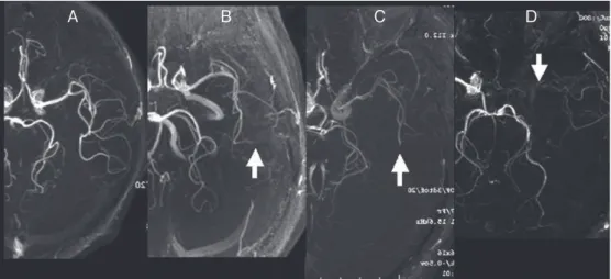
Image B demonstrating that pyoktanin staining has visualized previously unrecognized minute plaque debris captured on the same

Objective: To report a case of symptomatic internal carotid artery stenosis with a persistent proatlantal artery (PPA), which was treated by carotid artery stenting (CAS) with