要旨:症例は 76 歳女性である.左眼の一過性黒内障で発症し,眼科的診察で左眼の視力低下および血管新生緑内 障,虹彩ルベオーシスをみとめた.頸動脈エコー検査から左総頸動脈近位部に高度狭窄の存在が推定され,眼動脈 エコー上左眼動脈の血流速度は低下していた.脳血管撮影では左総頸動脈起始部の高度狭窄が確認され,左眼動脈 の描出は不良であった.左総頸動脈起始部狭窄による眼虚血症候群と診断した.眼動脈血流改善により眼虚血症候 群の増悪を防ぐため,左総頸動脈起始部にステントを留置した.術後,黒内障発作は消失,虹彩ルベオーシスは消 退し,視力低下の進行はなかった.本例では総頸動脈狭窄部位の推定や血行動態の評価に超音波検査が有用であっ た. (臨床神経 2011;51:114-119) Key words:頸動脈狭窄症,眼虚血症候群,ステント,頸動脈エコー検査,眼動脈エコー検査 はじめに 頸動脈あるいは眼動脈の狭窄性病変にともなう眼症状は眼 虚血症候群と呼ばれ,急性症状として一過性黒内障や網膜動 脈閉塞症を生じ,慢性期には網膜機能不全や血管新生緑内障 による視力障害を生じる.左総頸動脈起始部の高度狭窄病変 により眼虚血症候群を呈した症例に対し,ステント留置術を 施行し眼動脈血流の改善をえたので報告する. 症 例 症例:76 歳 女性 主訴:左眼の一過性視力障害 既往歴:高血圧にてオルメサルタン,フロセミド,脂質異常 症にてアトルバスタチン,心筋梗塞にてアスピリン,心房細動 にてワルファリンを内服. 生活歴:機会飲酒,75 歳まで喫煙(10 本!日×20 年間). 現病歴:某年 8 月頃より左眼視野が黒くなる発作が出現す るようになった.症状は臥位から座位になった際に出現する ことが多く,10 分ほど持続し 2∼3 回!日の頻度であった. 徐々に眼がみえにくくなっていると感じ眼科を受診したとこ ろ眼虚血症候群と診断され,精査のため当院へ入院した. 入院時身体所見:身長 147cm,体重 49kg,BMI 22.7.両側で 頸 部 血 管 雑 音 を 聴 取 し,上 肢 血 圧 に は 左 右 差(右 77!42 mmHg,左 59!49mmHg)があった.右上肢の臥位血圧は 70! 42mmHg,立位血圧は 80!57mmHg で臥位,立位ともに脈拍 55!分で不整をみとめた.両側橈骨動脈および両側足背動脈の 脈拍触知は不良だった. 神経学的所見:意識は清明.矯正視力は右(0.7),左(0.15), Goldmann 視野計では緑内障による左眼鼻側視野の狭窄をみ とめ,左眼底周辺部に小斑状出血をみとめた.網膜動脈には栓 子をみとめなかった.その他の脳神経系,運動系,感覚系,深 部腱反射,協調運動系所見に異常をみとめなかった. 眼科的所見:左眼に虹彩ルベオーシスと新生血管緑内障を みとめた. 一般検査所見:中性脂肪 189mg!dl と高値,HDL コレステ ロール 36mg!dl と低値である他に血算,生化学,尿検査に異 常をみとめなかった.凝固検査ではワルファリン内服中のた め PT-INR が 1.45 と高値であった.心電図は心房細動であっ た. 頸動脈エコー検査:左総頸動脈の内中膜複合体厚は全周性 に肥厚しており,収縮期最高血流速度(peak systolic veloc-ity:PSV)30.3cm!sec や拡張末期血流速度(end diastolic ve-locity:EDV)9.7cm!sec は低下,pulsatility index(PI)1.17, resistance index(RI)0.68 であり,立ち上がり時間 201.5 秒と * Corresponding author: 広南病院脳血管内科〔〒982―8523 宮城県仙台市太白区長町南 4―20―1〕 1) 広南病院脳血管内科 2) 同 血管内脳神経外科 3) 同 脳神経外科 (受付日:2010 年 6 月 15 日)
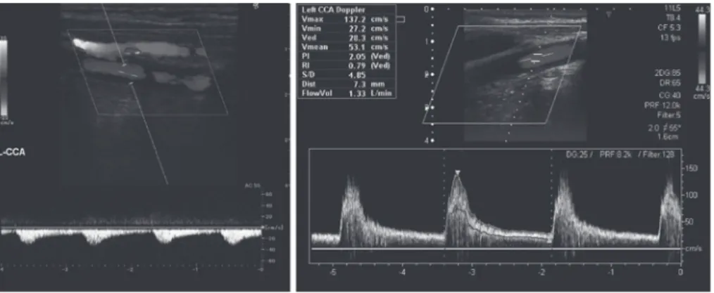
A: Before carotid artery stenting (CAS), the peak systolic and end diastolic flow velocities were decreased. The blunted waveform with prolonged acceleration time represents a post-stenotic pattern.
B: After CAS, the peak systolic flow velocity increased and the waveform was normalized.
Fig. 2 The Doppler spectrum within the left ophthalmic artery.
A: Before CAS, the waveform showed an antegrade flow with reduced peak systolic velocity. The waveform was blunted demonstrating a post-stenotic pattern.
B: After CAS, the peak systolic flow velocity increased significantly and the waveform was normalized.
A
B
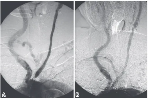
延長していた.総頸動脈の拡張末期血流速度の左右比は 2.4 と高値であった.左総頸動脈のドプラ血流波形は立ち上がり 時間が延長し,なだらかなカーブを描く狭窄後パターンを呈 しており,収縮期最高血流速度低下所見とあわせると左総頸 動脈近位部狭窄がうたがわれた(Fig. 1A).本例では心房細動 を合併していたため,眼動脈エコー検査もふくめエコー検査 における血流速度計測はすべて 3 心拍の平均値とした. 眼動脈エコー検査:両側とも順行性血流であったが,左眼 動脈では PSV 6.2cm!sec,EDV 1.4cm!sec といちじるしく低 下しており,PI 1.66,RI 0.78 であっ た.立 ち 上 が り 時 間 は 182.2 秒と延長し,ドプラ血流波形は狭窄後パターンであった (Fig. 2A). MRI:拡散強調像, T2強調像で脳梗塞をみとめなかった. 脳血管造影検査:左総頸動脈起始部に高度狭窄病変あり, 狭窄部より遠位に造影遅延をみとめた.左外頸動脈は描出さ れず,左眼動脈の描出は不良で retinal blush をみとめず,左 前大脳動脈は造影されなかったが,左中大脳動脈に異常な かった(Fig. 3A).右総頸動脈造影で前交通動脈を介して左前 大脳動脈,左中大脳動脈が描出された. 術前脳血流 SPECT:安静時脳血流は左中大脳動脈領域で 健側比 91% と低下しており,ダイアモックス負荷後の血管反 応性は左中大脳動脈領域で 1.3% と低下していた. 入院後経過:超音波検査,脳血管造影検査の結果から,眼虚 血症候群の原因は左総頸動脈起始部の高度狭窄病変による左 眼動脈の血流低下と診断した.左眼動脈の血流改善を目的に, 左総頸動脈起始部高度狭窄病変に対するステント留置術を施 行した.ステント留置後の脳血管造影検査では左眼動脈の描 出が改善し,左総頸動脈造影で術前は描出されなかった左外Fig. 3 Aortography (anteroposterior view).
A: Before CAS, aortography showed severe stenosis of the left common carotid artery at its orfice. The flow of the left common carotid artery was restricted.
B: After CAS, dilation of the stenosis and stenting were confirmed in the left common carotid artery.
A
B
頸動脈や左前大脳動脈が描出されるようになった(Fig. 3B, 4B, 5B).頸動脈エコー検査では,左総頸動脈 PSV 137.2cm! sec,EDV 27.2cm!sec と上昇,PI 2.05,RI 0.79 であり,立ち上 がり時間は 98.7 秒に短縮していた(Fig. 1B).また,眼動脈エ コー検査でも左眼動脈の血流速度上昇が確認され,PSV 62 cm!sec,EDV 10.9cm!sec,PI 2.05,RI 0.81 であり,立ち上が り時間が 147.5 秒と短縮したことにより,ドプラ血流波形の 狭窄後パターンも改善した(Fig. 2B).ステント留置術後過灌 流症候群の発症はなかったものの,術後第 1 病日の脳血流 SPECT では,左中大脳動脈領域の安静時血流が健側比 120% と上昇しており降圧療法を開始,第 5 病日には健側比 108% に安定し,術前とくらべ安静時血流増加をえた.ステント留置 術 5 週間後の眼科的検査では虹彩ルベオーシスの退縮をみと め,術後 2 年の現在まで視力,視野障害の悪化はなく,黒内障 発作の再発もない. 考 察 左総頸動脈起始部高度狭窄にともない一過性黒内障をくり かえした眼虚血症候群に対し,ステント留置術を施行し眼動 脈の血流改善をえ,眼虚血症候群の進行を抑制することがで きた.本例の総頸動脈狭窄部位の推定には頸動脈エコーが有 用であり,眼動脈血流の評価には眼動脈エコーが有用であっ た.検索しえたかぎり,眼虚血症候群を生じた総頸動脈起始部 狭窄病変に対しステント留置した報告はなく,本例はまれな 症例と考えられる. 本例では総頸動脈起始部狭窄が眼虚血症候群の原因であっ た.総頸動脈における 75% 以上の高度狭窄病変はまれであ り,脳卒中症例におけるその発生頻度は,内頸動脈高度狭窄が 29.8% であるのに対し総頸動脈高度狭窄は 0% であったと報 告されている1).総頸動脈狭窄の臨床症候についての報告は非 常に少ないが,総頸動脈狭窄では一過性黒内障を生じること が多いとされている.Hoya らは European Carotid Surgery Trial 法で 50% 以上の狭窄を有する症候性総頸動脈狭窄症例 の 50% に一過性黒内障をみとめ,同法で 50% 以上の狭窄を 有する症候性内頸動脈狭窄例における 13% とくらべ有意に 多かったと報告した2).また,Karacostas らは本例と同様,総 頸動脈閉塞により眼虚血症候群のみを呈した例を報告してい る3).総頸動脈や内頸動脈狭窄例における一過性黒内障の発現 機序は,微小栓子機序の頻度が高く,血行力学性機序は少ない とされる4).微小栓子機序は,総頸動脈または内頸動脈のアテ ローム病変で発生した血栓が遊離し,末梢の眼動脈に小塞栓 をおこすとともに,血栓が脳動脈を閉塞し大脳半球症状をみ とめることも多い.一方,血行力学性機序では,何らかの原因 で眼動脈が低灌流になることによりびまん性の網膜虚血が生 じ黒内障を発現するものの,側副血行により脳血流は保たれ 大脳半球症状を生じないことが多い.本例が血栓性塞栓機序 であったのか,血行力学性機序であったのか確定はできない が,大脳半球症状をともなわなかったことや臥位から座位へ の体位変換時に黒内障発作を生じることが多かったことか ら,後者の可能性が高いと推測する.内頸動脈狭窄による眼動 脈血流障害では,主に外頸動脈から側副血行路が形成され眼 動脈血流は維持されるばあいが多いが,本例では左総頸動脈 起始部に高度狭窄病変が存在したため,外頸動脈経由の側副
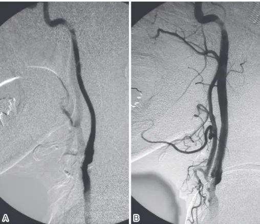
Fig. 4 Angiography of the cervical segment of the left common carotid artery (lateral view).
A: Before CAS, the left external carotid artery was not observed. The cervical segment of the left internal carotid artery showed mild stenosis.
B: After CAS, external carotid artery became visible.
A
B
Fig. 5 Angiography of the intracranial left common carotid artery (lateral view).
A: Before CAS, the left ophthalmic artery was faint and no collateral circulation from the left external carotid artery to the left ophthalmic artery was observed.
B: After CAS, the blood supply to the left ophthalmic artery was improved. The left anterior cerebral artery, which was previously not observed, became visible.
A
B
血行路が不十分で眼虚血を生じた.これは眼動脈エコー検査 所見や脳血管撮影所見からも確認できる.外頸動脈から眼動 脈へ側副血行が発達しているばあい,眼動脈エコー検査では 眼動脈血流が逆行性となることが多いが,本例では左眼動脈 血流は順行性であり(Fig. 2A),左外頸動脈からの側副血行が 不十分であったことを示している.また,ステント留置前脳血 管撮影では左外頸動脈は造影されておらず(Fig. 4A),術後左 総頸動脈撮影(Fig. 3B, 4B)を考え合わせると,左総頸動脈起 始部高度狭窄と左頸部外頸動脈狭窄により左外頸動脈血流が 低下していたことを示している.ステント留置前脳血管撮影部狭窄の存在を推定することができた.総頸動脈または内頸 動脈高度狭窄を有する患者における眼動脈エコー検査では, 眼動脈の収縮期血流速度の低下,pulsatility index の上昇をみ とめ5)6),ドプラ血流波形は逆流型,to and fro 型,アーチ型,
動脈硬化型などの異常波形を呈する7)8).Kawaguchi らは内頸 動脈高度狭窄を有する 38 症例において,ステント留置術の前 後で眼動脈エコー検査を施行し,ステント留置術後には有意 に眼動脈血流速度の増加をみとめ,慢性的な眼虚血症候群が 改善した例もあったと報告している9).Kawaguchi らの報告 と同様に,これまで頸動脈高度狭窄による眼虚血症候群に対 しステント留置術を施行した報告の多くは,頸部内頸動脈に 高度狭窄を有する例であった9)∼11).著者が検索したかぎり,本 例のように総頸動脈起始部高度狭窄により生じた眼虚血症候 群に対しステント留置術を施行した報告はなく,本例はまれ な症例である. 眼虚血症候群における視力予後は不良であり,眼科的には 主に網膜光凝固術がおこなわれるが,対症療法に過ぎず,多く のばあい新生血管緑内障に進行する.根治療法は眼虚血の改 善であるが,早期に実施しえないばあいは不可逆性の視機能 障害をきたす12)13).本例ではステント留置術後に眼動脈血流 の増加をみとめ,眼虚血の特徴的所見である虹彩ルベオーシ スの退縮を確認できたことから,今後の新生血管緑内障の改 善,視機能改善が期待される. 文 献
1)Mikael M, Labreuche J, Gongora-Rivera F, et al. Autopsy prevalence of proximal extracranial atherosclerosis in
pa-5)Hu HH, Sheng WY, Yen MY, et al. Color Doppler imaging of orbital arteries for detection of carotid occlusive dis-ease. Stroke 1993;24:1196-1203.
6)Mawn LA, Hedges TR 3rd
, Rand W, et al. Orbital color Doppler imaging in carotid occlusive disease. Arch Oph-thalmol 1997;115:492-496.
7)日本神経超音波学会 機関紙 Neurosonology 編集委員会, 編. 脳神経超音波マニュアル. 大阪: 報光社; 2006. p. 54-57. 8)Kouvidis GK, Benos A, Kyriakopoulou G, et al. Colour
Doppler ultrasonography of the ophthalmic artery: flow parameters in normal subjects. Int Angiol 2000 ; 19 : 319-325.
9)Kawaguchi S, Sakaki T, Iwahashi H, et al. Effect of ca-rotid artery stenting on ocular circulation and chromic ocular ischemic syndrome. Cerebrovasc Dis 2006;22:402-408.
10)鬼塚正成, 松屋直樹, 宮崎久弥. 頸動脈ステント留置術後に 改 善 を み た 眼 虚 血 症 候 群 の 1 例. 脳 と 神 経 2001;53:679-682.
11)Ho TY, Lin PK, Huang CH. White-centered retinal hemor-rhage in ocular ischemic syndrome resolved after carotid artery stenting. J Chin Med Assoc 2008;71:270-272. 12)Mizener JB, Podhajsky P, Hayreh SS. Ocular ischemic
syndrome. Ophthalmology 1997;104:859-864.
13)根木 昭. 眼虚血症候群. 石橋一樹, 安積 淳, 編. これなら わかる神経眼科. 東京: 文光堂; 2005. p. 339-341.
Herein, we present a case of amaurosis fugax in a 76-year-old female with high blood pressure, dyslipidemia, and chronic atrial fibrillation who frequently suffered from transient visual impairment of the left eye. Color Dop-pler imaging of the flow profile in the left ophthalmic artery and the left common carotid artery revealed stenosis in the proximal portion of the left common carotid artery. Aortography revealed severe stenosis of the left com-mon carotid artery at its orifice. The left external carotid artery was not observed as no collateral circulation from the left external carotid artery to the left ophthalmic artery was present. The presence of severe stenosis of the left common carotid artery at its orifice was considered to have caused the amaurosis fugax of the left eye, due to a reduction in the blood supply from both the left external carotid artery and the left internal carotid artery. Per-cutaneous transluminal angioplasty with stenting was successfully performed to dilate the stenosis at the orifice of the left common carotid artery with the aim of restoring visual function by improving the blood supply to the left ophthalmic artery. After stenting, angiography of the left common carotid artery showed improved blood sup-ply in the left ophthalmic artery. The patient was followed up for 2 years after CAS, and no recurrence of amauro-sis fugax or rubeoamauro-sis iridis were observed. In this case, color Doppler imaging was useful for diagnosing and evalu-ating flow dynamics.
(Clin Neurol 2011;51:114-119) Key words: carotid artery stenosis, ocular ischemic syndrome, carotid artery stenting, color Doppler imaging, ophthalmic