53:452
はじめに
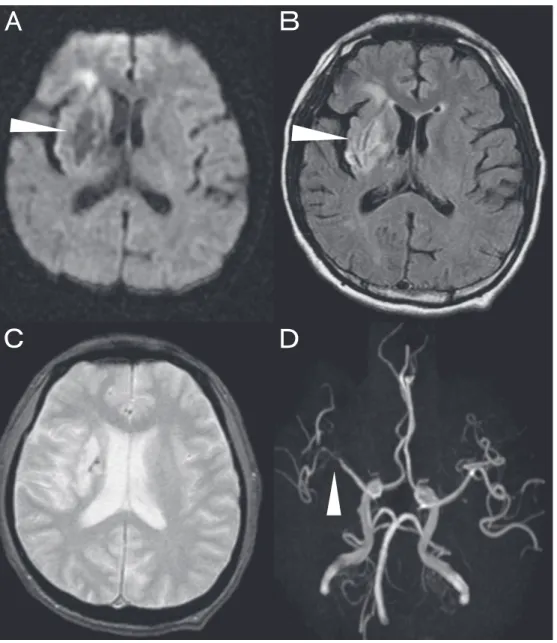
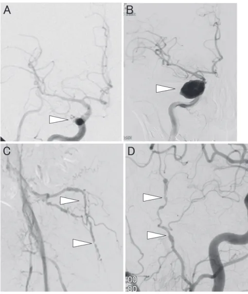
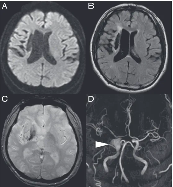
結節性多発動脈炎(polyarteritis nodosa; PAN)は中小型の 動脈に壊死性血管炎をみとめ,かつ細動脈炎,毛細血管炎を みとめない疾患と定義される1)2).PAN は 20∼30%に中枢 神経症状を合併し,13∼17%に脳梗塞がみられ,病型では ラクナ梗塞が多い3).脳動脈瘤の合併例の報告は散見される が4)∼8),瘤が巨大化した例は報告されていない.今回われわ れは,浅側頭動脈生検で診断しえた巨大内頸動脈瘤をともな う PAN の 1 例を経験した.その発症機序に関する文献的考 察もふくめて報告する. 症 例 患者:46 歳,男性 主訴:右眼の視力低下 生活歴:喫煙歴あり(1 日 30 本を 30 年間). 既往歴:36 歳時に左網膜中心動脈閉塞症,41 歳時に右中 大脳動脈狭窄による脳梗塞発症(Fig. 1).脳血管造影で右椎 骨動脈瘤(V4),右内頸動脈瘤(C4)がみられた(Fig. 2A). 44 歳時,1 ヵ月続く発熱と両側大腿筋肉痛で近医に入院した. CPK の一過性上昇はなく,原因不明だった.その時に施行 された脳血管造影検査で,右内頸動脈瘤は 17 mm に拡大し ていた(Fig. 2B). 現病歴:2012 年 3 月,起床時に右眼がみえにくくなり, その 30 分後,更に両眼がみえにくくなり,当院の救急外来 を受診した. 入院時現症:身長 170 cm,体重 66 kg,血圧 128/78 mmHg, 脈拍 100/ 分・整,体温 37.6C.頸部・眼窩部に血管雑音なし. 肺音,心音は特記所見なし.神経学的所見は意識清明,右眼 手動弁,眼底にさくらんぼ赤色班をみとめた.既往の左網膜 中心動脈閉塞症のため,左眼指数弁だった.左顔面麻痺,構 音障害,嚥下障害,左上下肢に MMT 3/5 の麻痺があり,こ れらは陳旧性脳梗塞による症状だった.感覚系,協調運動は 特記所見なし,腱反射は左右差なく,病的反射も陰性だった (NIHSS 3). 検査所見:採血所見は,血沈が 1 時間値 43 mm,WBC 値 は 4,330 l と正常で.ALT 56 U/l,AST 46 U/l と軽度上昇, CRP は陰性,D-dimmer 1.2 g/ml と上昇,甲状腺機能,リウ マチ定量は正常,抗核抗体,PR3-ANCA,MPO-ANCA,ルー プスアンチコアグラント,抗カルジオリピン 2GPI 抗体, 抗カルジオリピン IgG 抗体は陰性だった.脳脊髄液検査は 細胞数 0/l,蛋白 56 mg/dl,糖 54 mg/dl だった.心電図検査 は洞調律,経胸壁心エコー検査は EF 62.2%,脳波は基礎波 8 Hz で異常なく,頸部血管エコーは有意な狭窄性病変,動脈 壁肥厚はなかった.経頭蓋ドプラ検査は右左シャントなく, 経食道心エコーは異常所見なく,大動脈壁の肥厚はなかった. 入院時の頭部 MRI 拡散強調画像には新たな虚血病巣はなく, FLAIR 画像で右中大脳動脈領域に陳旧性出血性脳梗塞をみ とめた.頭部 MRA では右内頸動脈 C4 部に動脈瘤があった (Fig. 3A ∼ C).同日施行した脳血管造影検査にて,右内頸動
症例報告
巨大内頸動脈瘤をともなう結節性多発動脈炎の 1 例
植村 順一
1)* 井上 剛
1)青木 淳哉
1)佐治 直樹
1)芝崎 謙作
1)木村 和美
1) 要旨: 症例は 46 歳の男性である.右眼の視力低下で発症した.既往歴に左網膜中心動脈閉塞症,右中大脳動 脈領域の脳梗塞,巨大右内頸動脈瘤,右椎骨動脈瘤があった.右眼底にさくらんぼ赤色班があり,右網膜中心動 脈閉塞症と診断した.血沈の亢進をみとめるが,WBC 上昇なく,CRP 陰性,抗核抗体と ANCA は陰性,脳脊髄液 検査は正常だった.大動脈造影では,大血管に異常ないが内腸骨動脈,前腕動脈筋肉枝に数珠状変化があった. 脳血管造影で両側浅側頭動脈に狭窄,数珠状変化があり,その部位の生検にて内膜にフィブリノイド壊死をとも なう動脈炎をみとめ,結節性多発動脈炎と診断した.本例は結節性多発動脈炎に巨大内頸動脈瘤をみとめたまれ な症例である. (臨床神経 2013;53:452-457) Key words: 結節性多発動脈炎,巨大内頸動脈瘤,浅側頭動脈生検 *Corresponding author: 川崎医科大学脳卒中医学教室〔〒 701-0192 岡山県倉敷市松島 577 番地〕 1) 川崎医科大学脳卒中医学教室 (受付日:2012 年 10 月 23 日)脈 C4 部に 17 mm の動脈瘤,右椎骨動脈 V4 部に動脈瘤があっ た(Fig. 2D). 入院後経過:入院日に脳血管造影をおこない,眼動脈近傍 からウロキナーゼ 6 万単位動注した.動注後に右眼視力は指 数弁まで改善した.大動脈造影では,大動脈,腎動脈,腹部 大動脈に異常なく,内腸骨動脈筋肉枝,前腕動脈筋肉枝に狭 窄,数珠状変化があった(Fig. 2C).脳血管造影で,浅側頭動 脈に狭窄,数珠状変化があった(Fig. 2D).全身性血管炎を うたがい,病理的診断のために浅側頭動脈生検を施行した. 病理所見で,HE 染色は血管内腔の狭窄が一部にあり,病変 は分節状だった.内膜にフィブリノイド壊死をともなうリン パ球,好中球,マクロファージなどの炎症細胞浸潤があった. 多核巨細胞はなかった.血管内腔の狭窄が一部にあり,病変 は分節状だった.特殊染色では,SMA(smooth muscle actin) 染色,E-Masson 染色では内膜肥厚があり,中膜の不明瞭化 があり,内弾性板は破裂していた(Fig. 4A ∼ C).臨床所見, 血管造影所見,病理所見から 2006 年厚生労働省特定疾患難 治性血管炎班の PAN の診断基準より definite PAN と診断した. 診断後,プレドニン 60 mg(1 mg/kg)とエンドキサン 50 mg を開始した.その後,60 日間再発がなく,経過良好である. Fig. 1 Magnetic resonance imaging/angiography.
A: Diffusion-weighted image (1.5 T; b = 800 sec/mm2
, transverse view) shows high and low intensity lesions in the right middle cerebral artery area. B: FLAIR image shows high intensity lesions in the right middle cerebral artery area. C: T2* image shows low intensity lesions in the right middle cerebral artery area. D: Magnetic resonance angiographie
臨床神経学 53 巻 6 号(2013:6) 53:454 考 察 本例は若年発症の巨大内頸動脈瘤をきたした脳梗塞の 1 例 である.2 年前に,発熱,体重減少,筋肉痛が一過性にあっ た が 原 因 不 明 だ っ た. 今 回, 血 沈 の 亢 進 が あ る も の の, WBC,CRP は正常だった.ANCA は陰性で,大動脈造影で 中型動脈異常があり,浅側頭動脈生検でえられた組織所見が 内膜にフィブリノイド壊死をともなう動脈炎であったことか ら PAN と診断しえた.2006 年厚生労働省特定疾患難治性血 管炎班の診断基準では,PAN 診断の生検部位は腎,肺・気 管支,大腸が多いが,本例は浅側頭動脈生検により,PAN が 診断しえた最初の報告例であり貴重である.本例で浅側頭動 脈生検を施行しえたのは,巨大動脈瘤の経過観察のために施 行した脳血管造影検査で,外頸動脈系に数珠状の血管異常に たまたま気付いたことによる.これまでに PAN に合併した 脳動脈瘤の報告5)∼9)で,外頸動脈系の異常に着目したもの はない.今後,PAN の診断に浅側頭動脈生検は有効である と考えられる. PAN は 20∼30%に中枢神経症状を合併し,13∼17%に脳 梗塞がみられるが,脳梗塞の病型分類において,NINDSIII 分類はラクナ梗塞,TOAST 分類9)は small vessel disease が
多い4).本例の陳旧性脳梗塞は主幹動脈である右中大脳動脈
領域の梗塞で,T2* 像では出血性梗塞であったことから,塞
栓性機序により血管が再開通したと考えられた.
PAN は 70∼80%に中型動脈の狭窄性病変があることから1)
, Fig. 2 Cranial angiography.
A: Cranial angiography shows C4 portion intracranial aneurysm at 41 years. B: Cranial angiography shows C4 portion giant intracranial aneurysm at 44 years. C: Pelvic angiography shows that internal iliac artery had abnormality of stenosis and dilation at 46 years. D: Cranial angiography shows external carotid artery had abnormality of stenosis and dilation at 46 years.
Fig. 4 Superficial temporal artery biopsy.
A, B, C: Superficial temporal artery biopsy at H-E, E-Masson shows the arteritis with fibrinoid necrosis. Internal and external elastic laminae have completely diaappeared.
Fig. 3 Magnetic resonance imaging/angiography on admission. A, B, C: Diffusion-weighted image (1.5 T; b = 800 sec/mm2, transverse view), FLAIR and T
2* image did not show high
臨床神経学 53 巻 6 号(2013:6) 53:456 PAN の診断には全身血管評価が必要とされる.しかし,脳 血管撮影をされる例は少なく,脳動脈瘤の報告例はまれであ る4)∼8).Oomura ら4)は PAN で動脈瘤が形成された 9 例を報 告している.部位は 5 例が内頸動脈系,3 例が椎骨脳底動脈 系であり,1 例は多発していた.これらの瘤の最大径は 5 mm と比較的小さい.本例は 3 年の経過で内頸動脈に 17 mm の 巨大動脈瘤が形成されたはじめての報告である.Takahashi ら7)は剖検で破裂した脳動脈瘤の組織所見が血管壁に炎症 細胞の浸潤,血管内膜のフィブリノイド壊死,血管内弾性板 の破壊をともなっていたと報告している.本例も血管の内弾 性板は破裂していた.巨大内頸動脈瘤が形成された成因は, PAN による炎症性変化とそれによる血管壁脆弱性,血管内 弾性板破裂が考えられる.本例はステロイド,免疫抑制剤の 投与後の血管評価では内頸動脈瘤の大きさは変化なかった. PAN の標準治療はステロイド,エンドキサンとされてい る1)2)が,本例も診断後プレドニン 60 mg(1 mg/kg)とエン ドキサン 50 mg を開始した.本例は発熱,倦怠感などの炎症 所見に乏しく,採血では WBC,CRP などの炎症所見に乏しく, 血沈のみが上昇しており,治療の効果判定に苦慮した.リウ マチの炎症マーカーである PTX-3,MMP-3 値を測定したが, 上昇していなかった. 今回,われわれは若年発症の両側網膜中心動脈閉塞症,陳 旧性脳梗塞,巨大内頸動脈瘤があり,浅側頭動脈生検で結節 性多発動脈炎と診断した 1 例を経験した.今後,脳動脈瘤が あり血管炎がうたがわれる症例は,脳血管造影検査で外頸動 脈系に注目して,異常があれば浅側頭動脈生検を施行すべき である. ※本論文に関連し,開示すべき COI 状態にある企業,組織,団体 はいずれも有りません. 文 献
1) Guillevin L, Le Thi HDU, Godeau P, et al. Clinical findings and prognosis of polyarteritis nodosa and churg-strauss angiitis: A study in 165 patients. Rheumatology 1988;27:258-264.
2) Pagnoux C, Seror R, Henegar C, et al. Clinical features and outcomes in 348 patients with polyarteritis nodosa: A systematic retrospective study of patients diagnosed between 1963 and 2005 and entered into the French vasculitis study group database. Arthritis Rheum 2010;62:616-626.
3) Reichhart MD, Bogousslavsky J, Janzer RC. Early lacunar strokes complicating polyarteritis nodosa. Neurology 2000; 54:883-889.
4) Oomura M, Yamawaki T, Naritomi H, et al. Polyarteritis nodosa in association with subarachnoid hemorrhage. Intern Med 2006; 45:655-658.
5) Oran I, Memis A, Parildar M, et al. Multiple intracranial aneurysms in polyarteritis nodosa: MRI and angiography. Neuroradiology 1999;41:436-439.
6) Provenzale JM, Allen NB. Neuroradiologic findings in polyarteritis nodosa. AJNR Am J Neuroradiol 1996;17:1119-1126.
7) Takahashi JC, Sakai N, Iihara K, et al. Subarachnoid hemorrhage from a ruptured anterior cerebral artery aneurysm caused by polyarteritis nodosa. J Neurosurg 2002;96:132-134.
8) Thompson B, Burns A. Subarachnoid hemorrhages in vasculitis. Am J Kidney Dis 2003;42:582-585.
9) Goldstein LB, Jones MR, Matchar DB, et al. Improving the reliability of stroke subgroup classification using the Trial of Org 10172 in Acute Stroke Treatment (TOAST) criteria editorial comment: Classifying the mechanisms of ischemic stroke. Stroke 2001;32:1091-1097.
Abstract
A case of polyarteritis nodosa with giant intracranial aneurysm
Jyunichi Uemura, M.D.
1), Takeshi Inoue, M.D.
1), Junya Aoki, M.D.
1), Naoki Saji, M.D.
1),
Kensaku Shibazaki, M.D.
1)and Kazumi Kimura, M.D.
1)1)Stroke Medicine, Kawasaki Medical School