日呼吸誌 1(1),2012
緒 言
転移性心臓腫瘍は原発性心臓腫瘍の 20〜40 倍の頻度 とされ,原発巣としては乳癌,肺癌,悪性リンパ腫が比 較的多く,胃癌,食道癌,腎癌,悪性黒色腫などが続く1). 心臓への転移様式は直接浸潤がほとんどであり,右心系 への単独の転移はきわめてまれである.今回,血痰にて 発症し食道癌の右心系への転移により肺動脈腫瘍塞栓症 をきたし,肺梗塞を繰り返し呈した食道癌の 1 例を経験 したので報告する.
症 例 患者:65 歳,女性.
主訴:血痰.
既往歴:甲状腺機能低下症.
喫煙歴:5 本/日×44 年.
家族歴:次女に特発性血小板減少性紫斑病.
現病歴:2009 年 1 月頃より血痰を自覚し,4 月に前医 を受診した.血液検査では Hg 9.8 g/dl と貧血,CRP 1.6 mg/dlと軽度の炎症反応の上昇などを認めた.胸部X線,
CT 検査にて結節影,すりガラス影など認めたため(Fig.
1),前医にて気管支鏡検査を施行された.生検では有意 な所見を認めず,血性の気管支肺胞洗浄液であったため 特発性肺胞出血とされ,また無治療で陰影の軽減を認め たため経過観察となった.同年 9 月頃から再び血痰の出 現,労作時の呼吸困難も出現し,血小板数の低下(6×
104/μl)を認めたため前医に入院となり,肺病変の精査 目的のため当院に転院となった.転院時の血液検査にて 血小板減少,線溶系の亢進,cytokeratin 19 fragment
(CYFRA)の上昇などを認めた(Table 1).
入院時現症:体温 36.6℃,脈拍 84/min,血圧 106/70 mmHg,SpO2 94%(room air).頸部:リンパ節腫脹なし,
胸部:ラ音聴取せず,心雑音聴取せず.腹部:軟,圧痛 なし,下肢:軽度の浮腫あり.胸部 X 線:両肺に斑状影,
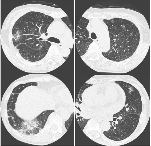
浸潤影,両側肺門部肺動脈拡大を認めた(Fig. 2).胸部 CT:両側肺に斑状影,モザイク状のすりガラス影,両 側胸水を認めた(Fig. 3).
転院後経過:入院後も徐々に酸素化の悪化を認め,心 不全も疑い第 9 病日に心臓超音波検査を施行した.三尖 弁基部に付着する腫瘤を認め(Fig. 4),高度な肺高血圧 連絡先:松尾 潔
〒701‑1192 岡山市北区田益 1711‑1
a国立病院機構岡山医療センター呼吸器科1
b同 消化器科
(E-mail: [email protected])
(Received 24 Mar 2011/Accepted 2 Sep 2011)
●症 例
血痰にて発症し右心転移から肺動脈腫瘍塞栓をきたし,
肺梗塞を繰り返した食道癌の 1 例
工藤健一郎a 松尾 潔a 山下 晴弘b 藤原 慶一a 米井 敏郎a 佐藤 利雄a
要旨:症例は 65 歳女性.2009 年 1 月頃から血痰を自覚し 4 月に前医を受診した.胸部 CT 検査にて多彩 な陰影を認め,前医にて気管支鏡検査を施行された.血性の気管支肺胞洗浄液であったため特発性肺胞出血 とされ,また無治療で陰影は消退傾向となったため経過観察となった.同年 9 月に再び血痰,労作時の息 切れが出現したため前医に入院となり,精査のため当院へ転院となった.心臓超音波検査にて三尖弁に付着 する腫瘤影を認めたため,心臓腫瘍摘出術,肺生検を施行した.また上部消化管内視鏡検査にて中部食道に 食道癌を認め,食道癌から右心系に転移し肺動脈腫瘍塞栓,肺梗塞をきたしたと考えられた.病理学的に pulmonary tumor thrombotic microangiopathy(PTTM)も伴っていた.docetaxel にて化学療法を施行し,
CT 上での陰影の改善や血液検査での線溶系異常の改善を認めたが,診断後約 2ヶ月半で死亡した.食道癌 の右心系への転移はきわめてまれであり,肺動脈腫瘍塞栓が血痰と肺野陰影の主因と考えられた.
キーワード:転移性心臓腫瘍,肺腫瘍塞栓,食道癌,肺梗塞,三尖弁置換術
Metastatic heart tumor,Pulmonary tumor embolism,Esophageal cancer,
Pulmonary infarction,Tricuspid valve replacement 20
肺動脈腫瘍塞栓による肺梗塞を呈した食道癌の 1 例
を伴っていた(推定収縮期肺動脈圧 75 mmHg).同日の 造影 CT にて肺動脈に塞栓像を認め(Fig. 5),肺野の多 彩な陰影は肺動脈塞栓による肺梗塞が疑われた.主肺動 脈への塞栓による突然死の可能性が危惧され,第 10 病 日に三尖弁置換術,心臓腫瘍摘出術,肺生検(右 S5)
を施行した.腫瘍は三尖弁に固着していたが脆弱であり,
中分化型扁平上皮癌と判明した(Fig. 6).原発巣精査の
Table 1 Laboratory findings on admission
[Hematology] [Chemistry]
RBC 450×104/μl T-Bil 0.7 mg/dl Hb 11.4 g/dl AST 29 IU/L WBC 9,900/μl ALT 17 IU/L
Nt 74% ALP 424 IU/L
Ly 19% LDH 503 IU/L
Mo 4% [Serology]
Eo 3% CRP 6.4 mg/dl
PLT 12.3×10⁴/μl CEA 2.6 ng/ml PT 14.2 s CYFRA 13.8 ng/ml
APTT 39 s BNP 187 pg/ml
Fbg 255 mg/dl PA-IgG 49.5 ng/107 cells FDP 67.5 μg/ml [Blood gas analysis]
D-dimer 30 μg/ml pH 7.41 ESR 20 mm/h PaCO2 31 Torr
PaO2 78 Torr BE −4.0 mEq/L HCO3− 19.3 mEq/L
Fig. 1 (A) Chest radiograph at previous hospital showing nodular and ground-glass shadows in both
lung fields. (B) Chest CT scan at previous hospital showing ground-glass opacity and nodular shadows in both lung fields.Fig. 2 Chest radiograph on admission showing
ground-glass shadows in both lung fields with enlargement of the pulmonary artery.21
日呼吸誌 1(1),2012
Fig. 3 Chest CT scan on admission showing ground-glass opacity and patchy shadows in both lung
fields.Fig. 4 Echocardiographic examination showed a tumor
attaching the right ventricle and tricuspid valve.Fig. 5 Chest-enhanced CT showing a defect in the
right lower pulmonary artery.22
肺動脈腫瘍塞栓による肺梗塞を呈した食道癌の 1 例
ため上部内視鏡検査を施行し中部食道に不正な隆起性病 変を認め,生検にて扁平上皮癌と診断され原発巣と考え られた(Fig. 7).第 20 病日には肝門部の転移リンパ節 による閉塞性黄疸をきたしたため,経皮経肝胆管ドレ ナージを施行し胆管ステントの留置を行った.その後,
第 42 病日から docetaxel(60 mg/m2)にて治療を開始 した.化学療法により呼吸困難や画像上,陰影の改善,
推定肺動脈圧の低下,血小板数の増加などを得た.第 54 病日に退院し,外来にて 2 コース目の化学療法を施 行したが労作時呼吸困難の増強あり,救急搬送された.
心タンポナーデをきたしており,緊急で心嚢ドレナージ を施行した.自覚症状は改善し小康状態を得たが,全身 状態は悪化しており化学療法の継続は困難であった.
徐々に呼吸状態は悪化し当院での全経過約 4ヶ月,発症 から約 1 年で永眠された.死後,病理解剖を行った.
病理解剖にて食道癌の浸潤は食道の固有筋層までの深 達であり,心外膜へは癌の浸潤を認めなかった.脆弱な 腫瘍が右心系へ転移したことにより,中小の肺動脈に腫 瘍塞栓をきたし(Fig. 8A)肺高血圧を併発していた.
また両肺はすべての葉に出血性壊死巣が散在し,末梢の 肺動脈内の腫瘍塞栓による肺梗塞巣と考えられた(Fig.
8B).
考 察
転移性心臓腫瘍は原発性心臓腫瘍の 20〜40 倍の頻度 とされている1).原発巣としては乳癌,肺癌,白血病,
悪性リンパ腫が比較的多く,胃癌,食道癌,腎癌などが 続く2).心転移の部位は,心外膜(60%以上),と心筋(約 30%)で 90%以上を占め,本症例のような心内膜(心 腔内)のみの転移は,非常にまれである3).さらに食道 癌の心臓への転移様式は解剖学的関係からも通常は直接 浸潤であるが,本症例は病理解剖にて,食道癌の浸潤は
Fig. 6 (A) Tumor attaching the tricuspid valve in open-heart surgery. (B) Pathological findings of the
tumor obtained by the operation, showing squamous cell carcinoma.
Fig. 7 A primary lesion was detected in the upper
thoracic esophagus by the gastrointestinal fiberscope.23
日呼吸誌 1(1),2012
食道の固有筋層までの深達であり,また心外膜にも癌の 浸潤はみられなかったことが判明している.したがって,
三尖弁への転移様式は以下の経路が疑われた.食道表面 から奇静脈へは多数の細静脈が連なっており,奇静脈を 介して大循環に入り右心系に転移したものと考えられた.
また心臓腫瘍の診断方法として,心臓超音波検査4), CT,MRI を組み合わせて行うことが多いが,本症例は,
造影 CT では心臓内の腫瘍は指摘できず,心臓超音波検 査が診断に有用であった.本症例のような病態を早期か ら疑うには,血液検査での FDP,D-dimer の上昇,心 臓超音波検査での肺高血圧の存在などが重要であり,ま た胸部 CT 検査にて寛解と増悪を繰り返す陰影を認めた 際には,肺動脈塞栓に伴う肺梗塞も鑑別に考える必要が あると思われた.Fig. 3のCTでは浸潤影,すりガラス影,
結節影を認め,病理学的には Fig. 8B でみられるように 肺梗塞による出血性壊死やそれらが吸収されて消退する 過程を示しているものと考えられた.また本症例でみら れた血痰は肺梗塞に伴う出血性壊死が原因と考えられた.
自然に陰影の軽減を認めた機序については,腫瘍により 末梢の肺動脈が塞栓を起こし肺塞栓がもたらされ,肺梗 塞に陥った部分が肉芽組織により置換された結果,収縮 傾向のある瘢痕となったためと考えられた5).
心転移の臨床症状は,一般に心房心室内を占拠したこ とによる心不全症状と刺激伝導障害によりもたらされる
が6),本症例では生前の手術病理所見および病理解剖所 見から,脆弱な腫瘍の右心系への転移のため中小の肺動 脈に腫瘍塞栓をきたし(Fig. 8A)肺高血圧を併発し,
肺梗塞を繰り返していたものと考えられた(Fig. 8B).
また,本症例では手術時の肺生検にて,中小の肺動脈腫 瘍塞栓に加え,小肺動脈には pulmonary tumor thrombo- tic microangiopathy(PTTM)の所見も合併していたこ とが示されている(Fig. 9).PTTM は主に胃癌などの 粘液産生性の腺癌に関連した特殊な肺動脈腫瘍塞栓であ り,悪性腫瘍剖検例の 0.9〜3.3%に認められたと報告さ れている7).その病態は,小肺動動脈壁への腫瘍細胞の 付着により,サイトカインの産生を介し血管内膜の線維 細胞性肥厚,局所での凝固機能の活性化による血栓など により,末梢肺動脈の狭小化,閉塞を生じるとされる7). 臨床的には急速に進行する肺高血圧,右心不全を呈し,
死に至る.その致死的な臨床経過にもかかわらず,病変 の主座が末梢肺動脈であるため画像所見に乏しく,また 低酸素血症や肺末梢血管抵抗増大による心不全兆候が出 現したときには,すでに全肺野に PTTM が及んでおり 症状出現後は短期間に死に至るため,生前診断がきわめ て困難な病態である.Von Herbay らは剖検例の検討で 21 例の PTTM を報告し,そのうち胃癌が 11 例と半数 を占め,ついで肺癌 3 例,乳癌 2 例と報告している8). 本邦を含めこれまでの報告では PTTM の組織型はほと
Fig. 8 (A) Lung biopsy revealed a tumor embolization in the small and middle pulmonary arteries. (B)
It also revealed a pulmonary infarction due to a tumor embolism on the pulmonary artery.
24
肺動脈腫瘍塞栓による肺梗塞を呈した食道癌の 1 例
んどが腺癌であり9),食道癌での報告は検索した範囲で は報告されていなかった.
以上,本症例は血痰にて発症し,右心系への転移性腫 瘍から肺動脈腫瘍塞栓,PTTM,肺梗塞をきたし,治 療として三尖弁置換術,心臓腫瘍摘出術,化学療法を施 行した.当初は悪性腫瘍による肺病変とは考えていな かったが,右心内腔への転移により多彩な肺病変が出現 し,その後の臨床経過も教訓的な症例と考え報告した.
本論文の主旨は,第 50 回日本呼吸器学会学術講演会 症例 検討会(2010 年 4 月 24 日,京都市)にて発表した.
引用文献
1)Prichard R W. Tumors of the heart. Arch Pathol 1951; 51: 98‑128.
2)King Y, Paul D, Alexander C, et al. Tumors of the Heart. Arch Pathol Lab Med 1993; 117: 1027‑31.
3)Nakamura A, Suchi T, Mizuno Y, et al. The effect of malignant neopalasms on the heart. Jpn Circ J 1975;
39: 531‑42.
4)Johnson MH, Soulen RL. Echocardiography of the cardiac metastasis. AJR Am J Roentgenol 1983; 141:
677‑81.
5)土居裕幸,新居英二,寺澤優代,他.経気管支肺生 検により診断した肺梗塞の 1 例.日呼吸会誌 1999;
37: 974‑8.
6)Faganello G, Belham M, Wilde P, et al. A case of primary cardiac lymphoma. Echocardiography 2007; 24: 889‑92.
7)田村厚久,松原 修.肺動脈腫瘍塞栓症.日胸疾患 会誌 1993; 31: 1269‑78.
8)Von Herbay A, Illes A, Waldherr R, et al. Pulmonary tumor thrombotic microangiopathy with pulmonary hypertension. Cancer 1990; 66: 587‑92.
9)Chine K, Kazutomo T, Ohkura Y, et al. Pulmonary tumor thrombotic microangiopathy caused by a gastric carcinoma expressing vascular endothelial growth factor and tissue factor. Pathol Int 2005; 55:
27‑31.
Fig. 9 A lung biopsy revealed thrombotic embolisms in the small pulmonary artery with fibrocellular
intimal proliferations in many sites.25
日呼吸誌 1(1),2012
Abstract
A case of esophageal cancer initially presenting with bloody sputum and metastasizing to the right side of the heart causing repeated episodes of
pulmonary infarction due to an arterial tumor embolism
Kenichiro Kudo a, Kiyoshi Matsuo a, Haruhiro Yamashita b, Keiichi Fujiwara a, Toshiro Yonei a and Toshio Sato a
a Department of Respiratory Medicine, Okayama Medical Center
b Department of Gastroenterology, Okayama Medical Center
A 65-year-old woman became aware of bloody sputum around January 2009 and visited a hospital. There she showed a nodular shadow and ground-glass opacity on chest CT and underwent a bronchoscopy. Because the bronchoalveolar lavage fluid was bloody, she was diagnosed with an idiopathic pulmonary hemorrhage. Also, a chest abnormal shadow was regressed gradually without treatment. In September of the same year, she again developed bloody sputum and exertional dyspnea, was admitted to the same hospital, and then transferred to our hospital for further evaluation. Because cardiac ultrasound showed a tumor mass attached to the tricuspid valve, it was resected, and lung biopsies were performed. Moreover, an upper gastrointestinal endoscopy revealed esophageal cancer in the middle thoracic esophagus, which suggested that the cancer had metastasized to the right side of the heart, causing pulmonary arterial tumor embolism and pulmonary infarction. These findings were accompanied by pathological evidence of pulmonary tumor thrombotic microangiopathy (PTTM).
Although docetaxel chemotherapy resulted in an improvement of shadows on the CT and of the coagulation and fibrinolytic system of blood tests, the patient died about 2 months after diagnosis. The metastasis of esophageal cancer to the right side of the heart is extremely rare, and a pulmonary arterial tumor embolism was presumed to be the main cause of bloody sputum and shadows in the lung fields.
26