Acta Med. Nagasaki 46 : 55-58
Case Report
Respiratory Bronchiolitis-associated Interstitial Lung Disease
Chieko SHIKUWA 1), Jun-ichi KADOTA 2), Hiroshi MUKAE 1), Towako NAGATA 1), Hideyuki KAIDA 1), Hiroshi ISHII 1), Yuji ISHIMATSU 1), Shigeru KOHNO 1)
1) Second Department of Internal Medicine, Nagasaki University School of Medicine 2) Second Department of Internal Medicine, Oita Medical University
We report a case of respiratory bronchiolitis-associated interstitial lung disease (RB-ILD). A 57-year-old man with a 74-pack-year smoking history, had cough, stridor, yellow purulent sputum and general fatigue for several days. The symptoms almost improved after treatment. However, chest computed tomography (CT) showed diffuse centrilobular ground glass opacities although the chest X-ray film showed no obvious opacities. Examination of bronchoalveolar lavage fluid showed relative lymphocytosis. Examination of lung biopsy obtained by video-assisted thoracoscopy allowed the diagnosis of RB- ILD. The opacities on the CT scan were improved spontane- ously without any treatment after cessation of smoking.
ACTA MEDICA NAGASAKIENSIA 46 : 55-58, 2001
Key Words: respiratory bronchiolitis-associated interstitial lung disease, computed tomography, smoking,
desquamative interstitial pneumonia, bronchoal-
veolar lavage, video-assisted thoracoscopic lung
biopsy
Introduction
Respiratory bronchiolitis was first reported as a pathologic lesion in young smokers, and is character- ized by accumulation of pigmented macrophages within respiratory bronchioles and mild thickening of peribronchiolar interstitium' >. Respiratory bronchiolitis- associated interstitial lung disease (RB-ILD) was clini- cally and pathologically defined by Myers et al.') and Yousem et al.') as respiratory bronchiolitis in heavy smokers characterized pathologically by thickening of peribronchiolar interstitium and centrilobular infiltra-
Address Correspondence: Hiroshi Mukae, M.D.
Second Department of Internal Medicine, Nagasaki University School of Medicine, 1-7-1 Sakamoto, Nagasaki 852-8501, Japan TEL: +81-95-849-7273 FAX: +81-95-849-7285
E-mail: [email protected]
tion of macrophages containing brown cytoplasm.
However, there are only two case reports of RB-ILD in Japan"". We describe here another Japanese patient who was diagnosed as RB-ILD by video-assisted thoracoscopic lung biopsy (VATS-LB).
Case Report
A 57-year-old fisherman who had been smoking two package of cigarettes per day for 37 years, visited the nearby clinic on May 5, 1999 because of 4-day history of cough, yellow sputum and general fatigue. He was admitted to a local hospital on the next day, where physical examination revealed the presence of stridor and signs of peripheral hypoxia. Laboratory tests on admission showed leukocytosis (WBC: 17,000/,u 1) and C-reactive protein (CRP) of 29 mg/dl. Chest X-ray film showed no abnormal shadows. Based on these fea- tures, the patient was treated with antibiotics, bronchodilator, and steroids. He was discharged on June 12 after improvement of symptoms although a definite diagnosis was not established. The attending physician referred the patient to another hospital for further management soon, after discharge. In that hos- pital, chest computed tomography (CT) scans revealed diffuse centrilobular, scanty nodular shadows or ground glass opacities in both lungs although the chest X-ray showed no obvious opacities. Since the opacities in the chest CT scans seemed to increase thereafter, he was transferred to our tertiary hospital on August 30, 1999 for further examination and man- agement.
On admission, the patient was 163 cm tall and weighed 63 kg. Body temperature was 36.6 °C , pulse rate 66 ppm, and arterial blood pressure 150/88 mmHg. Cardiac examination was normal and ausculta- tion revealed normal vesicular breath sound over both lungs. There were no abnormal findings of the abdo- men, lower extremities or nervous system. Laboratory
tests on admission showed hemoglobin (Hb) 12. 9g/dl, red blood cell (RBC) 391 X 10 4 /'U 1, WBC 5,400/,u 1, neutrophils 39%, lymphocytes 46%, monocytes 9%, basophils 1%, and eosinophils 5%, platelet count 24.6
X 10 4 /'U 1, total protein 6.9 g/dl, albumin 3.8g/dl, and CRP 0.1 l mg/dl. RA test, angiotensin-converting en- zyme (ACE), lysozome, anti-nucleus antibodies, anti- viral antibodies and anti-trichosporon antibodies were negative. Respiratory function tests showed obstruc- tive ventilatory impairment with vital capacity (VC) 3.39 1, %VC 97.8%, forced expiratory volume in 1 sec- ond (FEV i) 1.99 1, FEV i % (FEV 1 /forced vital capac- ity; FVC) 60.9%, and residual volume (RV)/total lung capacity (TLC) 69.4%. Arterial blood gas analysis under room air revealed; PaO 2 90.2 torr, PaCO 2 45.1 torr, pH 7.40.
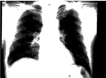
The chest X-ray film on admission was normal (Figure 1). High-resolution CT (HRCT) revealed diffuse centrilobular ground glass opacities in both lungs
(Figure 2). Cytological examination and culture of the sputum were both negative. Bronchoscopy was per- formed on day 3 after admission. Telangiectasis was seen between the trachea and bilateral main bronchus.
The fiberscope was wedged into rB 4 and bronchoal- veolar lavage (BAL) was performed. A 50-ml volume of sterile physiological saline was instilled three times, and 100 ml was recovered, representing a recovery rate of 66.7%. Analysis of BAL fluid (F) showed total cell count of 5.5 X 10 5 /ml, macrophages 53.2%, neutrophils 0.0%, lymphocytes 45.9%, eosinophi1g 0.9%, and CD4/CD8 ratio of 2.19.
Transbronchial lung biopsy (TBLB) was performed from rB 2 b, rB 1 b and rB 3 a. Histological examination showed no specific findings including fibrin and slight hemorrhage in the alveolar spaces, but there was nei- ther infiltration of inflammatory cells nor granulation tissue. From the analysis of BALF, sarcoidosis was highly suspected. He underwent scalenus lymph node
Figure 1. Chest X-ray film taken on admission. Note almost normal lung fields.
Figure 2. HRCT taken on admission. Note the diffuse
centrilobular ground glass opacities.
Figure 3. Histopathological findings of VATS-LB specimens obtained from 1S5. A) Note the presence of mild panlobular emphysema. Macrophages fill the terminal and respiratory bronchioles. Note also the centrilobular accumulation of macrophages and mild thickening of alveolar walls (HE stain, X 2.5). B) Macrophages contain light-brown cytoplasm and finely granular pigments (HE stain, X 50).
biopsy on day 25 after admission but the specimens were not of sufficient size to define the pathological diagnosis. He underwent VATS-LB on day 39 after ad- mission. Pathological findings of the specimens from IS 5 included mild centrilobular and panlobular em- physema, and mild thickening of alveolar walls but no granulomas, malignancy or necrosis. Macrophages packed the small airways and their distribution was centrilobular (Figure 3A). Macrophages contained light-brown cytoplasm with fine, dispersed and indis- tinct granules (Figure 3B). The granules stained posi- tively with Prussian blue. The final diagnosis was RB- ILD based on the above clinical and laboratory findings. The patient was placed under observation without steroid therapy since he had no symptoms and the opacities on HRCT were improved spontane- ously during the clinical course (Figure 4), following cessation of smoking after discharge on October 30, 1999. During the last follow-up visit in the outpatient department, 23 months after discharge from the hospi- tal, the patient was well and asymptomatic.
Figure 4. HRCT taken after discharge (January 21, 2000).
Note the improvement of the opacities.
Discussion
The differential diagnosis of our case included pul- monary sarcoidosis, drug-induced pneumonitis, or farmer's lung based on the initial clinical features, BALF analysis with relative lymphocytosis, the upper limit of normal range of CD4/CD8, and the finding on HRCT. In addition, desquamative interstitial pneumo- nia (DIP) and RB-ILD were also suspected from the findings on HRCT and his history of heavy smoking.
But these diseases are very rare", especially in Japan"').
In this case, we performed VATS-LB because we could
not make the definite diagnosis with any examination.
However, this case was pathologically confirmed as RB-ILD by surgical lung biopsy.
There are two case reports of RB-ILD in Japan pub- lished in 1994 and 19984''). Both cases were 48-year- old men with smoking histories, who presented with persistent dry cough for 6 months. The second case had clubbing of the fingers. Their chest X-ray films showed reticulonodular shadows and the chest CT scans showed ground glass opacities, accompanied with reticulonodular shadows (the first case), or low- attenuation areas (the second case). HRCT scans of our case showed diffuse ground glass opacities. This finding is non-specific in interstitial lung diseases',').
Ground-glass opacities, atelectasis, intralobular and in- terlobular interstitial thickening, emphysema, and pe- ripheral blebs are seen in HRCT of RB-ILD9'. On the other hand, a lack of HRCT findings cannot exclude RB-ILD9'. Analysis of BAL fluid of the first case re- vealed that macrophages formed 99.5% of total cells, and that of the second case showed relative eosinophilia. On the other hand, our case had no symptoms and no abnormal shadows of chest X-ray on admission, and the analysis of BAL fluid showed relative lymphocytosis. The clinical symptoms and re- sults of analysis of BAL fluid of the first case were more similar to those of the past cases of RB-ILD re- ported by Myers et al.') and Yousem et al.'). These findings indicate that it is often difficult to diagnose RB-ILD clinically.
Therefore, surgical lung biopsy is necessary for a definite diagnosis of RB-ILD. Specimens of peripheral zones including lobules, respiratory bronchioles and pulmonary parenchyma should be obtained for correct pathological diagnosis. Previous studies stressed the importance of distinguishing RB-ILD from DIP"'). RB- ILD is similar to DIP with regard to the following fea- tures; (i) all patients with RB-ILD are smokers and the majority of patients with DIP (91%) are smokers. (ii) Accumulation of macrophages and thickening of al- veolar septa are characteristically seen in both dis- eases. However, RB-ILD is different from DIP with re- spect to the following features; (i) RB-ILD patients are rather younger than those with DIP. (ii) Presence of milder symptoms in RB-ILD compared to DIP. (iii) Good prognosis in RB-ILD, while 32% of patients with DIP became worse or died during follow-up. (iv) Pathologically, the distribution of macrophages is centrilobular in RB-ILD but uniform and diffuse in DIP. (v) In RB-ILD, macrophages contain finely granu- lar, dispersed, indistinct, and golden-brown pigments positively stained with Prussian blue2' 3'
Moon et al.") recently proposed the term "smoking-
related interstitial lung disease", due to the presence of overlapping cases of pathological DIP and RB-ILD and that these two diseases are well associated with smoking. Katzenstein and Myers"' also indicated that DIP and RB-ILD more likely represent different ends of a spectrum of the same disease. More recently, Sadikot et al."' reported patients with RB-ILD who had severe symptoms and severe impairment of lung function in contrast to the mild symptoms and good outcome in previously reported cases including our case. Thus, further studies are necessary to investigate the independence of RB-ILD as a separate disease en- tity.
Acknowledgments
The authors thank Dr. Masanori Kitaichi,
Department of Laboratory Medicine, University of
Kyoto for the valuable advice and pathological diag- nosis, and Dr. F. G. Issa, Sydney, Australia, for his as- sistance in editing the manuscript.
References
1) Niewoehner D, Kleinerman J, Rice DB. Pathologic changes in the peripheral airways of young cigarette smokers. N Engl J Med 291:
755-758, 1974
2) Myers JL, Veal CF, Shin MS, Katzenstein AA. Respiratory
bronchiolitis causing interstitial lung disease. Am Rev Respir Dis 135: 880-884, 1987
3) Yousem SA, Colby TV, Gaensler EA. Respiratory bronchiolitis-
associated interstitial lung disease and its relationship to desquamative interstitial pneumonia. Mayo Clin Proc 64: 1373-
1380, 1989
4) Iwata M, Ida M, Kita T, Horiguchi T, Sato A. A case of respira- tory bronchiolitis-associated interstitial lung disease. Nihon
Kyoubu Shikkan Gakkai Zasshi 32: 803-808, 1994
5) Kurumagawa T, Kobayashi H, Kanoh S, et al. Respiratory bronchiolitis-associated interstitial lung disease. Nihon Kokyuuki
Gakkai Zasshi 36: 881-885, 1998
6) King Jr. TE, Costabel U, Cordier JF, et al. Idiopathic pulmonary fi- brosis: diagnosis and treatment. Am J Respir Crit Care Med 161:
646-664, 2000
7) Muler NL, Miller RR. Computed tomography of chronic diffuse infiltrative disease. Part 1. Am Rev Respir Dis 142: 1206-1215,
1990
8) Muler NL, Miller RR. Computed tomography of chronic diffuse infiltrative disease. Part 2. Am Rev Respir Dis 142: 1440-1448,
1990
9) Holt RM, Schmidt RA, Godwin JD, Raghu G. High resolution CT in respiratory bronchiolitis-associated interstitial lung disease. J
Comput Assist Tomogr 17: 46-50, 1993
10) Moon J, du Bois RM, Colby TV, Hansell DM, Nicholson AG.
Clinical significance of respiratory bronchiolitis on open lung bi-
opsy and its relationship to smoking-related interstitial lung dis- ease. Thorax 54: 1009-1014, 1999
11) Katzenstein AA, Myers JL. Idiopathic pulmonary fibrosis: clinical relevance of pathologic classification. Am J Respir Crit Care Med
157: 1301-1315, 1998
12) Sadikot RT, Johnson J, Lloyd JE, Christman JW. Respiratory bronchiolitis associated with severe dyspnea, exertional
hypoxemia, and clubbing. Chest 117 : 282-285, 2000