markedly inhibits cancer cell migration and
invasion in esophageal squamous cell
carcinoma.
著者
大迫 祐作
journal or
publication title
International Journal of Oncology
volume
49
number
6
page range
2255-2264
year
2016
ファイル(説明)
博士論文本文
最終試験結果の要旨
論文審査の要旨
博士論文要旨
別言語のタイトル
食道扁平上皮癌において、腫瘍抑制性microRNA-375
はMMP13を制御し腫瘍細胞の遊走能・浸潤能を著明
に抑制する。
学位授与番号
17701甲総研第496号
URL
http://hdl.handle.net/10232/00030533
doi: 10.3892/ijo.2016.3745INTERNATIONAL JOURNAL OF ONCOLOGY 49: 2255-2264, 2016
Abstract. Esophageal squamous cell carcinoma (ESCC) is
one of the most aggressive malignancies. Recently developed molecular targeted therapies are not available for patients with ESCC. After curative surgical resection, patients frequently suffer distant metastasis and recurrence. Exploration of novel ESCC metastatic pathways may lead to the development of new treatment protocols for this disease. Accordingly, we have sequentially identified microRNA (miRNA)-mediated metastatic pathways in several cancers. Our past studies of miRNA expression signatures have shown that microRNA-375 (miR-375) is frequently reduced in several types of cancers, including ESCC. In the present study, we aimed to investigate novel miR-375-mediated metastatic pathways in ESCC cells. The expression of miR-375 was downregulated in ESCC tissues, and ectopic expression of this miRNA markedly inhibited cancer cell migration and invasion, suggesting that
miR-375 acted as an antimetastatic miRNA in ESCC cells.
Our strategies for miRNA target searching demonstrated that matrix metalloproteinase 13 (MMP13) was directly regu-lated by miR-375 in ESCC cells. Overexpression of MMP13 was observed in ESCC clinical tissues, and the expression of MMP13 promoted cancer cell aggressiveness. Moreover, oncogenic genes, including CENPF, KIF14 and TOP2A, were shown to be regulated downstream of MMP13. Taken together, these findings demonstrated that the antitumor miR-375/onco-genic MMP13 axis had a pivotal role in ESCC aggressiveness.
These results provide novel insights into the potential mecha-nisms of ESCC pathogenesis.
Introduction
Esophageal squamous cell carcinoma (ESCC) is one of the most aggressive cancers and the major histological type of esophageal cancer in Japan and East Asia (1-3). ESCC cells frequently metastasize to the lymph nodes, liver, lungs and bone (2-4). Despite the use of multimodality therapies, the prognosis of patients with ESCC is still poor, with an overall 5-year survival rate of approximately 20-30% (2,4). Recently developed molecularly targeted therapeutics have not been shown to have beneficial effects in patients with ESCC (2). Additionally, the molecular pathogenesis of the aggressive phenotype in ESCC remains unclear. Thus, in order to improve disease outcomes in patients with ESCC, it is necessary to elucidate the molecular mechanisms of ESCC cell aggressive-ness using advanced genomic approaches.
The discovery of microRNAs (miRNAs) has resulted in major advancements in cancer research (5,6). miRNAs are small non-coding RNAs that function to fine tune the expression of protein coding/non-coding RNAs by repressing translation or cleaving RNA transcripts in a sequence-depending manner (7). The unique characteristic function of miRNAs is to regulate RNA transcripts in human cells. Therefore, dysregulated expression of miRNAs can disrupt tightly regulated RNA networks in cancer cells. Currently, numerous studies have shown that miRNAs are aberrantly expressed in several cancers, including ESCC (6,8). Using miRNA expression signature analyses, we have sequentially identified tumor-suppressive miRNAs and shown that these miRNAs mediate novel cancer networks (9-13).
Our miRNA expression signatures revealed that
microRNA-375 (miR-375) is frequently downregulated in
several types of squamous cell carcinoma (10,13,14). Moreover, our previous studies demonstrated that ectopic expression of miR-375 suppressed cancer cell aggressiveness in several types of cancer cells (15). In ESCC cells, several studies have
Regulation of MMP13 by antitumor microRNA-375
markedly inhibits cancer cell migration and invasion
in esophageal squamous cell carcinoma
YUSAkU OSAkO1, NAOhIkO SEkI2, YOShIAkI kITA1, kEIIChI YONEMORI1, kEIIChI kOShIzUkA2, AkIRA kUROzUMI2, ITARU OMOTO1, kEN SASAkI1, YASUTO UChIkADO1,
hIROShI kURAhARA1, kOSEI MAEMURA1 and ShOJI NATSUGOE1
1Department of Digestive Surgery, Breast and Thyroid Surgery, Graduate School of Medical Sciences,
kagoshima University, Sakuragaoka, kagoshima 890-8520; 2Department of Functional Genomics, Chiba University Graduate School of Medicine, Chuo-ku, Chiba 260-8670, Japan
Received August 8, 2016; Accepted September 28, 2016 DOI: 10.3892/ijo.2016.3745
Correspondence to: Dr Naohiko Seki, Department of Functional Genomics, Chiba University Graduate School of Medicine, 1-8-1 Inohana, Chuo-ku, Chiba 260-8670, Japan
E-mail: [email protected]
Key words: microRNA, miR-375, esophageal squamous cell carcinoma, matrix metalloproteinase 13, tumor suppressor
indicated that miR-375 has antitumor roles through targeting oncogenic genes (16,17). Moreover, miR-375-mediated cancer pathways are essential for cancer cell initiation, development and aggressiveness.
Accordingly, in the present study, we aimed to investigate the novel cancer networks regulated by miR-375 in ESCC cells. Our present data showed that matrix metalloproteinase 13 (MMP13) was directly regulated by miR-375 in ESCC cells. Overexpression of MMP13 was observed in ESCC clinical tissues, and knockdown of MMP13 expression markedly inhib-ited ESCC cell migration and invasion, indicating that MMP13 acted as a cancer-promoting gene in ESCC cells. Moreover, the oncogenic genes CENPF, KIF14 and TOP2 were found to function downstream of MMP13. Taken together, these results showed that the antitumor miR-375/oncogenic MMP13 axis had a pivotal role in ESCC aggressiveness.
Materials and methods
Clinical ESCC specimens and ESCC cell lines. Clinical
speci-mens were collected from 25 patients with ESCC. All patients underwent primary surgical treatment and were pathologically proven to have ESCC at the kagoshima University hospital from 2010 to 2014. The present study was approved by the Bioethics Committee of kagoshima University; written
prior informed consent and approval were obtained from all patients. The clinicopathological characteristics of the patients are shown in Table I.
We used two ESCC cell lines: TE-8, which was moderately differentiated; and TE-9, which was poorly differentiated. Both of these cells lines were provided by Riken BioResourse Center (Tsukuba, Japan).
Extraction of total RNA from clinical specimens and cell lines was performed using ISOGEN (Nippon Gene, Tokyo, Japan) according to the manufacturer's protocol. The quality of RNA was checked using an Agilent 2100 Bioanalyzer (Agilent Technologies, Santa Clara, CA, USA).
Quantitative real-time reverse transcription polymerase chain reaction (qRT-PCR). The procedure for PCR
quantifica-tion was previously described (13,18-20). The expression levels of miR-375 (assay ID: 000564; Applied Biosystems, Foster City, CA, USA) were analyzed by TaqMan qRT-PCR assays (TaqMan MicroRNA assays; Applied Biosystems) and RNU48 (assay ID: 001006) was used for normalization. TaqMan probes and primers for MMP-13 (assay ID: hs00233992_m1; Applied Biosystems), CENPF (assay ID: hs01118845_m1),
KIF14 (assay ID: hs00978236_m1) and GUSB (the internal
control; assay ID: hs00939627_ml; Applied Biosystems) were used for gene expression analysis.
Table I. Clinical features of patients with ESCC.
No. Age (years) Gender Differentiation T N M Stage ly v Recurrence 1 68 Male Poor 1b 2 0 IIIA 1 3 + 2 72 Male Moderate 1b 0 0 IA 0 1 3 69 Male Moderate 1b 0 0 IIIA 0 0 4 62 Male Well 3 2 0 IIIB 1 1 + 5 66 Male Moderate 3 0 0 IIA 1 1 6 74 Male Moderate 2 2 0 IIIA 3 1 + 7 56 Male Moderate 2 0 0 IB 0 1 8 79 Male Moderate 2 1 0 IIB 1 1 9 68 Male Moderate 1b 2 0 IIIA 1 1 -10 52 Male Poor 1b 0 0 IA 1 1 + 11 67 Male Well 3 2 0 IIIB 2 2 + 12 57 Male Poor 3 3 0 IIIC 1 1 + 13 70 Male Moderate 3 0 0 IIA 1 1 + 14 66 Male Moderate 3 0 0 IIA 1 1 -15 63 Male Well 3 3 0 IIIC 2 1 + 16 55 Male Moderate 3 2 0 IIIB 1 1 + 17 60 Male Well 1b 1 0 IIB 1 1 -18 78 Male Well 3 0 0 IIA 1 2 -19 71 Male Well 3 0 0 IIA 1 2 -20 75 Male Moderate 3 2 0 IIIB 1 1 + 21 60 Male Moderate 2 1 0 IIB 1 2 -22 62 Male Well 1a 1 0 IIB 0 0 -23 71 Male Moderate 1b 1 0 IIB 0 0 -24 69 Male Moderate 1b 0 0 IA 1 0 -25 84 Male Well 2 1 0 IIB 1 1
-INTERNATIONAL JOURNAL OF ONCOLOGY 49: 2255-2264, 2016 2257
Transfection with mature miRNAs and small interfering RNAs (siRNAs). The following mature miRNA was used:
Ambion Pre-miR miRNA precursor for hsa-miR-375 (product ID: PM10327; Applied Biosystems). The following siRNAs were used: Stealth Select RNAi siRNA, si-MMP13 (cat nos. hSS106637 and hSS106638; Invitrogen, Carlsbad, CA, USA), and negative control miRNA/siRNA (P/N: AM17111; Applied Biosystems). RNAs were incubated with Opti-MEM (Invitrogen) and Lipofectamine RNAiMax transfection reagent (Invitrogen), as previously described (13,18-20).
Cell proliferation, migration and invasion assays. TE-8
and TE-9 cells were transfected with 10 nM miRNAs or siRNAs by reverse transfection. Cell proliferation, migra-tion and invasion assays were performed as previously described (13,18-20).
Screening of miR-375 target genes using in silico analysis and gene expression data. To identify miR-375 target genes,
a combination of genome-wide gene expression and in silico analyses was conducted as previously described (13,18-20). The microarray data were deposited into the GEO repository under accession number GSE77790. Next, we selected puta-tive miRNA target genes using microRNA.org (August, 2010 release, http://www.microrna.org) databases. Our strategy for identification of miR-375 target genes is shown in Fig. 2.
Western blot analysis. Anti-human MMP-13 rabbit polyclonal
IgG (1:1,000; sc30073; Santa Cruz Biotechnology, Santa Cruz, CA, USA) was used as a primary antibody. Anti-human GAPDh mouse monoclonal IgG (1:5,000; 010-25521; Wako Pure Chemical Industries, Osaka, Japan) was used as an internal loading control. The membrane was washed and incu-bated with a horseradish peroxidase-conjugated secondary
antibody. Bands were visualized using Amersham ECL Prime Western Blotting detection reagent (GE healthcare Life Sciences, Uppsala, Sweden).
Immunohistochemistry. Tumor samples were fixed with 10%
formaldehyde in phosphate-buffered saline (PBS), embedded in paraffin and sectioned into 4-µm-thick slices. The sections were incubated with rabbit polyclonal anti-MMP-13 IgG (1:200; ab84594; Abcam, Cambridge, UK) at 4˚C overnight. The procedure for immunohistochemistry was previously described (21).
Plasmid construction and dual-luciferase reporter assays.
Partial wild-type sequences of the 3' untranslated region (UTR) of MMP13 containing the miR-375 target site (positions 100-113 of the MMP13 3' UTR) or sequences with a deleted
miR-375 target site were inserted between the XhoI and PmeI restriction sites in the 3' UTR of the hRluc gene in the
psiChECk-2 vector (product ID: C8021; Promega, Madison, WI, USA). TE-8 and TE-9 cells were transfected with 50 ng of the vector and 10 nM miR-375 using Lipofectamine 2000 (Thermo Fisher Scientific) in Opti-MEM (Thermo Fisher Scientific). The activities of firefly and Renilla luciferases were determined in lysates of transfected cells using a Dual-lucif-erase reporter assay system according to the manufacturer's recommendations (product ID: E1960; Promega). Data were normalized to firefly luciferase activity (ratio of Renilla/firefly luciferase activities).
Identification of downstream genes mediated by MMP13 in ESCC cells. Gene expression analyses of
si-MMP13-transfected TE-8 and TE-9 cells revealed molecular targets mediated by MMP13 in ESCC cells. This method is described in more detail in previous studies (13,18-20). Microarray
Figure 1. Expression levels of miR-375 and functional assays of miR-375 transfection in ESCC cell lines. (A) Expression levels of miR-375 in ESCC or normal esophageal tissues and ESCC cell lines. (B) Cell proliferation was determined by XTT assays. *P<0.0001, **P<0.05. (C) Cell migration activity was determined
results were deposited in the GEO database (accession number GSE82108).
Statistical analysis. Relationships between two or three
variables and numerical values were analyzed using the Mann-Whitney U test or the Bonferroni-adjusted Mann-Mann-Whitney test. Spearman's rank test was used to evaluate the correlations between the expression levels of miR-375 and MMP13. Expert StatView version 5.0 (SAS Institute, Inc., Cary, NC, USA) was used in these analyses.
Results
Expression levels of miR-375 in ESCC clinical specimens and cell lines. We evaluated the expression levels of miR-375 in
ESCC tissues (n=25), normal esophageal specimens (n=13), and ESCC cell lines (TE-8 and TE-9). The patient back-grounds and clinicopathological characteristics are shown in Table I. The expression levels of miR-375 were significantly downregulated in cancer tissues and ESCC cell lines compared with those in normal tissues (P<0.0001; Fig. 1A). Additionally, there were no significant relationships between the expression level of miR-375 and any of the clinicopathological parameters examined in this study (recurrence, T stage, N stage, vascular invasion, or survival rate).
Effects of miR-375 restoration on cell proliferation, migration and invasion in ESCC cell lines. To investigate the antitumor
functions of miR-375, we performed gain-of-function studies using mature miRNA transfection of TE-8 and TE-9 cells.
Cell proliferation was significantly suppressed by miR-375 transfection in TE-9 cells in comparison with that of mock or miR-control transfectants (Fig. 1B). however, no changes were detected in TE-8 cells (Fig. 1B).
Migration assays showed that cell migration activity was significantly inhibited by miR-375 transfection in TE-8 and TE-9 cells in comparison with that in mock or miR-control transfectants (Fig. 1C). Additionally, Matrigel invasion assays
demonstrated that cell invasion activity was significantly inhibited by miR-375 transfection in TE-8 and TE-9 cells in comparison with that in mock or miR-control transfec-tants (Fig. 1D).
Identification of putative target genes regulated by miR-375 in ESCC cells. To gain additional insights into the molecular
pathways regulated by antitumor miR-375 in ESCC cells, we used a combination of in silico and gene expression analyses. The strategy for identification of the miR-375-regulated genes in ESCC cells is shown in Fig. 2.
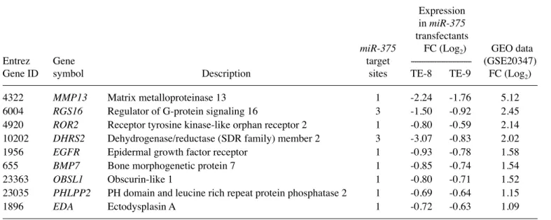
Table II. highly expressed genes putatively regulated by miR-375 in ESCC.
Expression
in miR-375
transfectants
miR-375 FC (Log2) GEO data
Entrez Gene target --- (GSE20347) Gene ID symbol Description sites TE-8 TE-9 FC (Log2)
4322 MMP13 Matrix metalloproteinase 13 1 -2.24 -1.76 5.12 6004 RGS16 Regulator of G-protein signaling 16 3 -1.50 -0.92 2.45 4920 ROR2 Receptor tyrosine kinase-like orphan receptor 2 1 -0.80 -0.59 2.14 10202 dHRS2 Dehydrogenase/reductase (SDR family) member 2 3 -3.07 -0.83 2.02 1956 EGFR Epidermal growth factor receptor 1 -0.93 -0.78 1.58
655 BMP7 Bone morphogenetic protein 7 1 -0.85 -0.74 1.54
23363 OBSL1 Obscurin-like 1 1 -0.80 -0.71 1.52 23035 PHLPP2 Ph domain and leucine rich repeat protein phosphatase 2 1 -0.69 -0.64 1.15 1896 EdA Ectodysplasin A 1 -0.72 -0.63 1.09
INTERNATIONAL JOURNAL OF ONCOLOGY 49: 2255-2264, 2016 2259
In gene expression analyses, 2,897 and 1,007 genes were downregulated (log2 ratio <-0.5) in TE-8 and TE-9 miR-375
transfectants, respectively, in comparison with that in control transfectants. Our present expression data were deposited in the Gene Expression Omnibus (GEO accession number GSE77790). Among these downregulated genes, we searched for genes having putative miR-375 binding sites in their 3' UTRs using the microRNA.org database. A total of 55 genes were identified as putative target genes of miR-375, and nine genes were upregulated in ESCC clinical specimens, as deter-mined using ESCC expression data (GEO accession number: GSE20347; Table II).
In this study, we focused on MMP13 because its expression was most upregulated in ESCC clinical specimens and most downregulated in miR-375 transfectants. Moreover, previous studies have shown that the activation of MMPs is associated with cancer cell aggressiveness (22).
Expression of MMP13 in ESCC clinical specimens. Next,
we validated the upregulation of MMP13 in the ESCC clinical specimens at both the mRNA and the protein levels. The expression of MMP13 was significantly upregulated in 25 ESCC specimens and ESCC cell lines compared with that in 13 normal specimens (P<0.0001; Fig. 3A). The
Figure 3. Expression levels of MMP13 mRNA and immunohistochemical staining of MMP13 protein in ESCC specimens. (A) Expression levels of MMP13 mRNA in ESCC or normal esophageal tissues and ESCC cell lines. (B) The expression of miR-375 and MMP13 mRNA was negatively correlated (r=-0.661 and P<0.0001). (C) Immunohistochemical staining of MMP13 in ESCC specimens. All ESCC tissues were stained positively, whereas normal esophageal tissues were stained negatively or weakly (left panel, MMP13 staining; right panel, hematoxylin-eosin staining; original magnification, x200).
Spearman's rank tests showed negative correlations between the expression of miR-375 and that of MMP13 (r=-0.661, P<0.0001; Fig. 3B).
Immunohistochemistry showed that MMP13 tended to be strongly expressed in ESCC lesions, whereas low expression was observed in normal esophageal epithelium (Fig. 3C).
direct regulation of MMP13 by miR-375 in ESCC cells. We
performed qRT-PCR to validate miR-375-mediated repression of MMP13 expression in ESCC cell lines. Our results showed that MMP13 mRNA was significantly reduced in miR-375
Figure 4. Direct regulation of MMP13 by miR-375 in ESCC cell lines. (A) Expression levels of MMP13 mRNA 72 h after transfection with miR-375. (B) MMP13 protein expression 72 h after transfection with miR-375. (C) Putative miR-375 binding sites in the 3' UTR of MMP13 mRNA. (D Luciferase reporter assay using vectors encoding putative miR-375 target sites at positions 100-113 for both wild-type and deletion-type constructs.
Renilla luciferase values were normalized to firefly luciferase values.
*P<0.0001, **P<0.05.
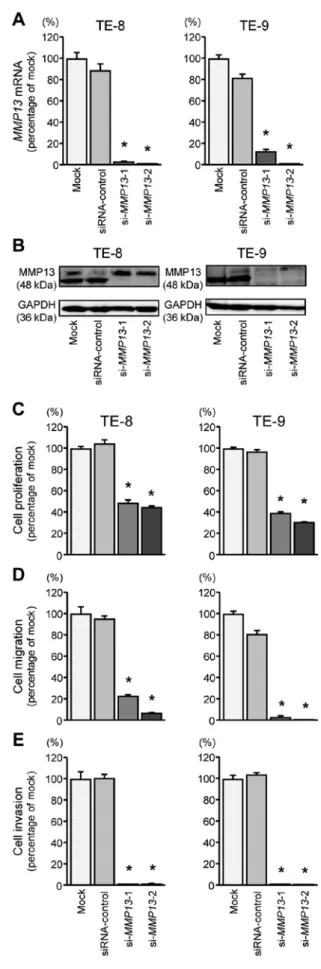
Figure 5. Loss of function studies using siRNAs. (A) Expression levels of
MMP13 mRNA after transfection with si-MMP13 in ESCC cell lines.
(B) MMP13 protein expression 72 h after transfection with si-MMP13. (C) Cell proliferation was determined by XTT assays. Inhibition of cell proliferation was observed in si-MMP13-transfected cell lines. (D) Cell migration activity was determined by migration assays. (E) Cell invasion was determined by Matrigel invasion assays. Inhibition of migration and invasion was observed in si-MMP13-transfected cell lines. *P<0.0001.
INTERNATIONAL JOURNAL OF ONCOLOGY 49: 2255-2264, 2016 2261
transfectants in comparison with that in mock or miR-control transfectants (P<0.0001; Fig. 4A). MMP13 protein expression was also repressed in miR-375 transfectants (Fig. 4B).
Next, we performed luciferase reporter assays using TE-8 and TE-9 cells to determine whether MMP13 had an actual target site for miR-375 binding. The microRNA.org database predicted that there was one putative target site in the 3' UTR of MMP13 (Fig. 4C). Compared with the miR-control, lumi-nescence intensity was significantly reduced by transfection with miR-375 at the miR-375 target site, positions 100-113, in the 3' UTR of MMP13 (Fig. 4D).
Effects of silencing MMP13 on proliferation, migration and invasion in ESCC cells. To investigate the functional roles of
MMP13 in ESCC cell lines, we performed loss-of-function assays by transfection of si-MMP13 into TE-8 and TE-9 cells.
First, we evaluated the knockdown efficiency of si-MMP13 transfection in ESCC cell lines. In the present study, we used two siRNAs targeting MMP13 (si-MMP13-1 and si-MMP13-2). According to qRT-PCR and western blot analyses, both siRNAs effectively downregulated MMP13 expression in both cell lines (Fig. 5A and B).
Cell proliferation, migration and invasion assays demon-strated that cell proliferation, migration, and invasion were inhibited in si-MMP13-transfected cells compared with those in mock- or siRNA-control-transfected cells (Fig. 5C-E).
Identification of downstream genes regulated by MMP13 in ESCC cells. To determine which downstream genes were
regu-lated by MMP13, genome-wide gene expression and in silico analyses were performed in TE-8 and TE-9 cells transfected with si-MMP13.
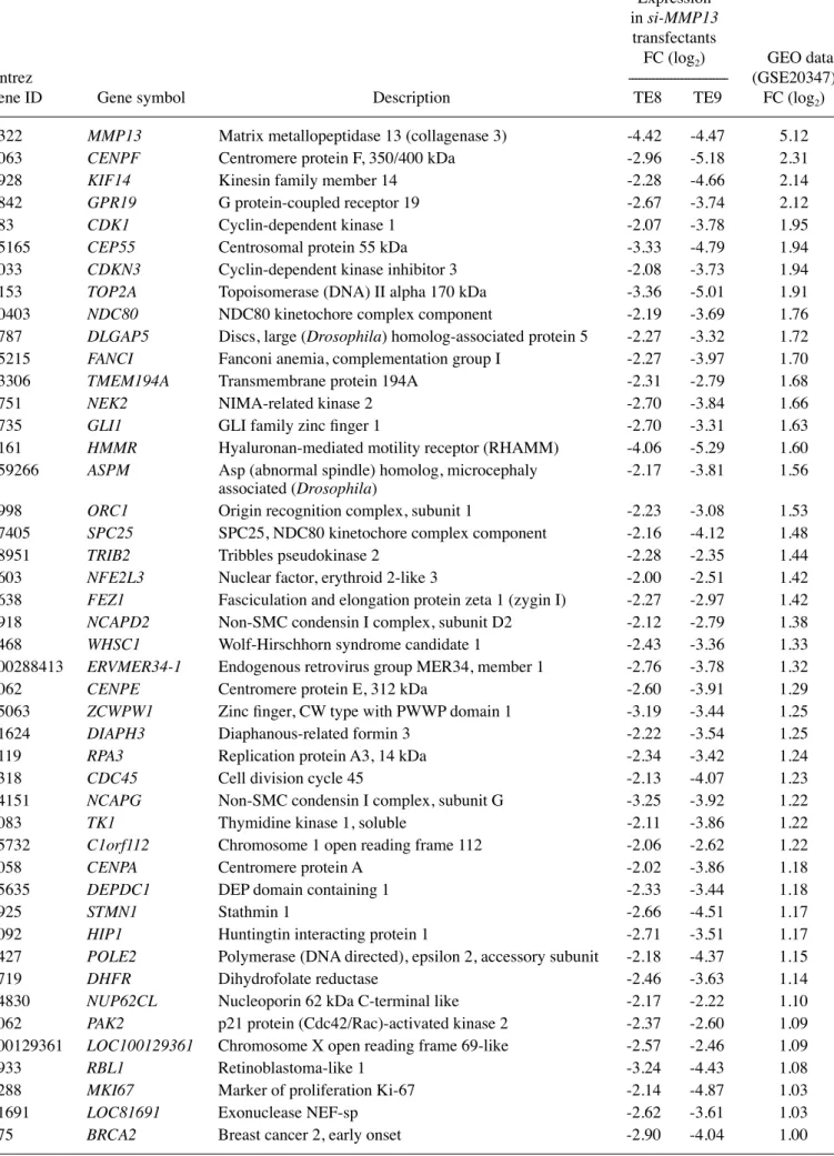
Our expression analysis showed that a total of 298 genes were commonly downregulated (log2 ratio <-2.0) in TE-8 and
TE-9 cells following si-MMP13 transfection. Among these genes, 52 were upregulated in ESCC clinical specimens, as determined using ESCC expression data (GEO accession number: GSE20347; Fig. 6 and Table III).
We then validated the upregulation of CENPF and KIF14 mRNAs in ESCC clinical specimens. The expression of
CENPF and KIF14 mRNAs was significantly upregulated
in 25 ESCC specimens and ESCC cell lines compared with that in 13 normal specimens (P<0.0001; Fig. 7A and C). The Spearman's rank tests showed correlations between the expres-sion of MMP13 and that of CENPF or KIF14 (CENPF: r=0.554, P=0.0007, Fig. 7B; kIF14: r=0.729, P<0.0001, Fig. 7D).
Figure 6. The strategy for analysis of MMP13 downstream genes.
Figure 7. mRNA expression levels of MMP13 downstream genes (CENPF and KIF14) in ESCC specimens. (A and C) Expression levels of CENPF and
KIF14 mRNA in ESCC or normal esophageal tissues and ESCC cell lines.
(B and D) The expression levels of MMP13/CENPF and MMP13/KIF14 mRNAs were positively correlated (P<0.0001).
Table III. Downregulated genes in si-MMP13-transfected ESCC cell lines.
Expression
in si-MMP13
transfectants
FC (log2) GEO data
Entrez --- (GSE20347) gene ID Gene symbol Description TE8 TE9 FC (log2)
4322 MMP13 Matrix metallopeptidase 13 (collagenase 3) -4.42 -4.47 5.12 1063 CENPF Centromere protein F, 350/400 kDa -2.96 -5.18 2.31 9928 KIF14 kinesin family member 14 -2.28 -4.66 2.14 2842 GPR19 G protein-coupled receptor 19 -2.67 -3.74 2.12
983 CdK1 Cyclin-dependent kinase 1 -2.07 -3.78 1.95
55165 CEP55 Centrosomal protein 55 kDa -3.33 -4.79 1.94 1033 CdKN3 Cyclin-dependent kinase inhibitor 3 -2.08 -3.73 1.94 7153 TOP2A Topoisomerase (DNA) II alpha 170 kDa -3.36 -5.01 1.91 10403 NdC80 NDC80 kinetochore complex component -2.19 -3.69 1.76 9787 dLGAP5 Discs, large (drosophila) homolog-associated protein 5 -2.27 -3.32 1.72 55215 FANCI Fanconi anemia, complementation group I -2.27 -3.97 1.70 23306 TMEM194A Transmembrane protein 194A -2.31 -2.79 1.68 4751 NEK2 NIMA-related kinase 2 -2.70 -3.84 1.66 2735 GLI1 GLI family zinc finger 1 -2.70 -3.31 1.63 3161 HMMR hyaluronan-mediated motility receptor (RhAMM) -4.06 -5.29 1.60 259266 ASPM Asp (abnormal spindle) homolog, microcephaly -2.17 -3.81 1.56
associated (drosophila)
4998 ORC1 Origin recognition complex, subunit 1 -2.23 -3.08 1.53 57405 SPC25 SPC25, NDC80 kinetochore complex component -2.16 -4.12 1.48 28951 TRIB2 Tribbles pseudokinase 2 -2.28 -2.35 1.44 9603 NFE2L3 Nuclear factor, erythroid 2-like 3 -2.00 -2.51 1.42 9638 FEZ1 Fasciculation and elongation protein zeta 1 (zygin I) -2.27 -2.97 1.42 9918 NCAPd2 Non-SMC condensin I complex, subunit D2 -2.12 -2.79 1.38 7468 WHSC1 Wolf-hirschhorn syndrome candidate 1 -2.43 -3.36 1.33 100288413 ERVMER34-1 Endogenous retrovirus group MER34, member 1 -2.76 -3.78 1.32 1062 CENPE Centromere protein E, 312 kDa -2.60 -3.91 1.29 55063 ZCWPW1 zinc finger, CW type with PWWP domain 1 -3.19 -3.44 1.25 81624 dIAPH3 Diaphanous-related formin 3 -2.22 -3.54 1.25 6119 RPA3 Replication protein A3, 14 kDa -2.34 -3.42 1.24 8318 CdC45 Cell division cycle 45 -2.13 -4.07 1.23 64151 NCAPG Non-SMC condensin I complex, subunit G -3.25 -3.92 1.22 7083 TK1 Thymidine kinase 1, soluble -2.11 -3.86 1.22 55732 C1orf112 Chromosome 1 open reading frame 112 -2.06 -2.62 1.22 1058 CENPA Centromere protein A -2.02 -3.86 1.18 55635 dEPdC1 DEP domain containing 1 -2.33 -3.44 1.18 3925 STMN1 Stathmin 1 -2.66 -4.51 1.17 3092 HIP1 huntingtin interacting protein 1 -2.71 -3.51 1.17 5427 POLE2 Polymerase (DNA directed), epsilon 2, accessory subunit -2.18 -4.37 1.15 1719 dHFR Dihydrofolate reductase -2.46 -3.63 1.14 54830 NUP62CL Nucleoporin 62 kDa C-terminal like -2.17 -2.22 1.10 5062 PAK2 p21 protein (Cdc42/Rac)-activated kinase 2 -2.37 -2.60 1.09 100129361 LOC100129361 Chromosome X open reading frame 69-like -2.57 -2.46 1.09 5933 RBL1 Retinoblastoma-like 1 -3.24 -4.43 1.08 4288 MKI67 Marker of proliferation ki-67 -2.14 -4.87 1.03 81691 LOC81691 Exonuclease NEF-sp -2.62 -3.61 1.03 675 BRCA2 Breast cancer 2, early onset -2.90 -4.04 1.00
INTERNATIONAL JOURNAL OF ONCOLOGY 49: 2255-2264, 2016 2263 Discussion
Numerous studies of miRNA expression signatures in ESCC have shown that miR-375 is frequently downregulated in cancer tissues and functions as an antitumor miRNA (14,23). In the present study, we confirmed that miR-375 was markedly downregulated in cancer tissues and that ectopic expression of miR-375 significantly suppressed cancer cell migration and invasion. Thus, we found that loss of miR-375 expression enhanced cancer cell aggressiveness in ESCC. Many previous studies have shown that the expression of miR-375 is mark-edly decreased in several types of cancers and that miR-375 functions as an antitumor miRNA (15,24). In contrast to these antitumor activities, miR-375 is upregulated in pedi-atric acute myeloid leukemia (AML) and prostate cancer, suggesting that miR-375 acts as an oncogenic miRNA in these diseases (25,26). The dual function of miR-375 is very unique; thus, it is important to identify miR-375-regulated pathways in various cancer types.
It is also important to elucidate novel RNA networks regu-lated by antitumor miR-375 in ESCC cells. Previous studies have shown that insulin-like growth factor 1 receptor (IGF1R), lactate dehydrogenase B (LdHB), and astrocyte elevated gene-1/metadherin (AEG-1/MTdH) are directly regulated by miR-375 in ESCC cells (16,17). These target genes are upregulated in ESCC clinical specimens and functioned as oncogenes in this disease. Another unique characteristic of miRNAs is that a single miRNA can regulate a large number of RNA transcripts in human cells (27,28). Thus, the contin-uous identification of miR-375-regulated oncogenes in ESCC cells is important for elucidation of the molecular pathogenesis of ESCC.
In this study, we identified MMP13 as a direct target of antitumor miR-375 in ESCC cells. MMP13 (also known as collagenase 3) is a member of the collagenase subfamily of MMPs and functions to degrade a wide range of extracellular matrix components, including tenascin C, fibronectin and type I-IV collagen (29). Thus, MMP13 has a wide range of proteolytic functions, suggesting that MMP13 is involved in several physiological and pathological processes (30). high expression of MMP13 has been reported in rheumatoid arthritis, osteoarthritis and several types of cancers (22). Previous studies have also shown that high expression of MMP13 is associated with vascular invasion and lymph node metastasis in ESCC (31). Our present data demonstrated that knockdown of MMP13 markedly reduced cancer cell migra-tion and invasion in ESCC cells.
The MMP13 gene has also been reported to be epigeneti-cally regulated by several other miRNAs, including miR-125b and miR-143, in cancer cells (32-34). Notably, our miRNA signatures have shown that miR-125b and miR-143 are down-regulated in ESCC and in oral and hypopharyngeal squamous cell carcinoma (12-14). Moreover, functional assays have indi-cated that these miRNAs act as tumor suppressors in several cancers, including ESCC cells (32-35). Loss of the expression of several antitumor miRNAs and activation of MMP13 may enhance cancer cell aggressiveness and metastasis. Thus, identification of miR-375/MMP13-mediated cancer pathways may facilitate the discovery of candidate therapeutic targets in ESCC.
Based on the above, we further investigated the downstream genes mediated by MMP13 in ESCC cells using genome-wide gene expression analysis. Our data showed that several centro-mere-associated proteins were regulated by MMP13-mediated genes, such as CENPF, CENPE, CENPA, CEP55, NdC80 and
SPC25. Moreover, cell cycle-promoting genes, e.g., KIF14, CdK1, TOP2A, CdC45 and PAK2, were also downregulated
by si-MMP13 in this study. Recent studies have reported that several genes encoding mitotic apparatus components are upregulated in cancer cells and contribute to cancer cell pheno-types (36,37). Therefore, overexpression of genes encoding mitotic apparatus components may represent a potential target for cancer drug development (38). Several compounds that inhibit centromere proteins and mitotic kinesins are being tested as potential cancer therapies in clinical trials (39).
Among these genes, we validated the overexpression of
CENPF and KIF14 in ESCC clinical specimens. Previous
studies have shown that CENPF is a master regulator of pros-tate cancer malignancy and that high expression of CEPNF is a prognostic indicator of poor survival and metastasis in patients with ESCC (40). KIF14 is a member of the kinesin superfamily of proteins and functions as a microtubule motor protein involved in cytokinesis and chromosome segrega-tion (41). Overexpression of KIF14 has been reported in several cancers, and its expression is associated with cancer cell phenotypes (42,43). An in-depth functional analysis of these genes in ESCC cells is necessary to further characterize these pathways. Identification of the downstream genes regulated by the miR-375/MMP13 axis may lead to a better understanding of ESCC aggressiveness.
In conclusion, downregulation of miR-375 was frequently observed in ESCC clinical specimens, and miR-375 was shown to function as an antitumor miRNA in ESCC cells. To the best of our knowledge, this is the first report demonstrating that
MMP13 is directly regulated by antitumor miR-375 and acts
to regulate several cell cycle promoting genes. The identifica-tion of novel molecular pathways and targets regulated by the
miR-375/MMP13 axis may lead to a better understanding of
ESCC molecular pathogenesis.
Acknowledgements
We wish to thank the Joint Research Laboratory, kagoshima University Graduate School of Medical and Dental Sciences, for the use of their facilities. The present study was supported by kAkENhI (C) grant 15k10801.
References
1. hongo M, Nagasaki Y and Shoji T: Epidemiology of esophageal cancer: Orient to Occident. Effects of chronology, geography and ethnicity. J Gastroenterol hepatol 24: 729-735, 2009.
2. Pennathur A, Gibson Mk, Jobe BA and Luketich JD: Oesophageal carcinoma. Lancet 381: 400-412, 2013.
3. Ohashi S, Miyamoto S, kikuchi O, Goto T, Amanuma Y and Muto M: Recent advances from basic and clinical studies of esophageal squamous cell carcinoma. Gastroenterology 149: 1700-1715, 2015.
4. Enzinger PC and Mayer RJ: Esophageal cancer. N Engl J Med 349: 2241-2252, 2003.
5. Lu J, Getz G, Miska EA, Alvarez-Saavedra E, Lamb J, Peck D, Sweet-Cordero A, Ebert BL, Mak Rh, Ferrando AA,
et al: MicroRNA expression profiles classify human cancers.
6. Calin GA and Croce CM: MicroRNA signatures in human cancers. Nat Rev Cancer 6: 857-866, 2006.
7. Bartel DP: MicroRNAs: Genomics, biogenesis, mechanism, and function. Cell 116: 281-297, 2004.
8. harada k, Baba Y, Ishimoto T, Shigaki h, kosumi k, Yoshida N, Watanabe M and Baba h: The role of microRNA in esophageal squamous cell carcinoma. J Gastroenterol 51: 520-530, 2016. 9. kikkawa N, hanazawa T, Fujimura L, Nohata N, Suzuki h,
Chazono h, Sakurai D, horiguchi S, Okamoto Y and Seki N: miR-489 is a tumour-suppressive miRNA target PTPN11 in hypopharyngeal squamous cell carcinoma (hSCC). Br J Cancer 103: 877-884, 2010.
10. Nohata N, hanazawa T, kikkawa N, Sakurai D, Fujimura L, Chiyomaru T, kawakami k, Yoshino h, Enokida h, Nakagawa M,
et al: Tumour suppressive microRNA-874 regulates novel cancer
networks in maxillary sinus squamous cell carcinoma. Br J Cancer 105: 833-841, 2011.
11. Nohata N, hanazawa T, kinoshita T, Inamine A, kikkawa N, Itesako T, Yoshino h, Enokida h, Nakagawa M, Okamoto Y,
et al: Tumour-suppressive microRNA-874 contributes to cell
proliferation through targeting of histone deacetylase 1 in head and neck squamous cell carcinoma. Br J Cancer 108: 1648-1658, 2013.
12. Fukumoto I, kinoshita T, hanazawa T, kikkawa N, Chiyomaru T, Enokida h, Yamamoto N, Goto Y, Nishikawa R, Nakagawa M,
et al: Identification of tumour suppressive microRNA-451a in
hypopharyngeal squamous cell carcinoma based on microRNA expression signature. Br J Cancer 111: 386-394, 2014.
13. Fukumoto I, hanazawa T, kinoshita T, kikkawa N, koshizuka k, Goto Y, Nishikawa R, Chiyomaru T, Enokida h, Nakagawa M,
et al: MicroRNA expression signature of oral squamous cell
carcinoma: Functional role of microRNA-26a/b in the modula-tion of novel cancer pathways. Br J Cancer 112: 891-900, 2015. 14. kano M, Seki N, kikkawa N, Fujimura L, hoshino I, Akutsu Y,
Chiyomaru T, Enokida h, Nakagawa M and Matsubara h: miR-145, miR-133a and miR-133b: Tumor-suppressive miRNAs target FSCN1 in esophageal squamous cell carcinoma. Int J Cancer 127: 2804-2814, 2010.
15. kinoshita T, hanazawa T, Nohata N, Okamoto Y and Seki N: The functional significance of microRNA-375 in human squamous cell carcinoma: Aberrant expression and effects on cancer pathways. J hum Genet 57: 556-563, 2012.
16. Isozaki Y, hoshino I, Nohata N, kinoshita T, Akutsu Y, hanari N, Mori M, Yoneyama Y, Akanuma N, Takeshita N,
et al: Identification of novel molecular targets regulated by tumor
suppressive miR-375 induced by histone acetylation in esopha-geal squamous cell carcinoma. Int J Oncol 41: 985-994, 2012. 17. kong kL, kwong DL, Chan Th, Law SY, Chen L, Li Y, Qin YR
and Guan XY: MicroRNA-375 inhibits tumour growth and metastasis in oesophageal squamous cell carcinoma through repressing insulin-like growth factor 1 receptor. Gut 61: 33-42, 2012.
18. Matsushita R, Yoshino h, Enokida h, Goto Y, Miyamoto k, Yonemori M, Inoguchi S, Nakagawa M and Seki N: Regulation of UhRF1 by dual-strand tumor-suppressor microRNA-145 (miR-145-5p and miR-145-3p): Inhibition of bladder cancer cell aggressiveness. Oncotarget 7: 28460-28487, 2016.
19. Goto Y, kojima S, Nishikawa R, kurozumi A, kato M, Enokida h, Matsushita R, Yamazaki k, Ishida Y, Nakagawa M,
et al: MicroRNA expression signature of castration-resistant
prostate cancer: The microRNA-221/222 cluster functions as a tumour suppressor and disease progression marker. Br J Cancer 113: 1055-1065, 2015.
20. Goto Y, kojima S, Nishikawa R, Enokida h, Chiyomaru T, kinoshita T, Nakagawa M, Naya Y, Ichikawa T and Seki N: The microRNA-23b/27b/24-1 cluster is a disease progression marker and tumor suppressor in prostate cancer. Oncotarget 5: 7748-7759, 2014.
21. kita Y, Nishizono Y, Okumura h, Uchikado Y, Sasaki k, Matsumoto M, Setoyama T, Tanoue k, Omoto I, Mori S, et al: Clinical and biological impact of cyclin-dependent kinase subunit 2 in esophageal squamous cell carcinoma. Oncol Rep 31: 1986-1992, 2014.
22. Brinckerhoff CE, Rutter JL and Benbow U: Interstitial colla-genases as markers of tumor progression. Clin Cancer Res 6: 4823-4830, 2000.
23. Yan JW, Lin JS and he XX: The emerging role of miR-375 in cancer. Int J Cancer 135: 1011-1018, 2014.
24. hui AB, Bruce JP, Alajez NM, Shi W, Yue S, Perez-Ordonez B, Xu W, O'Sullivan B, Waldron J, Cummings B, et al: Significance of dysregulated metadherin and microRNA-375 in head and neck cancer. Clin Cancer Res 17: 7539-7550, 2011.
25. Wang z, hong z, Gao F and Feng W: Upregulation of microRNA-375 is associated with poor prognosis in pediatric acute myeloid leukemia. Mol Cell Biochem 383: 59-65, 2013.
26. Szczyrba J, Nolte E, Wach S, kremmer E, Stöhr R, hartmann A, Wieland W, Wullich B and Grässer FA: Downregulation of Sec23A protein by miRNA-375 in prostate carcinoma. Mol Cancer Res 9: 791-800, 2011.
27. kinoshita T, Yip kW, Spence T and Liu FF: MicroRNAs in extracellular vesicles: Potential cancer biomarkers. J hum Genet: Jul 7, 2016 (Epub ahead of print). doi: 10.1038/jhg.2016.87. 28. Yonemori k, kurahara h, Maemura k and Natsugoe S:
MicroRNA in pancreatic cancer. J hum Genet: Jun 2, 2016. (Epub ahead of print). doi: 10.1038/jhg.2016.59.
29. knäuper V, López-Otin C, Smith B, knight G and Murphy G: Biochemical characterization of human collagenase-3. J Biol Chem 271: 1544-1550, 1996.
30. Vincenti MP and Brinckerhoff CE: Transcriptional regulation of collagenase (MMP-1, MMP-13) genes in arthritis: Integration of complex signaling pathways for the recruitment of gene-specific transcription factors. Arthritis Res 4: 157-164, 2002.
31. Etoh T, Inoue h, Yoshikawa Y, Barnard GF, kitano S and Mori M: Increased expression of collagenase-3 (MMP-13) and MT1-MMP in oesophageal cancer is related to cancer aggres-siveness. Gut 47: 50-56, 2000.
32. Xu N, zhang L, Meisgen F, harada M, heilborn J, homey B, Grandér D, Ståhle M, Sonkoly E and Pivarcsi A: MicroRNA-125b down-regulates matrix metallopeptidase 13 and inhibits cutaneous squamous cell carcinoma cell proliferation, migration, and invasion. J Biol Chem 287: 29899-29908, 2012.
33. Wu D, Ding J, Wang L, Pan h, zhou z, zhou J and Qu P: microRNA-125b inhibits cell migration and invasion by targeting matrix metallopeptidase 13 in bladder cancer. Oncol Lett 5: 829-834, 2013.
34. Osaki M, Takeshita F, Sugimoto Y, kosaka N, Yamamoto Y, Yoshioka Y, kobayashi E, Yamada T, kawai A, Inoue T, et al: MicroRNA-143 regulates human osteosarcoma metastasis by regulating matrix metalloprotease-13 expression. Mol Ther 19: 1123-1130, 2011.
35. Liu J, Mao Y, zhang D, hao S, zhang z, Li z and Li B: MiR-143 inhibits tumor cell proliferation and invasion by targeting STAT3 in esophageal squamous cell carcinoma. Cancer Lett 373: 97-108, 2016.
36. Yuen kW, Montpetit B and hieter P: The kinetochore and cancer: What's the connection? Curr Opin Cell Biol 17: 576-582, 2005.
37. Sagona AP and Stenmark h: Cytokinesis and cancer. FEBS Lett 584: 2652-2661, 2010.
38. Rath O and kozielski F: kinesins and cancer. Nat Rev Cancer 12: 527-539, 2012.
39. huszar D, Theoclitou ME, Skolnik J and herbst R: kinesin motor proteins as targets for cancer therapy. Cancer Metastasis Rev 28: 197-208, 2009.
40. Aytes A, Mitrofanova A, Lefebvre C, Alvarez MJ, Castillo-Martin M, zheng T, Eastham JA, Gopalan A, Pienta kJ, Shen MM, et al: Cross-species regulatory network analysis identifies a synergistic interaction between FOXM1 and CENPF that drives prostate cancer malignancy. Cancer Cell 25: 638-651, 2014.
41. zhu C, zhao J, Bibikova M, Leverson JD, Bossy-Wetzel E, Fan JB, Abraham RT and Jiang W: Functional analysis of human microtubule-based motor proteins, the kinesins and dyneins, in mitosis/cytokinesis using RNA interference. Mol Biol Cell 16: 3187-3199, 2005.
42. Corson TW, zhu CQ, Lau Sk, Shepherd FA, Tsao MS and Gallie BL: kIF14 messenger RNA expression is independently prognostic for outcome in lung cancer. Clin Cancer Res 13: 3229-3234, 2007.
43. Thériault BL, Pajovic S, Bernardini MQ, Shaw PA and Gallie BL: kinesin family member 14: An independent prognostic marker and potential therapeutic target for ovarian cancer. Int J Cancer 130: 1844-1854, 2012.