「単球系細胞の分化誘導および機能に及ぼす放射線の影響」
提 出 者 氏 名 : 吉 野 浩 教
所 属 : 医療生命科学領域 放射線生命科学分野
指 導 教 員 : 柏 倉 幾 郎
目 次
略語一覧 ... 2
序論 ... 4
Chapter 1 「 Differential induction from X-irradiated human peripheral blood monocytes to dendritic cells 」 ... 7
1. Introduction ... 8
2. Materials and methods... 9
3. Results ... 15
4. Discussion ... 23
Chapter 2「Impairment of mature dendritic cells derived from X-irradiated human monocytes depends on the type of maturation stimulus used」 ... 27
1. Introduction ... 29
2. Materials and methods... 30
3. Results ... 36
4. Discussion ... 44
Chapter 3 「 Ionizing radiation affects the expression of Toll-like receptors 2 and 4 in human monocytic cells through c-Jun N-terminal kinase activation 」 ... 47
1. Introduction ... 49
2. Materials and methods... 50
3. Results ... 55
4. Discussion ... 62
Conclusions ... 64
謝辞 ... 65
References ... 66
要旨 ... 74
論文一覧表 ... 76
1
略語一覧 CD: cluster of differentiation
DAMPs: damaged-associated molecular patterns DCs: dendritic cells
EDTA: ethylenediaminetetraacetic acid FITC: fluorescein isothiocyanate
G-CSF: granulocyte colony-stimulating factor
GM-CSF: granulocyte-macrophage colony-stimulating factor HLA: human leukocyte antigen
HO-1: heme oxygenase-1 iDCs: immature dendritic cells IFN: interferon
IL: interleukin
JNK: c-Jun N-terminal kinase LPS: lipopolysaccharide
LPS-mDCs: DCs stimulated by LPS mABs: monoclonal antibodies
MAP: mitogen-activated protein kinase MCP-1: monocyte chemoattractant protein-1 mDCs: mature dendritic cells
MFI: mean fluorescence intensity
MHC: major histocompatibility complex molecules MIP-1β: macrophage inflammatory protein-1β
MIX: a cytokine mixture (10-ng/mL rhTNF-α, 10-ng/mL rhIL-1β, 10-ng/mL rhIL-6, and 1-μg/mL PGE 2 )
MIX-mDCs: DCs stimulated by MIX MLR: mixed leukocytes reaction MMP-9: matrix metalloproteinase-9
2
MyD88: myeloid differentiation factor 88 PAMPs: pathogen-associated molecular patterns PBMCs: peripheral blood mononuclear cells PBS: phosphate-buffered saline
PE: phycoerythrin
PMA: phorbol 12-myristate 13-acetate PRRs: pattern recognition receptors rh: recombinant human
ROS: reactive oxygen species
RT-PCR: reverse transcription polymerase chain reaction TNF-α: tumor necrosis factor-α
Th: T helper
TLR: Toll-like receptor
TRIF: Toll/IL-1R domain-containing adaptor inducing IFN
3
序 論
単球は白血球の一種で,自然免疫を担当する.単球は末梢血中に存在するが,組 織で樹状細胞やマクロファージに分化し,より強力な免疫応答能を獲得する.
樹状細胞は抗原提示細胞の一つで,免疫システムに中心的な役割を果たす.樹状 細胞は骨髄細胞や単球などの前駆細胞から分化するが,未熟状態と成熟状態の2つ の状態に大別される.樹状細胞の前駆細胞は皮膚や粘膜などの末梢組織に移動し,
未熟樹状細胞として存在する.未熟樹状細胞は体内に侵入してきた病原体を感知し,
貪食するとともに病原体由来の抗原をプロセシングする.未熟樹状細胞は病原体成 分や炎症性サイトカインの刺激により成熟化する.この成熟化に伴い高い抗原提示能 を獲得し,それによりナイーブ T 細胞に効果的に病原体由来抗原を提示し獲得免疫 を誘導する.従って,樹状細胞は自然免疫と獲得免疫を繋ぐ必要不可欠な免疫細胞 である.
菌体成分(リポ多糖,ペプ
TNF-α,IL-1β,IL-6など
チドグリカンなど)炎症性サイトカイン
( )
細菌 皮膚・粘膜
前駆細胞 リンパ節
樹状細胞の高抗原提示能に着目し,樹状細胞を応用した抗腫瘍免疫誘導療法が 期待されている.一方で,化学療法やがん放射線治療の治療後の再発予防などの予 後を考える上で治療後の免疫力が非常に重要であるが,樹状細胞及びその前駆細 胞に与える放射線の影響に関する報告は少ない.近年の研究により,分化した樹状
成熟樹状細胞
(mature DCs: mDCs) T
T
T
・低抗原捕食能
・恒常性ケモカインへの高応答性
・高T細胞活性化能 未熟樹状細胞
(immature DCs: iDCs)
・高抗原捕食能
・炎症性ケモカインへの高応答性
・低T細胞活性化能
CD80/86 MHC II
CD40
CD28 TCR CD4 CD40L
Naïve T cells
樹状細胞4
細胞は放射線による細胞死に対して抵抗性を示すこと,また放射線曝露樹状細胞で は機能低下が起こることが明らかとなってきたが,樹状細胞の前駆細胞に及ぼす放射 線の影響については以前不明のままである.
本研究では,樹状細胞の前駆細胞のヒト末梢血単球に着目し,ヒト末梢血単球由来 樹状細胞の分化誘導における放射線の影響解明をすることで,がん放射線免疫療法 の有効性向上を目指して行った.さらに,樹状細胞をはじめとする自然免疫担当細胞 が病原体を認識する際に重要である病原体成分認識受容体の一種である Toll 様受 容体の発現に及ぼす放射線の影響解明を行った.研究に先駆け,ヒト末梢血単球を 用いた本研究は弘前大学大学院医学研究科倫理委員会より承認を得て実施した.本 論文は以下に述べる一章から第三章で構成される.
≪第一章≫「X 線曝露ヒト単球から樹状細胞への分化誘導」
樹状細胞の前駆細胞であるヒト末梢血由来単球に X 線 5 Gy を照射し,サイトカイン 存在下で未熟樹状細胞へ分化誘導を行ったところ,未熟樹状細胞に発現する細胞表 面発現抗原の発現や未熟樹状細胞の特徴である貪食能が確認された.また,腫瘍壊 死因子αで刺激し,成熟樹状細胞へ誘導したところ,成熟樹状細胞のマーカーである CD83 などの発現が観察された.しかしながら, X 線照射によって未熟樹状細胞の培 養上清中のマトリックスメタロプロテーゼ -9 量が減少し,さらに成熟樹状細胞の T 細胞 刺激能力が低下した.以上の結果より,放射線に曝露された単球からでも樹状細胞へ 誘導できるものの,一部の機能低下が起きることが示された.
≪第二章≫「成熟刺激に依存した X 線曝露ヒト単球由来成熟樹状細胞の機能障害」
X 線曝露ヒト末梢血単球から誘導した未熟樹状細胞をグラム陰性細菌細胞壁構成 成分のリポ多糖または炎症性サイトカインミックスで刺激し成熟樹状細胞へ誘導を行っ たところ,リポ多糖刺激では,X 線照射群においてサイトカイン産生及び T 細胞刺激能 力が著しく低下した一方で,炎症性サイトカインミックス刺激では X 線照射群での低下 は認められなかった.以上の結果より,X 線曝露ヒト単球由来成熟樹状細胞の機能障 害は成熟刺激に依存し,放射線によって外因性因子のリポ多糖に対する応答性は低
5
下するが,内因性因子の炎症性サイトカインミックスに対する応答性は比較的維持さ れていることが明らかとなった.
≪第三章≫「電離放射線は c-Jun N 末端キナーゼ活性を介してヒト単球系細胞株の Toll 様受容体 2 および 4 の発現を制御する」
病原体関連分子を認識する Toll 様受容体に及ぼす放射線の影響をヒト単球系 細胞 THP1 を用いて検討したところ,未分化 THP1 細胞では X 線曝露後に Toll 様受容体 2 及び 4 の発現増強が起きるとともに,それぞれのアゴニスト誘導性 の炎症性サイトカイン産生が増加した.一方で, THP1 由来マクロファージ様細 胞では X 線照射によって Toll 様受容体 2 及び 4 の発現が低下したが,アゴニス ト誘導性の炎症性サイトカイン産生は非照射細胞と同程度であった.以上の結 果より,放射線がヒト単球系細胞の Toll 様受容体の発現に及ぼす影響は細胞分 化に依存することが明らかとなった.さらに, THP1 細胞およびマクロファージ 様細胞の恒常的な Toll 様受容体発現に c-Jun N 末端キナーゼ(JNK)が関与し,
放射線が JNK の活性化制御を介して Toll 様受容体の発現を制御することが示唆 された.
6
Chapter 1
Differential induction from X-irradiated human peripheral blood monocytes to dendritic cells
Abstract
Dendritic cells (DCs) are a type of antigen-presenting cell which plays an essential role in the immune system. To clarify the influences of ionizing radiation on the differentiation to DCs, we focused on human peripheral blood monocytes and investigated whether X-irradiated monocytes can differentiate into DCs. The non-irradiated monocytes and 5 Gy-irradiated monocytes were induced into immature DCs (iDCs) and mature DCs (mDCs) with appropriate cytokine stimulation, and the induced cells from each monocyte expressed each DC-expressing surface antigen such as CD40, CD86 and HLA-DR. However, the expression levels of CD40 and CD86 on the iDCs derived from the 5 Gy-irradiated monocytes were higher than those of iDCs derived from non-irradiated monocytes. Furthermore, the mDCs derived from 5 Gy-irradiated monocytes had significantly less ability to stimulate allogeneic T cells in comparison to the mDCs derived from non-irradiated monocytes. There were no significant differences in the phagocytotic activity of the iDCs and cytokines detected in the supernatants conditioned by the DCs from the non-irradiated and irradiated monocytes. These results suggest that human monocytes which are exposed to ionizing radiation can thus differentiate into DCs, but there is a tendency that X-irradiation leads to an impairment of the function of DCs.
7
1. Introduction
Dendritic cells (DCs) are professional antigen-presenting cells which stimulate naïve T cells and regulate immune responses. 1-3) There are 3 stages of differentiation for DCs:
DCs precursors, immature DCs (iDCs), and mature DCs (mDCs). 1-3) iDCs locate in various tissues, where they capture antigens such as invading bacteria, viruses, or damaged tissue, and then process the antigens for presentation on major histocompatibility complex molecules (MHC). The pathogen-derived components or pro-inflammatory cytokines induce the DCs maturation. During the maturation process, the mDCs acquire a high antigen-presenting capacity instead of losing their phagocytotic activity, and vigorously stimulate T-cell responses. 2) This interaction results in immune activation or tolerance, depending on the phenotype and functional state of the involved DCs. In general, DCs are broadly classified into two types, plasmacytoid DCs and myeloid DCs. Plasmacytoid DCs are found primarily in blood and lymphoid organs and have the capacity to produce type I interferon (IFN). 3) On the other hand, myeloid DCs are found in many tissues.
Various applications of DCs to immunotherapy are now being investigated.
The most striking application of DCs is the immunotherapy for cancer. 4-7) This immunotherapy utilizes the immunostimulatory ability of DCs for inducing antitumor immunity. On the other hand, DCs have also been applied to induce graft tolerance in hematopoietic stem cell transplantation or to induce tolerance in patients with autoimmune disease. 3) The tolerogenic or regulatory DCs, which cause immune tolerance, have been used to treat acute graft-versus-host disease and systemic inflammatory response. 8-10)
For these applications, DCs are prepared from cluster of differentiation (CD) 34 + hematopoietic progenitors or CD14 + peripheral blood monocytes ex vivo. 11-16) Especially, CD14 + peripheral blood monocytes are relatively easy to obtain in large quantities. However, it appears that the DCs derived from the monocytes of cancer patients are phenotypically and functionally inefficient in comparison to the DCs
8
derived from the monocytes of healthy donors. 17, 18) This is crucial problem when DCs are prepared from cancer patients. On the other hand, although radiotherapy and chemotherapy have been established as effective cancer therapies, whether the differential process for DCs is affected by these therapies remains to be elucidated. 19) Some previous reports have described that DCs derived from peripheral blood monocytes of healthy donors are resistant to ionizing radiation-induced cell death, but the DCs exposed to ionizing radiation are functionally weakened. 20-23) However, the influences of ionizing radiation on the DCs precursors have not yet been reported. This issue is important to consider the immune system of patients undergoing radiotherapy and the prognosis of patients who have undergone radiotherapy. In addition, the influences of ionizing radiation on the DCs precursors may also involve in the efficacy of immunotherapy using monocyte-derived DCs when monocytes are prepared from patients who have already undergone radiotherapy.
In this study, in order to clarify the influence of radiation on the differentiation in DCs, we focused on peripheral blood monocytes and investigated whether or not X-irradiated monocytes can differentiate into normal DCs.
2. Materials and methods Reagents
The recombinant human granulocyte-macrophage colony-stimulating factor (rhGM-CSF), interleukin-4 (rhIL-4) and tumor necrosis factor-α (rhTNF-α) were purchased from PeproTech Inc. (Rocky Hill, NJ, USA). The fluorescence-labeled monoclonal antibodies (mABs), anti-human cluster of differentiation 1a-phycoerythrin (CD1a-PE), CD2-fluorescein isothiocyanate (CD2-FITC), CD40-FITC, human leukocyte antigen-ABC-FITC (HLA-ABC-FITC) and mouse IgG 2a -PE were purchased from Becton Dickenson (San Jose, CA, USA). Anti-human CD8-FITC, CD19-FITC, CD80-FITC, CD4-PE, CD14-PE, CD-11c-PE, CD83-PE, CD86-PE, mouse IgG 1 -FITC, mouse IgG 2b -PE were purchased from Beckman Coulter (Fullerton, CA, USA), and
9
anti-human CD3-PE, HLA-DR-PE were purchased from eBioscience (San Diego, CA, USA) to use for the flow cytometer analysis. FITC-dextran (40,000 MW) was purchased from Molecular Probes, Inc. (Eugene, OR, USA) for the analysis of phagocytosis.
Collection of Monocytes from Buffy-Coat
This study was approved by the Committee of Medical Ethics of Hirosaki University School of Medicine (Hirosaki, Japan). After obtaining informed consent from all of the normal human blood donors, peripheral blood was collected by the Aomori Red Cross Blood Center. The buffy-coat was prepared from whole-blood (400 ml) by the above-mentioned facility and was supplied to our research team. The peripheral blood mononuclear cells (PBMCs) were separated from the buffy-coat by centrifugation for 30 min at 400 × g on a cushion of Lymphosepar I (1.077 g/ml; Immuno-Biological Laboratories Co. Ltd., Takasaki, Japan). After centrifugation, the PBMCs were washed three times with phosphate-buffered saline (PBS) containing 5 mM ethylenediaminetetraacetic acid (EDTA). The monocytes were separated from the PBMCs according to the percoll density gradient-protocols published by Repnik et al. 24) or adhering. When the PBMCs were more than 2.0 × 10 8 cells, the monocytes were separated from the PBMCs by centrifugation for 15 min at 580 × g on a cushion of the hyper-osmotic percoll solution. 24) After centrifugation, the monocyte fractions were collected, and washed with PBS containing 5 mM EDTA. Thereafter, the number of these cells was counted. In order to remove any obstacle platelets and dead cells, the monocytes fraction were centrifuged for 15 min at 350 × g on a cushion of the iso-osmotic percoll solution. 24) After centrifugation, the monocytes were collected and washed with PBS containing 5 mM EDTA. The purity of CD14 + monocytes was 64.6 ± 7.6% by a flow cytometer. On the other hand, when the PBMCs were less than 2.0 × 10 8 cells, the PBMCs were resuspended in PBS and allowed to adhere plastic dish (up to 1.0
× 10 7 cells per 60 mm dish) at 37°C for 1.5 – 2 h in a humidified atmosphere containing
10
5% CO 2 . Thereafter, non-adherent cells were washed out twice with PBS.
The CD14 + monocytes or adherent monocytes were incubated in RPMI 1640 (GIBCO, Grand Island, NY, USA) supplemented with 2% heat-inactivated human AB serum (GEMINI BIO-PRODUCTS, Woodland, CA, USA) and 1%
antibiotic-antimycotic (which hereafter we refer to as the medium). The concentration of CD14 + monocytes was up to 2.0 × 10 6 cells/ml.
In Vitro Irradiation of Monocytes
The radiation of X-rays (150 kVp, 20 mA, 0.5 mm Al and 0.3 mm Cu filters) was performed with an X-ray generator (MBR-1520R, Hitachi Medical Corporation, Tokyo, Japan) at a distance of 45 cm from the focus at a dose rate of 80.0-98.0 cGy/min.
In Vitro Generation and Culture of Human DCs
The DCs were generated from the monocytes according to previously published protocols 14) with some modification. In brief, non-irradiated and X-irradiated monocytes were cultured in the medium at 37°C overnight in a humidified atmosphere containing 5% CO 2 . On the next day, 50 ng/ml rhGM-CSF and 50 ng/ml rhIL-4 were added to the medium and the monocytes were cultured to prepare the iDCs at 37°C for 5 days in a humidified atmosphere containing 5% CO 2 . Because there was a possibility that some factors induced by X-irradiation may be removed from the culture medium by changing the culture medium with new medium, the culture medium was not changed with new medium during this 5-day culture. After 5 days of culture, the cells were harvested and an aliquot of the culture supernatants were collected and kept frozen at -85°C for the cytokine and zymography assay. Half of the culture medium was changed with new medium and the cells were re-cultured in the presence of 50 ng/ml rhTNF-α at 37°C for an additional 4 days in a humidified atmosphere containing 5% CO 2 . After an additional 4 days of culture, the culture supernatants were also collected and kept frozen at -85°C for the cytokine and zymography assay.
11
Phenotypic Analysis
For the surface marker analysis of the iDCs and mDCs, the cells were stained with mABs conjugated to FITC or PE for 30 min at 4°C in the dark. The cells were also stained with corresponding FITC- or PE-conjugated isotype control mouse IgG. After 30 min, the cells were washed with cold PBS and analyzed by a flow cytometer (Epics XL, Beckman Coulter). The induced cells from monocytes could be distinguished from debris and lymphocytes using a region established by their high forward and side scatter signals. The cell surface phenotype of 1.0 × 10 4 cells within the region were analyzed.
The data were expressed as the percent of positive cells or the mean fluorescence intensity (MFI).
Phagocytosis Assay
In order to determine the phagocytotic activity of the iDCs, FITC-dextran was added in a final concentration of 91 μg/ml to the cells (1.0 × 10 6 cells/ml), and the cells were incubated for 60 min at 37°C. As a negative control, the cells were put on ice in the presence of FITC-dextran. After incubation, the cells were washed twice with cold PBS and the FITC-dextran uptake of the iDCs was analyzed by a flow cytometer.
Allogeneic Mixed Leukocytes Reaction
Allogeneic 1.0 × 10 5 CD4 + T cells (> 98% CD4 + T cells), which were purified from PBMCs of three different individuals with the use of Human CD4 T lymphocyte Enrichment Set-DM (BD Bioscience), were co-cultured in 96-well flat bottom microplates (Asahi Techno Glass Co. Ltd., Chiba, Japan) with different numbers of mDCs. In order to prevent the proliferation of mDCs, the mDCs were 20 Gy-irradiated with an X-ray generator prior to co-culture with allogeneic CD4 + T cells. Both cells were co-cultured for 3 days in RPMI 1640 supplemented with 2% heat-inactivated human AB serum at 37°C in a humidified atmosphere containing 5% CO 2 . After 3 days
12
of culture, the cells were incubated for additional 20 h in the presence of [ 3 H]-thymidine (1 μCi/well, specific activity, 5 Ci/mmol; Moravek Biochemicals Inc, CA, USA). To determine CD4 + T cells proliferation, the cells were harvested onto glass fiber filters (Whatman, England) with a semiautomatic harvester (Labo Mash, Labo Science, Tokyo, Japan), and then the amount of [ 3 H]-thymidine incorporation was measured by a liquid scintillation counter (LSC-5100, Aloka Co. Ltd., Tokyo, Japan). The CD4 + T cells alone were cultured as a negative control. The experiment was performed in triplicate wells.
Cytokines Measurements
The cytokines in the culture supernatants released from the iDCs and mDCs were measured using the Bio-Plex protein array system (Bio-Rad Laboratories, Hercules, CA, USA). The Bio-Plex cytokine assay is designed for the multiplexed quantitative measurement of multiple cytokines in a single well using as little as 50 μl of sample. 25) For the cytokine assays, we used premixed multiplex beads of the Bio-Plex human cytokine Th1/Th2 panel (Bio-Rad Laboratories), which included nine cytokines [IL-2, IL-4, IL-5, IL-10, IL-12 (p70), IL-13, GM-CSF, IFN-γ, TNF-α]. Samples were analyzed in duplicate wells. The data were analyzed using the Bio-Plex Manager 4.0 software (Bio-Rad Laboratories). The detectable concentration of each cytokine was 2.0 pg/ml – 32 ng/ml.
Analysis of secreted MMP-9
The total matrix metalloproteinase-9 (MMP-9) in the culture supernatants of the iDCs and mDCs was analyzed by substrate zymography. 26) The electrophoresis of each culture supernatant was carried out on a sodium dodecyl sulfate–polyacrylamide gel (10% acrylamide) containing 0.1% gelatin. Proteinases separated in the gels were renatured by gentle shaking in 50 mM Tris–HCl buffer (pH 7.5, containing 0.1 M NaCl and 2.5% Triton X-100) at room temperature for 1.5 h. The gels were then incubated in 50 mM Tris–HCl buffer (pH 7.5, containing 10 mM CaCl 2 ) at 37˚C for 15 h, and
13
stained with Coomassie brilliant blue R-250. The bands were quantified by Image J analysis (1.37).
Statistical Analysis
The data are expressed as the mean values ± SD. The comparisons of non-irradiation vs X-irradiation were performed by two-sided Mann-Whitney’s U-test. The p-value was considered to be statistically significant if p < 0.05. The statistical analysis was performed using the Excel 2003 software program (Microsoft, USA) with the add-in software Statcel 2. 27)
14
3. Results Phenotype of DCs
To clarify whether X-irradiated monocyte can differentiate into DCs, we attempted to induce iDCs from the human peripheral blood non-irradiated and 5 Gy-irradiated monocytes with a combination of rhGM-CSF plus rhIL-4. The cells derived from the non-irradiated monocytes had a typical dendritic morphology, which show a very large contact surface to their surroundings in comparison to the overall cell volume with the dendrites, under a microscope (data not shown). To analyze the cell surface phenotype of the iDCs, the induced cells were measured after gating on forward and side scatter by a flow cytometer (Fig. 1-[A]). The cells within this region showed no expression of any lineage-markers such as CD3 (T cells) and CD19 (B cells) (data not shown).
Furthermore, these cells expressed high levels of CD11c, which is a myeloid-DCs marker (Fig. 1-[B]). These cells also expressed high levels of CD40, CD86, and HLA-DR, low levels of CD1a, and trace levels of CD14 (Fig. 1-[B]). Based on these results, the induced cells were thus determined to not be monocytes/macrophages, but DCs. Furthermore, these DCs also expressed low levels of CD80 and showed no expression of CD83 (Fig. 1-[B]), thus showing the characteristics of iDCs. The cells derived from the 5 Gy-irradiated monocytes also showed similar phenotype to the cells derived from the non-irradiated monocytes (Fig. 1-[C]).
Next, both types of iDCs were induced to mDCs in the culture with rhTNF-α.
As shown in Fig. 2, both the cells induced from the non-irradiated and 5 Gy-irradiated monocytes expressed CD83, which is a DC’s maturation marker. 28) In addition, the expression of CD80, CD86 and HLA-DR on the stimulated cells were higher than those on the iDCs, showing the characteristics of mDCs. These results indicate that the induced cells from 5 Gy-irradiated monocytes are phenotypically iDCs and mDCs.
15
[A] Forward and side scatter panel
[B] 0 Gy [C] 5 Gy
Fig. 1 Phenotypic characterization of iDCs by a flow cytometer. The non-irradiated and 5 Gy-irradiated monocytes were cultured with rhGM-CSF plus rhIL-4 for 5 days. After gating as indicated in the forward scatter (FS) and side scatter (SS) panel [A], the surface molecule expression of the cells derived from the non-irradiated [B] and 5 Gy-irradiated [C] monocytes were analyzed by a flow cytometer. The values in each quarter are the positive percentages of cell populations. The representative cytograms are shown.
16
[A] 0 Gy [B] 5 Gy
Fig. 2 Phenotypic characterization of mDCs by a flow cytometer. The cells, which were cultured with rhGM-CSF plus rhIL-4 for 5 days, were re-cultured in the presence of rhTNF-α for an additional 4 days. For determination of surface molecule expression, the cells derived from the non-irradiated [A] and 5 Gy-irradiated [B] monocytes were analyzed by a flow cytometer after forward and side scatter gating as indicated in Fig.
1-[A]. The values in each quarter are the positive percentages of cell populations. The representative cytograms are shown.
17
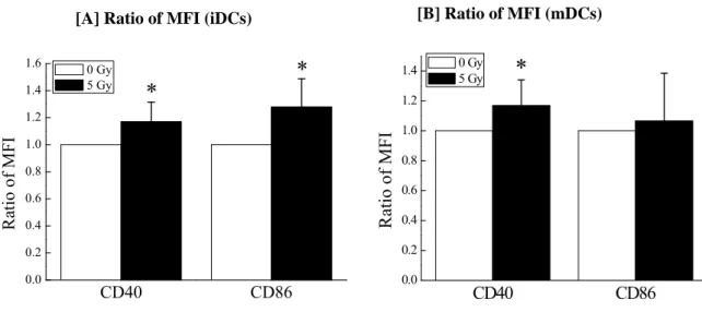
We investigated the influences of 5 Gy-irradiation to monocytes on the cell surface antigen expression in detail. As shown in Fig. 3-[A], the MFI of CD40 and CD86 on the iDCs derived from 5 Gy-irradiated monocytes was higher than that on the iDCs derived from non-irradiated monocytes. Although the MFI of CD40 on the mDCs derived from 5 Gy-irradiated monocytes was higher than that on the mDCs derived from non-irradiated monocytes, there was no significant difference in the CD86 expression on mDCs between non-irradiation and 5 Gy-irradiation (Fig. 3-[B]).
[B] Ratio of MFI (mDCs) [A] Ratio of MFI (iDCs)
CD40 CD86
0.0 0.2 0.4 0.6 0.8 1.0 1.2
1.4 0 Gy
5 Gy *
Ratio of MFI
CD40 CD86
0.0 0.2 0.4 0.6 0.8 1.0 1.2 1.4
1.6 *
Ra tio o f MF I
0 Gy
5 Gy *
Fig. 3 Differences in the expression levels of the cell surface antigens between the DCs derived from non-irradiated and 5 Gy-irradiated monocytes. Ratios of CD40 and CD86 MFI, respectively, were obtained by calculating a ratio of the MFI of DCs derived from 5 Gy-irradiated monocytes to that of DCs derived from non-irradiated monocytes (5 Gy/
0 Gy) in each individual. [A] The ratio of MFI on iDCs are expressed as the mean + SD of nine different individuals. [B] The ratio of MFI on mDCs are expressed as the mean + SD of 13 different individuals. * p< 0.05 by two-sided Mann-Whitney’s U-test.
Function of DCs
The functional characteristics of the DCs were analyzed. As one of the characteristics of iDCs, it is known that iDCs have a high phagocytotic activity which thus allows them to capture antigens. 1, 2) In the present study, the uptake of FITC-dextran was measured by a flow cytometer. The iDCs derived from non-irradiated monocytes showed an increase in
18
fluorescence intensity, thus demonstrating phagocytotic activity (Fig. 4-[A]). The iDCs derived from 5 Gy-irradiated monocytes showed less phagocytotic activity than the iDCs derived from non-irradiated monocytes in four out of six different individuals, and the large decrease (34% and 40%) was observed in two out of them (Fig. 4-[B]).
However, there was no statistically significant difference in the phagocytotic activity between the non-irradiation and 5 Gy-irradiation. These results suggest that X-irradiation to monocytes relatively attenuate the ability of iDCs to capture antigens.
[A] [B]
Fluorescence Intensity (FITC-Dextran)
Fig. 4. The phagocytotic activity of iDCs. Phagocytosis was measured by the uptake of FITC-dextran. [A] A representative cytogram is shown. The uptake of FITC-dextran by the iDCs derived from the non-irradiated (filled gray histogram) and 5 Gy-irradiated (broken line histogram) monocytes was quantified by a flow cytometer. The dotted line histogram represents the negative control. [B] The data of six different individuals are shown as mean fluorescence intensity (MFI).
Since mDCs can stimulate the proliferation of allogeneic leukocytes and this ability is often used as a surrogate marker of their activation state, we next investigated the ability of the mDCs in allogeneic mixed leukocytes reaction (MLR) in seven different individuals. In order to quantify the CD4 + T cells proliferation, we measured the incorporation of [ 3 H]-thymidine by CD4 + T cells. Both mDCs stimulated the proliferation of allogeneic CD4 + T cells (Fig. 5-[A]), although there was the large
10
010
110
210
3]
77
0 Gy on ice
5 Gy
0 Gy 5 Gy
2.2 2.4 2.6 2.8 3.0 3.2 3.4 3.6 3.8 4.0 4.2
FITC-de xtran MFI
19
difference in the incorporation of [ 3 H]-thymidine by CD4 + T cells among individuals.
Overall, the mDCs derived from 5 Gy-irradiated monocytes had about 32% less ability to stimulate allogeneic CD4 + T cells compared with mDCs derived from non-irradiated monocytes (Fig. 5-[B]) when mDCs were co-cultured with CD4 + T cells at a ratio of 1 : 10. These results suggest that X-irradiation to monocytes attenuate the interaction between mDCs and T cells.
Fig. 5 The stimulating ability of mDCs on the allogeneic lymphocytes in a MLR. As we described in the materials and methods, each mDC was co-cultured with allogeneic CD4 + T cells, and the proliferation of CD4 + T cells was determined by the incorporation of [ 3 H]-thymidine. [A] The representative result of seven different individuals is shown.
Values are the mean ± SD obtained from triplicate cultures. The incorporation of [ 3 H]-thymidine by only CD4 + T cells was less than 300 cpm. [B] A relative value of the proliferation (mean cpm obtained from triplicate cultures) was obtained by calculating the ratio of 5 Gy-irradiation to non-irradiation (5 Gy/ 0 Gy) in each individual. The data when mDCs were cultured with allogeneic CD4 + T cells at a ratio of 1: 10 are shown and expressed as the mean + SD of seven different individuals. *p < 0.05 by two-sided Mann-Whitney’s U-test.
Cytokines Production by DCs
mDCs produce various types of cytokines, which affect the T cell-mediated immune system. We thus next measured the cytokine production of the DCs in three different individuals. The cytokines released in the culture supernatants were assayed using
(100 : 1) (50 : 1) (10 : 1) 0
2 4 6 8 10 12 14 16 18
CPM (10 3 )
(T cells : mDCs)
0 Gy 5 Gy
[A] [B] (T cells : mDCs = 10 : 1)
0 Gy 5 Gy
0.0 0.2 0.4 0.6 0.8
1.0 *
R elative value
20
premixed multiplex beads, as described in the materials and methods. The culture supernatants of both iDCs contained large amounts of GM-CSF and IL-4 (data not shown). As shown in Fig. 6, IFN-γ, TNF-α and IL-10 were detected in the medium conditioned by both iDCs and mDCs derived from the non-irradiated monocytes. The medium conditioned by both iDCs and mDCs derived from the 5 Gy-irradiated monocytes also contained those cytokines (Fig. 6). The concentration of TNF-α in the culture supernatants of mDCs was more than 32 ng/ml (data not shown). Although IL-12 was not detected in the culture supernatants of the iDCs, it was detected in the culture supernatants of both mDCs (Fig. 6-[B]). However, no significant difference in the all cytokines concentrations tested in this study was observed between the non-irradiation and the 5 Gy-irradiation. These results indicate that DCs derived from 5 Gy-irradiated monocytes retain the capacity to produce cytokines tested here.
[A] iDC [B] mDC
Fig. 6 Cytokines concentration detected in the culture supernatants of iDCs and mDCs.
Each cytokine concentration in the culture supernatants of the iDCs [A] and mDCs [B]
was measured using the Bio-Plex cytokine protein array system. The data are represented as the mean + SD of three different individuals. n.d. =not detectable (< 2 pg/ml).
IL-10 IL-12 IL-13 IL-2 IFN- γ TNF- α 1
10 100
C onc en tr at io n ( pg /m l) n.d.
0 Gy 5 Gy
n.d.
IL-10 IL-12 IL-13 IL-2 IFN- γ
1 10 100 1000
C on cen tr at on ( pg /m l)
0 Gy 5 Gy
Total MMP-9 in the Culture Supernatants of DCs
It is reported that DCs express several MMPs including MMP-9. 29, 30) MMP-9, which is gelatinase B, cleaves type IV collagen and contributes to DCs migration through the
21
extracellular matrix. 29, 31) We therefore investigated total MMP-9 in four different individuals. As shown in Fig. 7-[A], MMP-9 was presented in the culture supernatants of each DC. In agreement with previous reports, 29) the total MMP-9 increased in the culture supernatants of the mDCs in comparison to the iDCs (Fig. 7-[B]). There was about 19% less total MMP-9 in the culture supernatants of iDCs derived from 5 Gy-irradiated monocytes compared with non-irradiation (Fig. 7-[B]). However, when the iDCs were induced to mDCs with rhTNF-α, this reduction disappeared.
Cell-free condition
medium 0 Gy 5 Gy 0 Gy 5 Gy
[A] iDCs mDCs
MMP-9
[B]
0 Gy 5 Gy 0 Gy 5 Gy
0.0 0.2 0.4 0.6 0.8 1.0 1.2 1.4 1.6
*
*
mDCs
Relative valu e
iDCs
Fig.7 Total MMP-9 in the culture supernatants of iDCs and mDCs. The total MMP-9 in the culture supernatants of iDCs and mDCs derived from non-irradiated and 5 Gy-irradiated monocytes was analyzed by substrate zymography. [A] The data are representative of similar results obtained from four different individuals. [B]
Quantification of bands was performed using Image J analysis. Each of data was divided by the value of iDCs derived from non-irradiation monocytes, respectively.
The data are represented as the mean + SD of four different individuals. * p< 0.05 by two-sided Mann-Whitney’s U-test.
22
4. Discussion
In this study, we evaluated the influences of X-irradiation on the differentiation from peripheral blood monocytes to DCs. We showed that X-irradiated monocytes can differentiate into iDCs and mDCs. We also showed that iDCs derived from 5 Gy-irradiated monocytes expressed higher levels of CD40 and CD86 compared with iDCs derived from non-irradiated monocytes (Fig. 3). Although few reports have described the influences of irradiation on the differential pathway from monocytes to DCs, previous studies have reported that ionizing radiation to iDCs caused a change in the expression of the surface antigens. 21-23) Cao et al. 21) and Reuben et al. 22) demonstrated that gamma irradiation reduced the expression of the CD86, CD80 and HLA-DR molecules on iDCs induced from human peripheral blood monocytes. On the other hand, Merrick et al. reported that the CD86 expression of iDCs was significantly up-regulated after gamma irradiation. 23) Our results are consistent with Merrick’s report regarding the up-regulation of co-stimulatory molecule expression, although there is a difference whether irradiation to monocytes or to iDCs. McBride et al. suggest the possibility that the “danger” signals induced by ionizing radiation may cause the maturation of DCs. 19) Their suggestion led us to consider that the various “danger”
signals, which were induced either in the irradiated DCs-precursors or released from the irradiated bystander cells (including DCs-precursors and others), might influence the differentiation into DCs. TNF-α is an inflammatory cytokine and is one of the danger signals. Krivenko et al. reported that irradiation increased TNF production by human monocytes. 32) One possibility is that X-irradiation performed on peripheral blood monocytes may induce TNF-α production , which directly influences the differentiation into DCs from monocytes. However, there is no significant difference between the tested cytokines including TNF-α in the supernatant conditioned by the non-irradiated and the 5 Gy-irradiated monocytes (Fig. 6-[A]), thus indicating no involvement of TNF-α. Further studies regarding other “danger” signals, for example reactive oxygen species produced by irradiated-cells 19) and the high-mobility-group box 1 protein
23
released from dying tumor cells 33) , are needed to clarify the mechanisms involved in the relationship between X-irradiation and the up-regulation of co-stimulatory molecule expression on iDCs. In this study, we used rhTNF-α as the maturation stimuli, and the mDCs derived from 5 Gy-irradiated monocytes expressed higher levels of CD40 compared with mDCs derived from non-irradiated monocytes (Fig. 3-[B]). However, this up-regulation of CD40 expression by 5 Gy-irradiation was not observed when we used lipopolysaccharide (LPS) as maturation stimuli in our progressing experiments (data not shown). These results indicate that the influences of X-irradiation to monocytes on the maturation of DCs depend on the types of maturation stimulus. It is thought that X-irradiation may influence the intracellular signal transduction induced by maturation stimuli, because the receptor of LPS is Toll-like receptor 4 and different from TNF receptor. Further studies regarding other maturation stimuli including LPS are now in progress.
In a functional analysis of the monocyte-derived DCs, no significant difference in the phagocytotic activity was observed between the non-irradiation and 5 Gy-irradiation, although this activity decreased by 5 Gy-irradiation in four out of six different individuals (Fig. 4-[B]). The result that the reduction of phagocytotic activity was not observed in all individuals may be owing to individual difference in the radiosensitivity. Regarding the stimulating ability of the mDCs on allogeneic T cell proliferation, although there was no difference in the MHC class II (HLA-DR) expression, which is involved in allogeneic MLR, 28, 34) the T cell proliferation by mDCs induced from the irradiated monocytes was reduced (Fig. 5). This result is consistent with previous studies that the stimulating capacity of DCs on T cell proliferation is reduced by applying ionizing radiation to the DCs, 21, 23) although there is a difference whether irradiation to monocytes or to DCs. Merrick et al. indicated that irradiated DCs were less effective in a MLR, and upon maturation produced significantly less IL-12 in comparison to the non-irradiated controls, while IL-10 secretion was maintained. 23) On the other hand, Chauveau et al. 35) demonstrated that human iDCs express heme
24
oxygenase-1 (HO-1), which is an intracellular antioxidant enzyme to degrade heme and inhibit immune responses, 36) and that HO-1 expression is down-regulated by maturation stimuli. In addition, the induction of HO-1 not only inhibited the allostimulatory capacity of DCs but also suppressed IL-12 secretion of DCs, while it didn’t affect IL-10 secretion of DCs. 35) These two reports led to a hypothesis that oxidative stress such as ionizing radiation induce HO-1 in the monocytes and/or iDCs, and HO-1 suppress IL-12 secretion and allostimulatory ability of DCs. However, no detectable change in the IL-12 production by 5 Gy-irradiation was observed in our study (Fig. 6-[B]).
Therefore, we think that other factors such as chemokine may be controlled by X rays-induced HO-1 in this case. 37) In order to clarify these precise mechanisms, further experiments about the involvement of HO-1 are now in progress. On the other hand, zymographic analysis revealed that 5 Gy-irradiation induced a decrease of total MMP-9 during differentiation into iDCs from monocytes (Fig. 7). In contrast, 5 Gy-irradiation increased the ratio of mDCs to iDCs in the total MMP-9 activity. Lu et al. reported that addition of H 2 O 2 to LPS-activated monocytes, but not naïve monocytes, caused a significant enhancement of MMP-1 production. 38) We think that the reactive oxygen species generated by ionizing radiation enhance production of MMP-9 in the TNF-α-stimulated mDCs. However, it is unclear how X-irradiation induced a decrease of total MMP-9 during the differentiation into iDCs from monocytes.
In conclusion, the present data demonstrate that human peripheral blood monocytes exposed to ionizing radiation can differentiate into DCs, but some differences in their characteristics are observed between the DCs from non-irradiated monocytes and those from irradiated monocytes. Since the DCs derived from the monocytes of patients are phenotypically and functionally inefficient in comparison to healthy donors, 17, 18) the immune system in many cancer sufferers is thus considered to possibly be more severely damaged by radiotherapy or other therapies, such as chemotherapy during or after therapy. Furthermore, the efficacy of immunotherapy using monocyte-derived DCs against malignant tumors may be attenuated after
25
undergoing radiotherapy. Further understanding about the influences of ionizing radiation on immune cells including DCs will allow combination of radiotherapy and immunotherapy. 39) At the start of radiotherapy or chemotherapy, the degree of damage to the DCs or DC precursors by various types of oxidative stress, including ionizing radiation, should therefore be monitored.
26
Chapter 2
Impairment of mature dendritic cells derived from X-irradiated human monocytes depends on the type of maturation stimulus used
Abstract
Dendritic cells (DCs) play an essential role in the immune system. We have showed that X-irradiated monocytes, precursors of DCs, can differentiate into DCs and then mature in terms of surface antigen expression after tumor necrosis factor-α stimulation while showing reduced functionality. DCs can mature in response to various types of maturation stimuli. Therefore, this study investigated whether DCs from monocytes exposed to ionizing radiation can adequately respond to pathogen-derived components and proinflammatory cytokines. Human monocytes separated from buffy coats were exposed to x-rays and were then differentiated into immature DCs (iDCs). iDCs were stimulated by lipopolysaccharide (LPS) or proinflammatory cytokine mixture (MIX).
The DCs from nonirradiated and X-irradiated monocytes showed maturation after LPS and MIX stimulation as confirmed by findings of surface antigen expression. Upon LPS stimulation, however, the expression levels of CD80 and CD83 on the DCs from the X-irradiated monocytes were lower than those of the DCs from the nonirradiated monocytes. Such reductions were not observed upon MIX stimulation. Similarly, an impairment of matrix metalloproteinase-9 and cytokine production was observed in the LPS-stimulated DCs from the X-irradiated monocytes, whereas these impairments were not observed upon MIX stimulation. The ability of DCs to stimulate T cells was lower in the irradiated group compared with the nonirradiated group despite the type of maturation stimulus. Thus, the present results suggest that the influence of X-irradiation
27
on the maturation of DCs depends on the type of maturation stimulus used and that X-irradiation especially impairs the response of DCs to LPS.
28
1. Introduction
Dendritic cells (DCs) play key roles in immune regulation as professional antigen-presenting cells. They serve as a link between innate and adaptive immunity because they stimulate naive T cells more powerfully than other antigen-presenting cells.
The development of DCs involves three stages of differentiation: DC precursors, immature DCs (iDCs), and mature DCs (mDCs). 1-3) iDCs are present in various tissues, and they migrate to inflammatory sites. At inflammatory sites, iDCs capture antigens, such as invading bacteria and viruses, and then process the antigens for presentation on major histocompatibility complex molecules. Pathogen-derived components or proinflammatory cytokines can induce maturation of DCs. mDCs acquire a high antigen-presenting capacity and thereby vigorously stimulate T-cell responses. The powerful antigen-presenting capacity of DCs makes them useful for immunotherapy against cancer. 40,41)
The different classes of specific immune responses are driven by the biased development of pathogen-specific effector helper T cell subsets ― that is, T helper 1 (Th1) and Th2 cells, that activate different components of cellular and humoral immunity. The polarization of Th-cell by DCs depends on the maturation stimuli that the DCs are exposed to. 42) Recent reports propose that various types of stimuli including pathogen-derived components and proinflammatory cytokines can lead to the maturation of DCs. 43,44) As a representative pathogen-derived component, lipopolysaccharide (LPS) has been widely studied. 45-47) LPS is a major component of the outer membrane of Gram-negative bacteria and is recognized by the Toll-like receptor 4 (TLR4), which is a pattern recognition receptor. 48) LPS strongly activates DCs, and activated DCs induce a Th1 immune response, which results in cell-mediated immunity including activation of cytotoxic T cells. 45-47) On the other hand, a combination of proinflammatory cytokines and prostaglandin E 2 (PGE 2 ), which mimics monocyte-conditioned medium, has been used as a maturation stimulus for processing DCs for cancer immunotherapy. 49) However, it is controversial whether this
29
combination can induce the Th1 immune response because DCs stimulated by this combination produce low levels of interleukin-12 (IL-12). 50) In general, it is considered that DCs stimulated by this combination induce a Th2 immune response from naive T cells, which results in a humoral immune response. 51)
It is well known that T and B lymphocytes, which compose the adaptive immune system, are highly sensitive to ionizing radiation. On the other hand, DCs are relatively resistant to ionizing radiation-induced cell death, although DCs exposed to ionizing radiation are functionally weakened. 21-23) We have previously showed that X-irradiated human monocytes, which are precursors of DCs, can differentiate into iDCs and then mature in terms of surface antigen expression after tumor necrosis factor-α (TNF-α) stimulation. However, the functions of mDCs derived from X-irradiated human monocytes, such as the stimulation of allogeneic T cells, are attenuated when compared with mDCs derived from nonirradiated monocytes. 52) It remains unknown whether X-irradiation of human monocytes influences the maturation of DCs by stimuli such as pathogen-derived components and a proinflammatory cytokine mixture. This is an important issue to consider in an immune system that has been exposed to ionizing radiation. Furthermore, it is also important to determine the efficacy of immunotherapy using monocyte-derived DCs when monocytes are prepared from patients who have already undergone radiotherapy. Therefore, the present study investigated whether DCs derived from monocytes exposed to ionizing radiation can adequately respond to LPS and a proinflammatory cytokine mixture.
2. Materials and methods Reagents
Recombinant human granulocyte-macrophage colony-stimulating factor (rhGM-CSF), rhIL-1β, rhIL-4, rhIL-6, and rhTNF-α were purchased from PeproTech Inc. (Rocky Hill, NJ, USA). LPS (Escherichia coli 055:B5) and PGE 2 were purchased from Sigma Aldrich (St Louis, Mo, USA). The fluorescently labeled monoclonal antibodies (mABs)
30
anti-human cluster of differentiation 40-fluorescein isothiocyanate (CD40-FITC) and mouse IgG 2a -phycoerythrin (PE) were purchased from Becton Dickenson (San Jose, CA, USA). Anti-human CD19-FITC, CD80-FITC, CD11c-PE, CD14-PE, CD83-PE, CD86-PE, human leukocyte antigen-ABC-FITC (HLA-ABC-FITC), mouse IgG 1 -FITC, and mouse IgG 2b -PE were purchased from Beckman Coulter (Fullerton, CA, USA), and the anti-human CD3-PE, HLA-DR-PE, and TLR4-PE for the flow cytometery were purchased from eBioscience (San Diego, CA, USA). The fluorescent probe 2′,7′-dichlorodihydrofluorescein diacetate (H 2 DCFDA) was purchased from Molecular Probes, Invitrogen Corporation (Eugene, Oregon, USA).
Collection of human monocytes from buffy coat
This study was approved by the committee of medical ethics of the Hirosaki University Graduate School of Medicine (Hirosaki, Japan). After obtaining informed consent from all of the healthy human blood donors, peripheral blood was collected by the Aomori Red Cross Blood Center. Peripheral blood mononuclear cells (PBMCs) were separated from the buffy coats by centrifugation with Lymphosepar I (1.077 g/mL;
Immuno-Biological Laboratories Co. Ltd., Takasaki, Japan). The PBMCs were collected and washed at least three times with PBS(−) containing 5-mM EDTA. To obtain the monocyte fraction, the Human Monocyte Enrichment Set-DM (BD Bioscience, San Jose, CA, USA) was used for the negative selection of monocytes from PBMCs according to the manufacturer’s instructions. The purity of CD14 + monocytes was > 70.0%, as determined by flow cytometry. To purify the monocyte fraction, the collected monocytes were resuspended in PBS(−) and allowed to adhere to a plastic dish at 37°C for 1.5 – 2 h in a humidified atmosphere containing 5% CO 2 . Thereafter, nonadherent cells were removed by washing the dish twice with PBS(−).
The adherent monocytes were cultured in RPMI 1640 (Gibco, Grand Island, NY, USA) supplemented with 2% heat-inactivated human AB serum (Gemini Bio-products, Woodland, CA, USA) and 1% antibiotic-antimycotic (Gibco) (hereafter
31
referred to as the medium). The concentration of the monocytes was approximately 1.0
× 10 6 cells/mL.
In vitro irradiation of human monocytes
Irradiation with x-rays (150 kVp, 20 mA, 0.5-mm Al, and 0.3-mm Cu filters) was performed with an x-ray generator (MBR-1520R, Hitachi Medical Corporation, Tokyo, Japan) at a distance of 45 cm from the focus and at a dose rate of 98.0 – 104.0 cGy/min.
In vitro generation and culture of human DCs
DCs were generated from monocytes according to a previously published protocol with some modifications. 53) Briefly, 20 h after X-irradiation of monocytes, 50 ng/mL rhGM-CSF and 50 ng/mL rhIL-4 were added to the medium, and the monocytes were cultured at 37°C for 5 days in a humidified atmosphere containing 5% CO 2 to prepare iDCs. On day 3 of this 5-day culture, half of the culture medium was changed with new medium containing rhGM-CSF and rhIL-4 (50 ng/mL). After 5 days of culture, the cells were harvested and counted by the trypan blue dye exclusion method.
For the zymography assay, an aliquot of the culture supernatant was collected and kept frozen at −85°C. After 5 days of culture, half of the medium was changed with new medium and the cells were recultured in the presence of an LPS (1 μg/mL) or a cytokine mixture (10-ng/mL rhTNF-α, 10-ng/mL rhIL-1β, 10-ng/mL rhIL-6, and 1-μg/mL PGE 2 : MIX) for 48 h. 54) After 48 h, the cells were harvested and counted by the trypan blue dye exclusion method. Furthermore, the culture supernatants were also collected and kept frozen at −85°C for the zymography and cytokine assays. In some cases, the cells were stimulated for the indicated period.
Cell surface staining
For surface marker analysis of the iDCs and the mDCs, the cells were stained with mAbs conjugated to FITC or PE for 30 min at 4°C in the dark. The cells were also
32
stained with corresponding FITC- or PE-conjugated isotype control mouse IgGs. After 30 min, the cells were washed with cold PBS(−) and analyzed by a flow cytometer (Epics XL, Beckman Coulter). The cells induced from monocytes could be distinguished from debris and lymphocytes using a region established by their high forward and side scatter signals as shown in a previous report. 53)
Analysis of matrix metalloproteinase-9 activity
Total matrix metalloproteinase-9 (MMP-9) in the culture supernatants of iDCs and mDCs was analyzed by substrate zymography. 52) Each culture supernatant was subjected to electrophoresis on a sodium dodecyl sulfate polyacrylamide gel (10%
acrylamide) containing 0.1% gelatin. Proteinases separated in the gels were renatured by gentle shaking in 50-mM Tris-HCl buffer (pH 7.5, containing 0.1-M NaCl and 2.5%
Triton X-100) at room temperature for 1.5 h. The gels were then incubated in 50-mM Tris-HCl buffer (pH 7.5, containing 10-mM CaCl 2 ) at 37°C for 15 h and stained with Coomassie Brilliant Blue R-250. The bands were quantified by an Image J analysis (1.37).
Measurement of intracellular reactive oxygen species production
The fluorescent probe H 2 DCFDA was used for the measurement of intracellular reactive oxygen species (ROS). The cells were incubated for 15 min at 37°C, with 5-μM H 2 DCFDA in PBS(−). After incubation, the cells were washed twice with PBS(−) and analyzed by flow cytometry.
Allogeneic mixed leukocyte reaction
Allogeneic CD4 + T cells numbering 1.0 × 10 5 cells (> 98% CD4 + T cells), which were purified from PBMCs with the Human CD4 T lymphocyte Enrichment Set-DM (BD Bioscience), were co-cultured in 96-well flat bottom microplates (Asahi Techno Glass Co. Ltd., Chiba, Japan) with 1.0 × 10 4 mDCs. The mDCs were 20 Gy-irradiated with an
33
X-ray generator before co-culture with allogeneic CD4 + T cells. Both cells were co-cultured for 4 days in RPMI 1640 medium supplemented with 2% heat-inactivated human AB serum. After 4 days of culture, the cells were incubated for an additional 20 h in the presence of [ 3 H]-thymidine (1 μCi/well, specific activity, 5 Ci/mmol; Moravek Biochemicals Inc, CA, USA). To determine CD4 + T cell proliferation, the cells were harvested onto glass fiber filters (Whatman, England) with a semiautomatic harvester (Labo Mash, Labo Science, Japan), and then the amount of [ 3 H]-thymidine incorporation was measured by a liquid scintillation counter (LSC-5100, Aloka Co. Ltd., Japan).
Cytokine measurements
The cytokines in the culture supernatants were measured using the Bio-Plex protein array system (Bio-Rad Laboratories, Hercules, CA, USA). The Bio-Plex cytokine assay is designed for the multiplexed quantitative measurement of multiple cytokines in a single well. 52) For the cytokine assays, premixed multiplex beads of the Bio-Plex human cytokine 17-Plex panel (Bio-Rad Laboratories), which includes 17 cytokines [IL-1β, IL-2, IL-4, IL-5, IL-6, IL-7, IL-8, IL-10, IL-12 (p70), IL-13, IL-17, granulocyte colony-stimulating factor (G-CSF), GM-CSF, interferon-γ (IFN-γ), monocyte chemoattractant protein-1 (MCP-1), macrophage inflammatory protein-1β (MIP-1β), and TNF-α], were used. The samples were analyzed in duplicate. The data were analyzed using the Bio-Plex Manager 4.0 software (Bio-Rad Laboratories). The concentration of IL-5 was lower than the lowest detectable concentration (3.0 pg/mL).
Statistical analysis
The data are expressed as mean ± SD values. The comparisons between the nonirradiation and X-irradiation conditions were performed by two-sided Mann-Whitney’s U-test or a two-sided paired t-test. A p < 0.05 was considered to be statistically significant. The statistical analysis was performed using the Excel 2003
34
software program (Microsoft, USA) with the add-in software Statcel 2. 27)
35
3. Results
X-irradiation impairs the response of DCs to LPS in terms of cell surface antigen expression
To generate iDCs, nonirradiated or X-irradiated monocytes were cultured in the presence of rhGM-CSF plus rhIL-4. The iDCs derived from the nonirradiated monocytes expressed high levels of CD40, CD86, and HLA-DR (Fig. 8-[A]). However, they expressed no or trace levels of the co-stimulatory molecule CD80 and the DC maturation marker CD83, 28) thus showing the characteristics of iDCs. When the iDCs from the nonirradiated monocytes were stimulated by LPS or MIX, the expressions of CD80 and CD83 were largely upregulated, suggesting that the cells had matured into mDCs (Fig. 8-[A]). Hereafter, we refer to DCs stimulated by LPS and MIX as LPS-mDCs and MIX-mDCs, respectively. Although no significant difference in number or viability of DCs was observed between the nonirradiation and X-irradiation conditions (Fig. 8-[B], [C]), the expressions of CD80 and CD83 in the LPS-mDCs from the X-irradiated monocytes were lower in a dose-dependent manner as compared with those of the LPS-mDCs from the nonirradiated monocytes (Fig. 8-[D]). On the other hand, no significant difference in the expression of these antigens between nonirradiation and X-irradiation was observed in the MIX-mDCs. These results show that X-irradiation impairs the response of DCs to LPS in terms of the expressions of maturation-related cell surface antigens.
Figure 8 [A]
CD86 HLA-ABC HLA-DR
Fluorescence Intensity
90
90
iDCs
LPS
MIX
Ce ll Nu m b er 1.2
CD40
90
90
3.1
90
100 101 102 103
3.3
90
90
90
0.7 CD80
7.6
7.8
100 101 102 103
0.6 CD83
100 101 102 103
8.0
11.4
80
80
80
37.4
68.1
87.2
100 101 102 103 100 101 102 103 110
110
110
8.1 51.3
135
189
12.7
18.0
90
90
90
100 101 102 103 90


![Figure 8 [A] CD86 HLA-ABC HLA-DR Fluorescence Intensity9090iDCsLPSMIXCell Number1.2CD4090903.1901001011021033.39090900.7CD807.67.81001011021030.6CD831001011021038.011.480808037.468.187.2100101102103100101102103110110110 8.151.3135189 12.7 18.0909090100101](https://thumb-ap.123doks.com/thumbv2/123deta/5921304.2055164/37.892.160.775.871.1123/figure-hla-abc-hla-fluorescence-intensity-idcslpsmixcell-number.webp)
![Fig. 8. Expression of cell surface antigens in the DCs stimulated by LPS and MIX. [A]](https://thumb-ap.123doks.com/thumbv2/123deta/5921304.2055164/38.892.139.826.136.742/fig-expression-cell-surface-antigens-dcs-stimulated-lps.webp)


