緒 言
後大脳動脈瘤(posterior cerebral artery aneurysms:
PCA動脈瘤)は全脳動脈瘤の0.7~3%と比較的稀であ
る3,5,6).Zealら16)は後大脳動脈をP1~P4に分類し,P
2 segmentを後交通動脈との合流部位からPCAが四丘体 槽に入るまでの部位としている.
P2 segmentにはPCA動脈瘤のうち約40%~60%が発 生し,その特徴として紡錘状動脈瘤または巨大動脈瘤が 他の部位と比較して多いとされる1,3,4,5,14).
治療法に関しては外科治療から血管内治療によるもの と多岐にわたる報告がある.外科治療についてはクリッ ピングやバイパスを併用したアプローチがとられるが,
いまだに治療困難な動脈瘤の一つである2,9).一方,近
年の血管内治療の発達に伴いPCA動脈瘤に対しては血 管内治療が施行される場合も多く,瘤内塞栓あるいはバ ルーン,コイルを用いた親動脈の閉塞が行われ良好な治 療成績が得られている1,3,4,6,8).
今回,PCAのP2 segmentに発生した動脈瘤(PCA P 2 動脈瘤)の7例に対して血管内治療を施行したので その治療法,問題点について検討し,報告する.
対象と方法
対象は2002年8月から2008年1月までPCA P2動脈 瘤に対して血管内治療を施行した7例である.
年齢は18歳~82歳(平均55歳),男性1例,女性6例で,
くも膜下出血(subarachnoid hemorrhage;SAH)で発 症したもの3例,mass effectで発症したもの1例,破裂
後大脳動脈P2部動脈瘤に対する血管内治療
須山武裕 永島宗紀 我妻敬一 乾 敏彦 長谷川洋 富永紳介
Endovascular treatment for P2 segment aneurysms of the posterior cerebral artery
Takehiro SUYAMA Munenori NAGASHIMA Keiichi AZUMA Toshihiko INUI Hiroshi HASEGAWA Shinsuke TOMINAGA
Department of Neurosurgery, Tominaga Hospital
●Abstract●
Purpose: To evaluate treatment outcome of P2-segment aneurysms of the posterior cerebral artery by coil embolization.
Subjects: Seven patients (three with subarachnoid hemorrhage due to ruptured aneurysms and four with unruptured ones) underwent endovascular treatment. 4 patients presented with unruptured aneurysms, 1 presented with mass effect, 1 was associated with another ruptured cerebral aneurysm, and 2 were diagnosed during a routine examination for headache.
Results: Endosaccular coil embolization was performed for the saccular aneurysms (3 ruptured and 2 unruptured cases) and parent vessel occlusion was performed for the fusiform aneurysm (1 unruptured case) and thrombosed aneurysm (1 unruptured case). No apparent complications were noted perioperatively or during the follow-up periods of 2 years to a maximum of 5 years and 5 months, average: 3.6 years.
Retreatment was not required for any case.
Conclusion: Outcomes of endovascular treatments for P2-segment aneurysms of the posterior cerebral artery were good. If endosaccular coil embolization is not possible, it is advisable to evaluate collateral circulation with an occlusion test, checking the presence of neurological ischemic symptoms, and perform parent artery occlusion. However, each case requires individual consideration.
●Key Words●
cerebral aneurysm, endovascular surgery, parent artery occlusion, posterior cerebral artery
(Received May 14, 2010:Accepted July 1, 2010)
富永病院 脳神経外科
<連絡先:須山武裕 〒556-0017 大阪市浪速区湊町1-4-48 E-mail:[email protected]>
脳動脈瘤に合併して発見されたもの1例,頭痛の精査に て発見されたもの2例であった.
当院では後方循環の脳動脈瘤,特に後大脳動脈瘤につ いては開頭手術の困難さから血管内治療を第一選択とし ている.コイル塞栓術に際してはできる限り親動脈を温 存した瘤内塞栓術を施行した.親動脈温存が不可能な動 脈瘤であればPCAのballoon occlusion test(BOT)によ る神経症状の有無や脳血管撮影による側副血行路により PCA閉塞の可否を判断し,瘤内および親動脈閉塞術
(parent artery occlusion;PAO)を施行した.
全例で経大腿動脈アプローチで手技を施行した.破裂 脳動脈瘤では全身麻酔下にて手技を行い,first coil留置 後よりヘパリンを投与,activated clotting time(ACT)
をコントロール値の約2~2.5倍に維持した.治療後は ヘパリンの中和は施行せず,治療直後より抗血小板薬1 剤を胃管より注入した.術後スパイナルドレナージを留 置し,翌日まで全身麻酔下に血圧管理を行い,頭部CT にて再出血が無いことを確認し覚醒させた.抗血小板薬 は術後3ヵ月間投与した.
未破裂脳動脈瘤は全例,局所麻酔下にて手技を施行し た.治療1週間前より抗血小板薬2剤を併用し,術中は ヘパリンにてACTをコントロール値の約2~2.5倍に維 持し,術後は抗トロンビン剤を2~6日間点滴した.抗 血小板薬は術後2週間は2剤を投与し,その後3ヵ月間 は1剤のみ投与した.
PAOを施行した場合は術後の血栓塞栓症予防のため 抗トロンビン剤を6日間使用した.また術後7日間は低 灌流予防のため塩酸ドブタミンを5μg/kg/minから 10μg/Kg/minまで使用し術前の血圧に対して10~20%
の昇圧を行った.
退院後は頭部単純レントゲン,頭部magnetic resonance angiography(MRA),脳血管撮影などにより経過観察 を施行した.
結 果
(Table 1)嚢状動脈瘤の破裂脳動脈瘤3例,未破裂脳動脈瘤2例 は瘤内塞栓術を施行し,いずれもtight packingを行い完 全閉塞が得られた.血栓化脳動脈瘤(Case 4)および 紡錘状脳動脈瘤(Case 6)ではPAOを施行した.治療 に伴う合併症は認めなかったが,破裂脳動脈瘤の1例
(Case 5)で脳血管攣縮により脳梗塞を生じ,modified Rankin Scale(mRS)が入院時grade0から退院時grade 2へ低下した.Case 2において1年後にneck付近のコ イル形態の変形を認めたがその後は進行していない.
2~5年5ヵ月(平均3.6年)の観察期間に再破裂は 認めず,動脈瘤の増大による再治療は行っていない.
症例呈示
1.症例1
82歳,女性.自宅にて倒れているところを発見され近
5 18/M SAH/IVH
HK Ⅲ saccular 5×5 Coil
occlusion 2 37
6 64/F headache fusiform 6×5 Coil PAO 0 30
7 60/F SAH HK Ⅱ saccular 5×5 Coil
occlusion 1 24
AN:aneurysm,HK:Hunt & Kosnik grade,IVH:intraventricular hemorrhage,mRS: modified Rankin Scale,
PAO:parent artery occlusion,SAH:subarachnoid hemorrhage
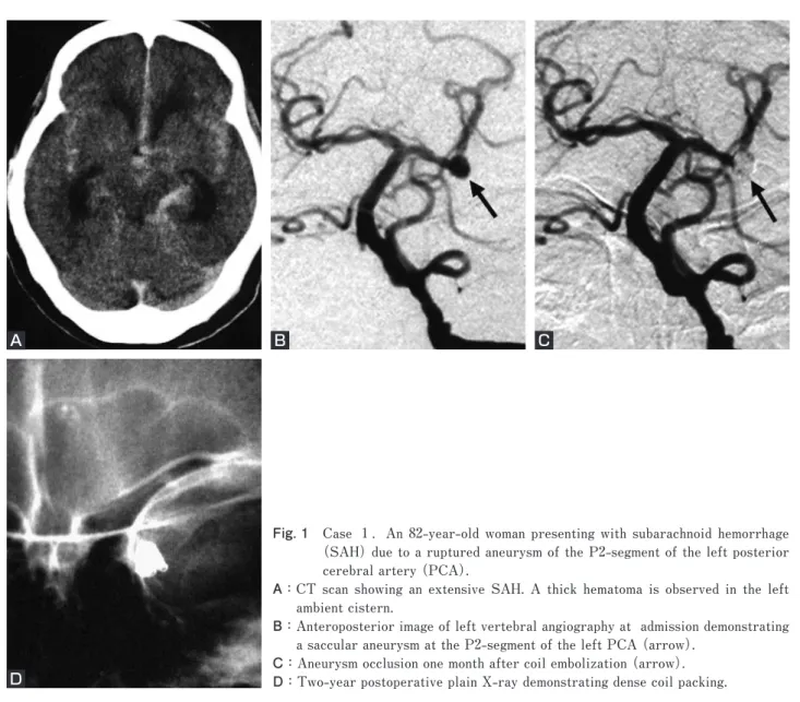
医に搬送され,頭部CTにてFisher group 3のSAHを認 めたため当院へ転送された(Fig. 1A).
発症時はGlasgow Coma Scale 8(E:2,V:3,M:
4),Hunt & Kosnik grade Ⅲ,World Federation Neurological Surgeons(WFNS)gradeⅣであった.脳 血管撮影にて左PCA P2 segmentにblebを伴う嚢状動脈 瘤を認めた(Fig. 1B).急性水頭症に対する脳室ドレナ ージの後,血管内治療を施行した.
6Fr ENVOY®(Cordis Neurovascular, Miami, FL, USA)
を左椎骨動脈に留置し,FasTracker10(Boston Scientific, Natick, MA, USA),SilverSpeed10®(eV3 Neurovascular, Irvine, CA, USA)を瘤内へ誘導し,Guglielmi Detachable Coil® 10(GDC10)(Boston Scientific, Natick, MA, USA)
を用い瘤内塞栓を行った.
術後経過は良好で神経学的な異常はなかった.発症1 ヵ月に施行した脳血管撮影で動脈瘤の増大は認めなかっ た(Fig. 1C).治療約2ヵ月後に独歩退院した.2年後 の頭蓋単純撮影でもコイルの形態変化は認めなかった
(Fig. 1D).治療4年10ヵ月後に呼吸器疾患にて死亡した.
2.症例2
27歳女性.頭痛の精査目的でmagnetic resonance image
(MRI),MRAを施行したところ脳動脈瘤が指摘された.
来院時には神経学的な異常はなく,脳血管撮影にて右 PCA P2 segmentに10mm×12mmの脳動脈瘤が認めら れた(Fig. 2A).未破裂動脈瘤ではあるが破裂および増 大予防のために血管内治療を行った.
6Fr ENVOYR留置後ExcelsiorSL-10(Boston Scientific, ® Natick, MA, USA)をSilverSpeed10®にて瘤内へ誘導し,
Fig. 1 Case 1.An 82-year-old woman presenting with subarachnoid hemorrhage (SAH) due to a ruptured aneurysm of the P2-segment of the left posterior cerebral artery (PCA).
A: CT scan showing an extensive SAH. A thick hematoma is observed in the left ambient cistern.
B: Anteroposterior image of left vertebral angiography at admission demonstrating a saccular aneurysm at the P2-segment of the left PCA (arrow).
C:Aneurysm occlusion one month after coil embolization (arrow).
D:Two-year postoperative plain X-ray demonstrating dense coil packing.
A B C
D
GDC10を用いて瘤内塞栓術を行った(Fig. 2B).術後特 に合併症なく退院した.
治療1年後の脳血管撮影,頭部単純レントゲン撮影で,
ネック部の軽度の描出およびcoil compactionが認められ た(Fig. 2C, D)が経過観察した.
術後約5年経過したがMRA,頭部単純レントゲン撮 影では大きな変化は認めていない(Fig. 3E, F).
3.症例4
57歳女性.左上下肢の脱力を自覚し他院にて頭部 MRI,MRAが施行され,右後大脳動脈血栓化動脈瘤と 診断されたが経過観察されていた.その後,左上下肢脱 力が増悪し,歩行困難を生じたため,半年後に当院を受 診した.
来院時には意識レベル清明で左不全片麻痺(MMT4/5)
が認められた.MRI T1,T2強調画像にて層状を呈し Fig. 2 Case 2.A 27-year-old woman presenting with an unruptured aneurysm of the P2-segment of the right posterior
cerebral artery.
A:Anteroposterior images of right vertebral angiography demonstrating a large saccular aneurysm at the P2-segment.
B:Angiography taken immediately after intra-aneurysmal coil embolization showing almost complete aneurysm occlusion.
C: Left vertebral angiography taken one year after coil embolization showing a slight re-opening at the neck of the aneurysm (arrow).
D:One-year postoperative plain X-ray demonstrating a slight coil compaction at the neck of the aneurysm (arrow).
E, F:Although a five-year postoperative MRA(E) and lateral plain X-ray(F) reveals a slight re-opening at the neck of the aneurysm (arrows), there are no dramatic changes as compared to other images taken annually.
A B C
D E F
Fig. 3 Case 4.A 57-year-old women presenting with left hemiparesis due to a thrombosed aneurysm of P2-segment of the right posterior cerebral artery.
A, B:Axial MRI (T1-weighted image (A), T2-weighted image (B)) showing internally heterogeneous mass without cerebral edema compressing the right cerebral peduncle (arrows).
C: An anteroposterior image of left vertebral angiography showing a saccular aneurysm at the P2-segment of the right posterior cerebral artery (PCA)(arrow).
D:A non-subtracted image during balloon occlusion test at the P2-segment (arrows).
E: A right internal carotid artery (lateral view) showing a leptomeningeal anastomosis to the right PCA territory (arrows).
F:Left vertebral angiography after parent artery coil occlusion (arrows).
G, H:A three-year postoperative MRI (T1-weighted image (G), T2-weighted image (H)) revealing a reduction of aneurysm size (arrows).
A B C
D E
H G
F
脈 瘤 をGDC10,GDC18を 用 い て 閉 塞 し た(Fig. 3E). 術直後の右内頚動脈撮影では右中大脳動脈からの側副血 行路は良好であった(Fig. 3F).新たな症状の出現や左 片麻痺の悪化は認めなかった.その後リハビリテーショ ンを行い独歩退院した.術後3年経過しているが左片麻 痺は改善し,MRIでは動脈瘤は縮小しmass effectは軽減 した(Fig. 3G, H).
考 察
PCA動脈瘤は全脳動脈瘤の0.7~3%とされ,椎骨脳 底動脈系の動脈瘤の中では約15%が後大脳動脈領域に発
生する2,3,5,8,9,12).Zeal16)らの分類によるP1-P2に87%,
P3以降に13%が発生し,約20~40%は紡錘状動脈瘤,
特に巨大動脈瘤が他の部位よりも多いとされる2,3,12).80
%がSAHで発症するが,巨大脳動脈瘤では動眼神経麻 痺などの圧迫症状や視床領域の虚血症状で発症するもの も認められる8,15).
PCA動脈瘤に対するクリッピングはその発生部位に よりさまざまなapproachがとられる.主なものとしては P1 segment に 発 生 す る も の に 対 し て は pterional approach あ る い は subtemporal approach が,P1P2 segment,P2 segment に発生するものにはsubtemporal approachが行われる.特にP1-P2 segmentの動脈瘤に 対するsubtemporal approachでは側頭葉の圧排により脳 挫傷を起こすことがあり,クリッピングに際しては最も 注意する必要がある6,12,13).特にこの部位の動脈瘤に巨 大動脈瘤の発生が多いことを考えるとクリッピングは比 較的難易度の高い手術である.
一方,近年は血管内治療の進歩がめざましくPCA動 脈瘤に対して瘤内塞栓術や,この部位に多いとされる巨 大脳動脈瘤や,紡錘状脳動脈瘤に対しては親血管を動脈 瘤と共にバルーンやコイルにて閉塞し良好な治療成績が 得られている1,3,4,6-9,12,13,15).
クロカテーテル単独では治療不可能なワイドネック動脈 瘤や瘤内塞栓術のみではcoil compactionが生じ,治療効 果が不十分である巨大脳動脈瘤,あるいは瘤内塞栓術を 施行しても血栓へのcoil migrationによりcoil compaction が高率に生じる血栓化脳動脈瘤に対してはPAOを検討 すべきである.内頚動脈巨大脳動脈瘤に対するPAOで は瘤内を含めて塞栓術を行う方法が用いられmass effect の改善や神経症状の改善にも効果がある11,14).
後大脳動脈のPAOはその豊富な側副血行路を期待し て行われる.Ciceri3)によると側副血行路は①外側後脈 絡叢動脈と前脈絡叢動脈との吻合,②P1 segmentから 発生する長回旋枝と上小脳動脈との吻合,③splenial arteryとposterior pericallosal arteryとの吻合,④後大 脳 動 脈 の inferior temporal artery と 中 大 脳 動 脈 の superior temporal arteryとの吻合がある.以上のような 多くの側副血行路が存在するため後大脳動脈領域の巨大 脳動脈瘤などに対するPAOは比較的安全とされる4,15). しかし12%~17%の虚血性合併症が報告され,それらに は皮質領域の虚血症状として同名半盲や,穿通枝の閉塞 による運動麻痺や知覚障害の発生があり1,3,6-8),特に問 題となるのは穿通枝による脳幹梗塞である.
Zeal16)らによるとPCAからの脳幹への穿通枝にはP 1 segmentからはdirect perforating branch,circumflex branchが,P2 segmentからはthalamogeniculate artery およびpeduncular perforating arteryが存在し,PAOに 伴う穿通枝の閉塞によって大脳脚や視床などに梗塞が生 じ知覚障害や片麻痺を起こす.そのため,あらかじめ後 大脳動脈のBOTを施行して側副血行路や閉塞時の虚血 症状を観察した上でPAOの可否を判断する必要がある.
後大脳動脈の閉塞に際してバルーンを使用し閉塞試験を 行い,single photon emission computed tomography(SPECT)
にて血流量の変化を確認後,親動脈の閉塞を試みた報告
もある8,13).しかし,後大脳動脈閉塞の可否については
皮質領域の虚血症状は判定可能であるものの穿通枝領域 の虚血症状の出現は予測困難とされる1,6,7,15).SPECTに 関しては施設の条件などから制限があり,当科では血管 撮影の側副血行路と臨床症状で閉塞の可否を判断してい る.虚血症状を防ぐため血行再建を施行後,開頭術での proximal ligationや動脈瘤を含めた親血管の閉塞を奨め る報告もあるが2,6),前述したとおり,皮質領域の血流 は維持できても穿通枝領域の虚血の出現は予測困難であ る.血管内治療による穿通枝領域の虚血症状出現を予防 する方法として,できる限り脳幹への穿通枝が存在する 部位でのコイル留置を避けるほうが良いとする報告もあ る3).
血栓塞栓症発生の予防には周術期における抗凝固療 法,抗血小板療法そして低灌流予防が重要と考えられる.
当科ではPAOを施行する際には2剤の抗血小板療法お よび抗トロンビン剤の併用に加えて,術後7日間は塩酸 ドブタミンを5μgから10μg/kg/minまで使用し術前の 血圧に比較して10~20%の昇圧を行い,低灌流予防を行 った.穿通枝閉塞や虚血症状を予防するための抗血小板 薬や抗凝固療法の使用法については今後も症例の蓄積と 検討を要すると考えられた.
PCA P2動脈瘤の血管内治療に際しては,原則とし て親動脈を温存した瘤内塞栓術を行うことが望まれる が,この部には紡錘状動脈瘤または巨大動脈瘤が他の部 位よりも多く発生するため,瘤内塞栓術が不可能な場合 にはBOTを施行した上でPAOを考慮するべきである.
文 献
1) Arat A, Islak C, Saatci I, et al: Endovascular parent artery occlusion in large-giant or fusiform distal posterior cerebral artery aneurysms. Neuroradiology 44:700-705, 2002.
2) Chang HS, Fukushima T, Miyazaki S, et al: Fusiform posterior cerebral artery aneurysm treated with excision and end-to-end anastomosis: case report. J Neurosurg 64:501-504, 1986.
3) Ciceri EF, Klucznik RP, Grossman RG, et al: Aneurysms of the posterior cerebral artery: classification and endovascular treatment. AJNR 22:27-34, 2001.
4) Coley SC, Hodgson TJ, Jakubowski J, et al: Coil embolization of giant serpentine aneurysms: report of two cases arising from the posterior cerebral artery. Br J Neurosurg 16:43
-47, 2002.
5) Ferrante L, Acqui M, Trillò G, et al: Aneurysms of the posterior cerebral artery: do they present specific characteristics? Acta Neurochir (Wien) 138:840-852, 1996.
6) Hallacq P, Piotin M, Moret J: Endovascular occlusion of the posterior cerebral artery for the treatment of P2 segment aneurysms: retrospective review of a 10-year series. AJNR 23:1128-1136, 2002
7) 石黒友也,小宮山雅樹,松阪康弘,他:血管内治療によ る親血管閉塞後に Dejerine-Roussy 症候群を来した未破 裂 後 大 脳 動 脈 瘤(P2部 ) の1例.No Shinkei Geka 30:1217 -1221,2002.
8) Kon H, Ezura M, Takahashi A, et al: Giant multilocular fusiform aneurysm of the posterior cerebral artery.
Report of a case treated by parent artery occlusion together with intra-aneurysmal embolization. Intervent Neuroradiol 3:319-324, 1997.
9) Jayakumar PN, Desai S, Spikanth SG, et al: Relevance of occlusion test in endovascular coiling of posterior cerebral artery (P2 segment) aneurysms. Intervent Neuroradiol 10:235-248, 2004.
10) Moret J, Cognard C, Weill A, et al:“The remodeling technique”in the treatment of wide neck intracranial aneurysms: angiographic results and clinical follow up in 56 cases. Intervent Neuroradiol 3:21-35, 1997.
11) Russell SM, Nelson PK, Jafar JJ, et al: Neurological deterioration after coil embolization of a giant basilar apex aneurysm with resolution following parent artery clip ligation. Case report and review of the literature. J Neurosurg 97:705-708, 2002.
12) Roh HG, Kim SS, Han H, et al: Endovascular treatment of posterior cerebral artery aneurysms using detachable coils. Neuroradiology 50:237-242, 2008.
13) Taylor CL, Kopitnik TA Jr, Samson DS, et al: Treatment and outcome in 30 patients with posterior cerebral artery aneurysms. J Neurosurg 99:15-22, 2003.
14) Ushikoshi S, Kikuchi Y, Houkin K, et al: Aggravation of brainstem symptoms caused by a large superior cerebellar artery aneurysm after embolization by Guglielmi detachable coils-case report. Neurol Med Chir (Tokyo) 39:524-529, 1999.
15) van Rooij WJ, Sluzewski M, Beute GN, et al: Endovascular treatment of posterior cerebral artery aneurysms. AJNR 27:300-305, 2006.
16) Zeal A, Rhoton A: Microsurgical anatomy of the posterior cerebral artery. J Neurosurg 48:534-559, 1978.