Title
Studies on subcellular localization of a moonlighting protein,
enolase, and its foci formation( Dissertation_全文 )
Author(s)
Miura, Natsuko
Citation
Kyoto University (京都大学)
Issue Date
2013-03-25
URL
http://dx.doi.org/10.14989/doctor.k17620
Right
Type
Thesis or Dissertation
Textversion
author
Studies on subcellular localization of a moonlighting protein,
enolase, and its foci formation
Natsuko MIURA
2013
CONTENTS
GENERAL INTRODUCTION
CHAPTER I Searching for secretory pathway of enolase and discovery of
enolase foci-forming region
CHAPTER II Foci-formation of enolase under hypoxia Section 1 Determination of foci-forming region of enolase
Section 2 Discovery of foci-formation of full-length enolase under hypoxia Section 3 Regulatory mechanisms of foci-formation of enolase
Section 4 Biological roles of the enolase foci
CHAPTER III Development of a novel method and an instrument to validate intracellular roles of extracellular moonlighting proteins GENERAL CONCLUSION ACKNOWLEDGEMENTS PUBLICATIONS 1 26 58 67 73 80 85 94 108 109 110
1
GENERAL INTRODUCTION
In the post-genomic era, it is becoming increasingly important to analyze how molecules in a single cell play individual roles on each specific occasion. Proteins, one of the cellular components, were previously believed to have only a single function. Therefore, other molecules, such as RNAs or peptides, were believed to have multiple functions and to compensate for the small numbers of protein-coding genes in a cell. However, it is now clear that some proteins have more than one function. The discoveries in the 1980s that the lens protein crystallin is similar to glycolytic enzymes opened up the potentiality of proteins once again. The examples of multifunctional proteins, called “moonlighting proteins” by Jeffery in 1999, are increasing year by year. It is now reported that 10 of 10 glycolytic enzymes and 7 of 8 TCA cycle enzymes, as well as chaperones and histone proteins, are moonlighting proteins. Moonlighting proteins have different functions depending on their time or location of production. Therefore, analyzing changes in the localization of moonlighting proteins is highly important. Revealing how moonlighting proteins perform more than one function will disclose the hidden living machinery of the cell. Although some moonlighting proteins are reported to be functional outside the cell, their secretion machineries are not known in all organisms. Determining the translocation pathway of glycolytic enzymes will be a feasible approach to analyze the molecular basis of moonlighting. In addition, development of suitable cultivation methods for analyzing intercellular proteins is necessary for further research.
Moonlighting proteins―history, molecular mechanisms, and evolution
The word “moonlighting” had been used to mean “commit crimes at night” in 19th-century Ireland1,2, and now, this term is used as an informal intransitive verb to describe taking on a second job 2. The compound term moonlighting proteins was first defined by Jeffery in 1999 (Jeffery 1999). According to Jeffery, moonlighting proteins are proteins with more than one function. These proteins are not cleaved or post-translationally modified, but they perform different functions in the same or in different locations (Jeffery 1999, Fig. 1).
1moonlight. (n.d.). Online Etymology Dictionary. Retrieved August 26, 2012, from Dictionary.com website:
http://dictionary.reference.com/browse/moonlight
2moonlight. (n.d.). Collins English Dictionary - Complete & Unabridged 10th Edition. Retrieved August 26, 2012,
2
Fig.1 Moonlighting proteins
The first examples of moonlighting proteins were lens crystallins, three of which were found to be identical to metabolic enzymes, namely lactate dehydrogenase, enolase, and aldolase (Wistow and Piatigorsky 1987, Piatigorsky and Wistow 1989, Piatigorsky 1998, Wistow et al. 1988). Surprisingly, these enzymes were purified from lens that retained enzymatic activity, suggesting that these proteins function as both structural proteins and metabolic enzymes (True and Carroll 2002, Graw 2009). A number of proteins have been subsequently found to have more than one function. A remarkable feature of moonlighting proteins is that, as a primary function, they often take part in central cellular processes such as transcription, translation, signaling, and metabolism (Fig. 2, Pancholi 2001). These proteins are also essential in the synthetic minimal genome created by Glass and colleagues (Glass et al. 2006).
Fig. 2 Previously-known roles of moonlighting proteins (modified from Sriram et al. 2005)
3
Two important points arise when considering the importance of moonlighting proteins. First, because of the presence of moonlighting proteins, the living system can reduce the number of protein-coding genes. It is known that Escherichia coli has fewer number of protein-coding genes than the number of biological processes needed for survival (Thiele et al. 2009). With the help of moonlighting proteins, organisms can fill the gap and save energy to maintain a large number of gene sets. Second, these proteins possibly reflect the primitive form of proteins. The moonlighting abilities of proteins may have evolved over generations and diverged as homologs in the late stages of life (Piatigorsky et al. 2003). Indeed, most mammalian proteins have homologs that have different functions or localization sites in the cell. In some cases, the number of homologs is greater in “later” organisms, such as mammals, than in “earlier” organisms, such as prokaryotes (Jensen 1976, Parsot et al. 1987). Investigating how moonlighting proteins could have more than one function may reveal the features of polypeptides necessary to form organisms, to evolve, and to stand the test of immense amount of time.
The reason why moonlighting proteins can have more than one function or the molecular basis of moonlighting proteins is not completely understood. Some insights have been gained from the following two examples: tau protein and moonlighting peptides (Rodríguez et al. 2012). Tau protein, which is unfolded in its native state (Jeganathan et al. 2008), is known to change its conformation to bind to neuronal axons or form aggregates that cause neuronal diseases (Kolarva et al. 2012). In addition, it has been demonstrated that a single amino acid residue can govern the folding of tau (Margittai and Langen, 2006). Rodríguez and colleagues (2012) demonstrated that a part of proteins (in their case, peptides) govern multiple functions. From these examples, it might be said that a single protein is likely to regulate multiple functions by changing its conformation to change the exposed surface of individual domains.
Some researchers consider that moonlighting is not a special feature. Proteins can change their three-dimensional folding to change their interactions with other proteins. In association with proper proteins, the moonlighting protein can play a role in some biological events (Tompa et al. 2005, Sugase et al. 2007). These proteins are sometimes called “intrinsically unstructured proteins (IUPs)” (Dunker et al. 2001) and can be considered as a subset of moonlighting proteins, although the differences between IUPs and moonlighting proteins are under debate (Hernández et al. 2012). It may be said that changing protein structure to accommodate associated biomolecules (Sinthuvanich et al. 2012) is the molecular mechanism of moonlighting. In that case, a part of the moonlighting protein domain can be attributed to moonlighting properties, and the domain can change its conformation easily. The important question here is whether the specific amino acid sequence, which participates in a certain function exists.
4
Fig. 3 Glycolysis and the associated pathways
Violet: Glycerol synthetic pathway, Black: Glycolytic pathway, Green: Pentose-5-phosphate pathway, Red: Ethanol or acetate fermentation, Orange: TCA cycle.
5 Glycolytic enzymes and their moonlighting functions
Glycolytic enzymes are conserved proteins in most biological species. They are also present in
Achaea, Mycoplasma (free living organism with the smallest genome), cyanobacteria, and algae.
Glycolytic enzymes are an important class of proteins that produce energy from carbon sources under anaerobic conditions. The glycolytic pathway is connected to and crossed with important metabolic pathways such as the pentose phosphate pathway, TCA cycle, amino acid synthesis, and lipid metabolism (Fig. 3). Thus, they are vital cellular components.
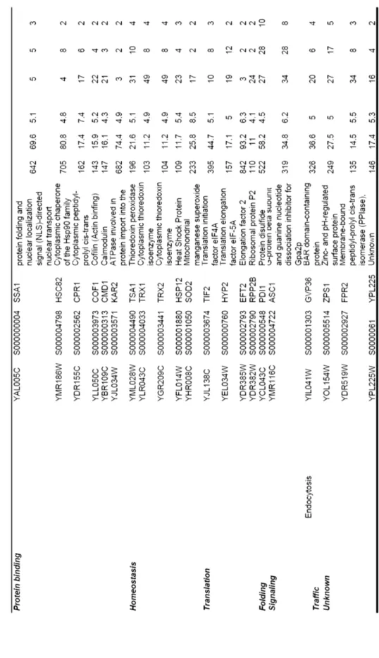
Of the 43 moonlighting proteins reported before 2005, 47% of them were previously known as glycolytic enzymes (Pancholi 2001, Sriram et al. 2005). It is known today that all glycolytic enzymes are moonlighting proteins (Table 1). The moonlighting functions of glycolytic enzymes are often related to important cellular machineries such as transcription, translation, signal transduction, cell movement, and trafficking.
Table 1 Glycolytic enzymes and examples of their moonlighting functions
Moonlighting glycolytic enzymes often need to change their location to perform their moonlighting functions. For example, enolase has at least seven moonlighting functions (Table 1) both inside and outside the cell. Extracellular enolase, which is a glycolytic enzyme, is a virulence factor in Candida albicans and some parasites (Jong et al. 2003, Avilan et al. 2011). Enolase has been found in small vesicles outside the cell (Oliveira et al. 2010, Oliveira et al. 2010) and in the cell
6
wall (Edwards et al. 1999). In addition, enolase is secreted in a sequence-dependent manner (Lopez-Villar et al. 2006, Yang et al. 2011) and is present in the cell wall with no enzymatic activity, but it binds to plasminogen and helps pathogens invade (Swenerton et al. 2011). Enolase is also found in viral particles (Bechtel et al. 2005, Chertova et al. 2006, Shaw et al. 2008) and is required for transcription in Sendai virus (Ogino et al. 2001). Therefore, enolase is a therapeutic target for many diseases, including candidiasis (van Deventer et al. 1996, Capello et al. 2011). Another extracellular glycolytic enzyme, phosphoglucose isomerase, enhances the motility of tumor cells (Dobashi et al. 2006) and acts like a cytokine (Torimura et al. 2001), although it possesses no enzymatic activity outside the cell (Tsutsumi et al. 2003). However, the secretory pathway of glycolytic enzymes such as enolase and phosphoglucose isomerase remains to be revealed. This pathway appears to be unconventional because glycolytic enzymes have no known secretion signals. Therefore, in this study, we analyze the secretory pathway of glycolytic enzymes.
Conventional secretion pathways of proteins have been extensively studied using Saccharomyces
cerevisiae (Schekman 2010). In addition, we may reveal unknown secretion pathways of proteins;
however, it remains challenging because the trafficking patterns inside the organism are not completely known.
Secretion pathways of S. cerevisiae
S. cerevisiae is a model organism for determining the secretion pathways of proteins and lipids
because it has known gene sets that work in various protein transport pathways. Schekman (Novick and Schekman 1979, Novick et al. 1980, Schekman 2010), Ohsumi (Nagatogawa et al. 2009, Mizushima et al, 2011), and numerous other researchers (Bryant and Stevens 1998, Hua et al. 2002, Gall et al. 2002) have developed various temperature-sensitive and/or knockout mutants of S.
cerevisiae to investigate protein transport pathways. The outline of S. cerevisiae secretion pathways
is shown in Fig. 4.
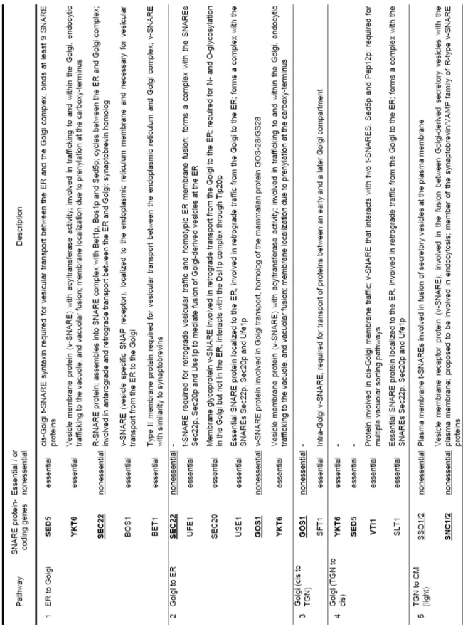
Protein transport mediated by membrane cargoes is regulated by various membrane-associated proteins or protein complexes (Whyte and Munro 2002, Bröcker et al. 2010). Among the proteins involved in transport machineries, soluble N-ethylmaleimide-sensitive factor attachment protein receptor (SNARE) proteins are the most extensively studied (Ungar and Hughson, 2003, Duman and Forte 2003, Jahn and Scheller 2006, Table 2). As shown in Table 2, 13 of 23 SNAREs are essential, and at least 9 proteins are used in more than two of the pathways described in Fig. 4. Of these, pathways 5 and 6 are considered conventional secretion pathways, while the others (17 and secretion via early endosome) are unconventional. The pathway used by unconventionally secreted moonlighting glycolytic enzymes remains unknown.
7
Fig. 4 Overview of protein secretion pathways in S. cerevisiae (modified figure from Brocker et al. 2010 and Muthusamy et al. 2009) N: Nucleus, ER: Endoplasmic reticulum, TGN: Trans-Golgi network, V: Vacuole, EE: Early endosome, LE: Late endosome, MVB: Multi vesicular vody, A: Autophagosome. See also Table 2 for detailed names of pathways numbered.
Spatial arrangement of glycolytic enzymes
Fluorescent protein tags have been used to determine the subcellular localization of proteins (Phillips 2001, Rudner and Losick 2010, Chudakov et al. 2010), especially in S. cerevisiae. Dr. Erin O'Shea and Dr. Jonathan Weissman at UCSF generated a collection of S. cerevisiae open reading frames that were tagged at the carboxy terminal using the coding sequence of Aequorea victoria GFP (S65T) (Huh et al. 2003). A database of GFP-fused protein localization (yeast GFP localization database, http://yeastgfp.yeastgenome.org/) is now available. In addition, many other databases for the subcellular localization of proteins are available (LOCATE, subcellular localization database for mouse and human, http://locate.imb.uq.edu.au/; eSLAB, a database of protein subcellular localization annotation for eukaryotic organisms, http://gpcr.biocomp.unibo.it/esldb/; Organelle DB, a database of organelle proteins and subcellular structures/complexes, http://organelledb.lsi.umich.edu/; locDB, collection of experimental annotations for the subcellular localization of proteins in human and weed, http://www.rostlab.org/services/locDB/). Apart from its property to accumulate in the nucleus to some extent (Seibel et al. 2007) and that its fluorescence intensity is affected by oxygen concentration (Yang et al. 1996, Takahashi et al. 2006), GFP is useful
8
and is one of the frequently used fluorescent proteins. Accumulation, aggregation, and association of proteins inside the cell often indicate some cellular machineries or protein functions (Kanda et al. 1998, Bence et al. 2001, Tilsner and Oparka 2010). Therefore, protein localization in response to certain stimuli has been extensively researched to discover novel cellular machineries (Sakai et al. 1997, Dastoor and Dreyer 2001, An et al. 2008, Narayanaswamy et al. 2009, Noree et al. 2010).
Spatial rearrangement of moonlighting proteins, including glycolytic enzymes, is highly important for their various functions. In several organisms and cells, some glycolytic enzymes have been reported to associate with the cytoskeleton (Masters 1984, Stephan et al. 1986), erythrocyte membrane (Campanella et al. 2005), or muscle (Brooks and Storey 1988), or to associate with each other (Mowbray and Moses 1976, Anderson et al. 1995, Mazzola and Sirover 2003). In a few species of protozoa, including Trypanosoma brucei, glycolytic enzymes are contained in a membrane-enclosed organelle called glycosome (Hannaert and Michels 1994, Bakker et al. 2000). Association of glycolytic enzymes is believed to facilitate metabolism (Beeckmans et al. 1990, Amar et al. 2008). In addition, changes in the localization of glycolytic enzymes suggest other moonlighting functions (Dastoor and Dreyer 2001, Decker and Wickner 2006).
The intracellular assembly of glycolytic enzymes has been observed in mammalian cells; one of the glycolytic enzymes, i.e., GAPDH conjugated with GFP, was found to form fluorescent foci under hypoxia (Agbor et al. 2011). Agbor and colleagues (2011) demonstrated that the spatial rearrangement was dependent on modification by small ubiquitin-like modifier (SUMOylation). However, its function and sensing machineries involved in the initiation of spatial reorganization of the glycolytic enzyme under hypoxia remain known. It is important to determine the location of the foci because GAPDH has been reported to translocate into the nucleus under hypoxia (Stannard et al. 2004). Moreover, according to the S. cerevisiae database (yeast GFP localization database, http://yeastgfp.yeastgenome.org/), subcellular localization of glycolytic enzymes fused with GFP (GFP clones, Invitrogen) is uniform in the cytoplasm. Therefore, it is uncertain whether glycolytic enzymes change their localization in response to hypoxia, especially in yeast cells. When the spatial rearrangement of glycolytic enzymes occurs under hypoxia, the relocalization of enzymes may affect cell physiology.
9 T able 2 SNARE pr ot eins inv ol ve d in secr etio n pa thwa y
10 T able 2 SNARE pr ot eins inv ol ve d in secr etio n pa thwa y (co n tinu ed) N : N uc leus , ER: En do plas m ic retic ul um , TG N : T rans -G olgi n etw or k, V : V acu ol e, EE : E arly e nd osom e, LE: La te en dos om e, MVB: M ul ti ve sicu lar bo dy , A: Autop ha go so m e, CM: Ce ll m em brane. Under l in e: un essent ial . B ol d: used in sev eral pat hway s
11 Cell physiology under hypoxia
Hypoxia is a condition in which the cell is deprived of adequate oxygen supply. Many studies define hypoxia at ≤2 mg/L of dissolved oxygen (DO) in an aqueous environment (Eby et al. 2002, Buzzelli et al. 2002). In cultured mammalian cells, 1% atmospheric oxygen is regarded to be a hypoxic state, while 21% is regarded to be the normal oxygen concentration (normoxia) (Hagen et al. 2003, Frezza et al. 2011). A hypoxic state for mammalian cells often occurs in vivo when the oxygen supply is limited (Denko 2008). Hypoxia is reported to correlate with many diseases including heart attack, cancer, and stroke (Lyer et al. 1998). Some tumor cells are known to response to hypoxia and obtain increased metastatic activity (Zhong et al. 1999), radiation resistance (Eyler and Rich 2008), and drug resistance (Teicher 1994). Baker’s yeast, S. cerevisiae is also well known to response to hypoxia during fermentation (Simeonidis et al. 2010). When sufficient amounts of nutrients are supplied, S. cerevisiae produces CO2 in metabolic processes, which decreases the oxygen concentration in the medium (Rosenfeld et al. 2003). The hypoxic responses of yeast cells have attracted attention because researchers have proved that these responses have some roles in infection by pathogenic fungi including C. albicans (Grahl and Cramer, 2010) and Aspergillus fumigatus (Grahl et al. 2011, Fller and Rhodes, 2012).
The hypoxic responses of mammalian cells and yeasts are common to some extent (Fig. 5). Because molecular oxygen is required for heme and sterol biosynthesis, the production of these molecules is reduced under hypoxia (Hickman et al. 2011, Siso et al. 2012). In addition, oxygen deprivation triggers the release of reactive oxygen species (ROS) from mitochondria (Chandel et al. 1998, Chandel et al. 2000, Blokhina et al. 2003, Guzy et al. 2005, Bell et al. 2007, Murphy 2009) by unknown mechanisms (Guzy and Schumacker 2006). These primary hypoxic responses trigger the following secondary responses. In mammalian cells, cytosolic ROS stabilize hypoxia-inducible factor 1α (HIF-1α) (Guzy and Schumacker 2006), which is a major regulator for the hypoxic response. ROS (Gillespie et al. 2009, Ruchko et al., 2009, Poyton et al. 2009, Gillespie et al. 2010), and HIF-1α (Ortiz-Barahona et al. 2010, Tanimoto et al. 2010, Schödel et al. 2011, Liu et al. 2012) oxidizes or binds several specific bases in hypoxia-responsive elements (HRE). Genes containing HRE in their promoters include those that encode aldolase, enolase, and lactate dehydrogenase (Semenza et al. 1996). In mammalian cells, transcription of PKM2, a gene that encodes one of the glycolytic enzymes, is activated by HIF-1α (Luo et al. 2011). Interestingly, Pkm2p interacts directly with the HIF-1α subunit and acts as a coactivator (Luo et al. 2011). In addition to HRE oxidization, mitochondria-generated ROS trigger AMP-activated protein kinase signaling (Jung et al. 2008, Emerling et al. 2009, Mungai et al. 2011, Kim et al. 2011) through several reaction steps (Mungai et al. 2011). Yeast cells have no HIF-1α homologs (Rytkönen and Storz 2011); however, yeast has HRE clusters in the promoters of TDH2, ALD6, and genes involved in amino acid metabolism (Ferreira et al. 2007). It is also reported that hexose transporters are affected under a hypoxic condition,
12
accelerating the uptake of extracellular hexoses (Rintala et al. 2008). Because of the resemblance of responses to hypoxia between S. cerevisiae and mammalian cells, some researchers regard S.
cerevisiae as one of the model organisms for studying a hypoxic response (Netzar and Breitenbach
2010). Mitochondrial ROS production can also be measured in yeast cells. Using dihydroethidium and MitoSOX Red, cellular and mitochondria-specific reactive oxygen can be measured in vivo (Quaranta et al. 2011). 2′, 7′-dichlorofluorescin diacetate can also be used as a cytosolic indicator of ROS (Gomes et al. 2005, Bonini et al. 2006, Al-Mehdi et al. 2012).
Hypoxic response mechanisms have been extensively studied by culturing yeast cells in a media depleted in oxygen by sparging with N2 (Kwast et al. 2002, Lai et al. 2008). Kwast and colleagues (2002) have shown that N2-induced hypoxia triggers global changes in metabolic gene induction. Interestingly, these two researches do not report enolase gene induction, in contrast to that in mammalian cells (Kwast et al. 2002, Lai et al. 2008). In S. cerevisiae, a decrease in heme and sterol levels induces the activation of transcription by Upc2p, while a decrease in only heme levels inhibits Rox1p and Mot3p to repress hypoxic genes, thus inducing hypoxia-responsive genes (Grahl and Cramer, 2010). The connection between sterol- and heme-regulated responses to hypoxia, and their correlation with mitochondrial ROS production have not been described.
Fig. 5 Outline of similar hypoxic responses in mammalian and yeast cells
In response to hypoxia, cultured mammalian cells produce large amounts of lactate, alanine (Brecht and Groot 1994, Chateil et al. 2001), and acetate. On the other hand, S. cerevisiae cells grown under hypoxic conditions are known to produce ethanol, glycerol, succinic acid, and alanine (Chico et al. 1978, Gleason et al. 2011) as end products of glycolysis. These changes in metabolites, especially overproduction of alanine under hypoxia, are also known in flies (Feala et al. 2007) and
13
plants (Rocha et al. 2010). In rat heart, aspartate production is significantly increased under hypoxia relative to normoxia (Rumsey et al. 1999). Alanine, aspartate, and other amino acids are considered to protect cells from hypoxic injury (Weinberg et al. 1990, Brecht and Groot 1994). In yeasts, conversion of pyruvate to oxaloacetate and aspartate is a part of gluconeogenesis, which enables yeast cells to grow on non-sugar carbon sources such as ethanol, glycerol, or peptone (Foy and Bhattacharjee 1977, Eschrich et al. 2002).
In case of gluconeogenesis in yeast, acetyl-CoA carboxylase produces malonyl-CoA under regulation by SNF1, which is a yeast functional homolog of mammalian AMP kinase (Woods et al. 1994). Malonyl-CoA is the first precursor of long fatty acids (Fig. 6). Without acetyl-CoA carboxylase, yeasts need fatty acids to survive and arrest the G2/M phase of the cell cycle (Al-Feel et al. 2003). Acetyl-CoA carboxylase is also vital in mammalian cells, as RNAi of acetyl-CoA carboxylase inhibits the growth of prostate cancer cells and mouse embryos (Abu-Elheiga et al. 2005, Brusselmans et al. 2005).
Fig. 6 Fatty acid synthesis from pyruvate in S. cerevisiae
There had been several reports demonstrating that cobalt and other metal ions induce cellular responses, which resemble the hypoxic response. However, reports also suggest that there are some differences between these two stimuli. For example, overproduction of alanine is observed under hypoxia but not in the presence of cobalt (Gleason et al. 2011). In addition, ROS generation by hypoxia and by CoCl2 addition is differently inhibited by adding mitochondria-inhibiting agents to mammalian cells (Chandel et al. 2000).
Although the role of the spatial relocalization of glycolytic enzymes under hypoxia is not revealed, there is a report suggesting the importance of spatial localization of cellular components. Recently, Al-Mehdi and colleagues (2012) have revealed that mitochondria localize near the nucleus
14
under hypoxia to translocate ROS into the nucleus and oxidize guanine nucleotides of specific promoter DNA sequences in order to induce the expression of the vascular endothelial growth factor-encoding gene (VFGF) (Murphy 2012). The results suggest that regulating intracellular localization of mitochondria has an important role in the hypoxic response. If glycolytic enzymes change their localization in the cell, the role this relocation plays in cellular metabolism and the mechanisms that regulate this translocation should be investigated.
Culture instruments for cultivation of yeast cells
Suitable culture vials are needed to study hypoxia in yeast cells. A novel culture device, which can separate and co-cultivate different types of cells at the same time, is needed to investigate unknown functions of unconventionally secreted proteins.
Because yeast cells experience a hypoxic environment during fermentation, vials for fermentation can be used to investigate their hypoxic responses. Glass vials have been developed by Matsumoto et al. (2002) for laboratory-scale fermentation. A small pump can be used to create normoxia (sufficient oxygen concentration) in these vials.
Several different culture vials, including Millicell culture inserts (Millipore, Germany), IdMOC (Kurabo, Osaka, Japan), and Alvetex (TaKaRa Bio, Otsu, Japan), have been developed for the investigation of intercellular proteins. Because these chambers developed are for mammalian cells, only a small volume of microbial cells can be cultured using these chambers. These chambers can be used for co-cultivation of different cells, but first, a model system to investigate the intercellular function of extracellular proteins should be constructed. Following which, large-scale culture vials for separated co-cultivation should be developed.
To determine the molecular machineries that enable proteins to perform moonlighting functions, investigating the mechanism regulating the localization of moonlighting proteins is a plausible approach. In this regard, the following questions arise:
Is there a specific amino acid sequence that participates in certain localizations? Through which transporting pathway is the change in localization achieved?
By which cellular mechanism or sensing pathway is the change in localization regulated? Does the change in localization correlate with cell physiology?
These issues were addressed in this study using the moonlighting glycolytic enzyme, enolase, as a model.
15 REFERENCES
1. Abu-Elheiga L, Matzuk MM, Kordari P, Oh W, Shaikenov T, Gu Z, Wakil SJ (2005) Mutant mice lacking acetyl-CoA carboxylase 1 are embryonically lethal. Proc. Natl. Acad. Sci. USA. 102:12011-12016.
2. Agbor TA, Cheong A, Comerford KM, Scholz CC, Bruning U, Clarke A, Cummins EP, Cagney G, Taylor CT (2011) Small ubiquitin-related modifier (SUMO)-1 promotes glycolysis in hypoxia. J. Biol. Chem. 286:4718-4726.
3. Al-Feel W, DeMar JC, Wakil SJ (2003) A Saccharomyces cerevisiae mutant strain defective in acetyl-CoA carboxylase arrests at the G2/M phase of the cell cycle. Proc. Natl. Acad. Sci. USA. 100:3095-3100.
4. Al-Mehdi AB, Pastukh VM, Swiger BM, Reed DJ, Patel MR, Bardwell GC, Pastukh VV, Alexeyev MF, Gillespie MN (2012) Perinuclear mitochondrial clustering creates an oxidant-rich nuclear domain required for hypoxia-induced transcription. Sci. Signal. 5:ra47. 5. Amar P, Legent G, Thellier M, Ripoll C, Bernot G, Nystrom T, Saier MH Jr, Norris V (2008)
A stochastic automaton shows how enzyme assemblies may contribute to metabolic efficiency. BMC Syst. Biol. 2:27.
6. An S, Kumar R, Sheets ED, Benkovic SJ (2008) Reversible compartmentalization of de novo purine biosynthetic complexes in living cells. Science. 320:103-106.
7. Anderson LE, Goldhaber-Gordon IM, Li D, Tang XY, Xiang M, Prakash N (1995) Enzyme-enzyme interaction in the chloroplast: glyceraldehyde-3-phosphate dehydrogenase, triose phosphate isomerase and aldolase. Planta 196:245-255.
8. Arnold H, Pette D (1968) Binding of glycolytic enzymes to structure proteins of the muscle. Eur. J. Biochem. 6:163–171.
9. Avilán L, Gualdrón-López M, Quiñones W, González-González L, Hannaert V, Michels PA, Concepción JL (2011) Enolase: a key player in the metabolism and a probable virulence factor of trypanosomatid parasites: perspectives for its use as a therapeutic target. Enzyme Res. 2011:932549.
10. Bakker BM, Mensonides FI, Teusink B, van Hoek P, Michels PA, Westerhoff HV (2000) Compartmentation protects trypanosomes from the dangerous design of glycolysis. Proc. Natl. Acad. Sci. USA. 97:2087-2092.
11. Bechtel JT, Winant RC, Ganem D (2005) Host and viral proteins in the virion of Kaposi’s sarcoma-associated herpesvirus. J. Virol. 79:4952-4964.
12. Beeckmans S, Van Driessche E, Kanarek L (1990) Clustering of sequential enzymes in the glycolytic pathway and the citric acid cycle. J. Cell Biochem. 43:297-306.
13. Bell EL, Klimova TA, Eisenbart J, Moraes CT, Murphy MP, Budinger GR, Chandel NS (2007) The Qo site of the mitochondrial complex III is required for the transduction of
16
hypoxic signaling via reactive oxygen species production. J. Cell Biol. 177:1029-1036. 14. Bence NF, Sampat RM, Kopito RR (2001) Impairment of the ubiquitin-proteasome system
by protein aggregation. Science. 292:1552-1555.
15. Berry MD, Boulton AA (2000) Glyceraldehyde-3-phosphate dehydrogenase and apoptosis. J. Neurosci. Res. 60:150–154.
16. Blokhina O, Virolainen E, Fagerstedt KV (2003) Antioxidants, oxidative damage and oxygen deprivation stress: a review. Ann. Bot. 91:179-194.
17. Bonini MG, Rota C, Tomasi A, Mason RP (2006) The oxidation of 2',7'-dichlorofluorescin to reactive oxygen species: a self-fulfilling prophesy? Free Radic. Biol. Med. 40:968-975. 18. Brecht M, Groot H (1994) Protection from hypoxic injury in cultured hepatocytes by glycine,
alanine, and serine. Amino Acids 6:25-35.
19. Bröcker C, Engelbrecht-Vandré S, Ungermann C (2010) Multisubunit tethering complexes and their role in membrane fusion. Curr. Biol. 20:R943-952.
20. Brooks SP, Storey KB (1988) Subcellular enzyme binding in glycolytic control: in vivo studies with fish muscle. Am. J. Physiol. 255:R289-94.
21. Brusselmans K, De Schrijver E, Verhoeven G, Swinnen JV (2005) RNA interference-mediated silencing of the acetyl-CoA-carboxylase-alpha gene induces growth inhibition and apoptosis of prostate cancer cells. Cancer Res. 65:6719-6725.
22. Bryant JA, Brice DC, Fitchett PN, Anderson LE (2000) A novel DNA-binding protein associated with DNA polymerase-alpha in pea stimulates polymerase activity on infrequently primed templates. J. Exp. Bot. 51:1945-1947.
23. Buzzelli CP, Luettich RA, Powers SP, Peterson CH, McNinch JE, Pinckney JL, Paerl HW (2002) Estimating the spatial extent of bottom-water hypoxia and habitat degradation in a shallow estuary. Mar. Eco. Prog. Seri. 230:103-112.
24. Campanella ME, Chu H, Low PS (2005) Assembly and regulation of a glycolytic enzyme complex on the human erythrocyte membrane. Proc. Natl. Acad. Sci. USA. 102:2402-2407. 25. Chandel NS, Maltepe E, Goldwasser E, Mathieu CE, Simon MC, Schumacker PT (1998)
Mitochondrial reactive oxygen species trigger hypoxia-induced transcription. Proc. Natl. Acad. Sci. USA. 95:11715-11720.
26. Chandel NS, McClintock DS, Feliciano CE, Wood TM, Melendez JA, Rodriguez AM, Schumacker PT (2000) Reactive oxygen species generated at mitochondrial complex III stabilize hypoxia-inducible factor-1alpha during hypoxia: a mechanism of O2 sensing. J. Biol. Chem. 275:25130-25138.
27. Chaput M, Claes V, Portetelle D, Cludts I, Cravador A, Burny A, Gras H, Tartar A (1988) The neurotrophic factor neuroleukin is 90% homologous with phosphohexose isomerase. Nature 332:454–455.
17
28. Chateil J, Biran M, Thiaudière E, Canioni P, Merle M (2001) Metabolism of [1-13C]glucose and [2-13C]acetate in the hypoxic rat brain. Neurochem. Int. 38:399-407.
29. Chertova E, Chertov O, Coren LV, Roser JD, Trubey CM, Bess JW Jr, Sowder RC 2nd, Barsov E, Hood BL, Fisher RJ, Nagashima K, Conrads TP, Veenstra TD, Lifson JD, Ott DE (2006) Proteomic and biochemical analysis of purified human immunodeficiency virus type 1 produced from infected monocyte-derived macrophages. J. Virol. 80:9039 -9052.
30. Chico E, Olavarría JS, Núnez de Castro I (1978) L-Alanine as an end product of glycolysis in
Saccharomyces cerevisiae growing under different hypoxic conditions. Antonie. Van.
Leeuwenhoek 44:193-201.
31. Chopade BA, Shankar S, Sundin GW, Mukhopadhyay S, Chakrabarty AM (1997) Characterization of membrane-associated Pseudomonas aeruginosa Ras-like protein Pra, a GTP-binding protein that forms complexes with truncated nucleoside diphosphate kinase and pyruvate kinase to modulate GTP synthesis. J. Bacteriol. 179:2181-2188.
32. Chudakov DM, Matz MV, Lukyanov S, Lukyanov KA (2010) Fluorescent proteins and their applications in imaging living cells and tissues. Physiol. Rev. 90:1103-1163.
33. Commichau FM, Rothe FM, Herzberg C, Wagner E, Hellwig D, Lehnik-Habrink M, Hammer E, Völker U, Stülke J (2009) Mol. Cell. Proteomics 8:1350-1360.
34. Dastoor Z, Dreyer JL (2001) Potential role of nuclear translocation of glyceraldehyde-3-phosphate dehydrogenase in apoptosis and oxidative stress. J. Cell Sci. 114:1643-1653.
35. Decker BL, Wickner WT (2006) Enolase activates homotypic vacuole fusion and protein transport to the vacuole in yeast. J. Biol. Chem. 281:14523-14528.
36. Denko NC (2008) Hypoxia, HIF1 and glucose metabolism in the solid tumor. Nat. Rev. Cancer 8:705-713.
37. Dobashi Y, Watanabe H, Matsubara M, Yanagawa T, Raz A, Shimamiya T, Ooi A (2006) Autocrine motility factor/glucose-6-phosphate isomerase is a possible predictor of metastasis in bone and soft tissue tumours. J. Pathol. 208:44 -53.
38. Dunker AK, Lawson JD, Brown CJ, Williams RM, Romero P, Oh JS, Oldfield CJ, Campen AM, Ratliff CM, Hipps KW, Ausio J, Nissen MS, Reeves R, Kang C, Kissinger CR, Bailey RW, Griswold MD, Chiu W, Garner EC, Obradovic Z (2001) Intrinsically disordered protein. J. Mol. Graph. Model. 19:26-59.
39. Duman JG, Forte JG (2003) What is the role of SNARE proteins in membrane fusion? Am. J. Physiol. Cell Physiol. 285:C237-249.
40. Eby LA, Crowder LB (2002) Hypoxia-based habitat compression in the Neuse River Estuary: context-dependent shifts in behavioral avoidance thresholds. Can. J. Fish. Aquat. Sci. 59:952-965.
18
41. Edwards SR, Braley R, Chaffin WL (1999) Enolase is present in the cell wall of
Saccharomyces cerevisiae. FEMS Microbiol. Lett. 177:211-216.
42. Emerling BM, Weinberg F, Snyder C, Burgess Z, Mutlu GM, Viollet B, Budinger GR, Chandel NS (2009) Hypoxic activation of AMPK is dependent on mitochondrial ROS but independent of an increase in AMP/ATP ratio. Free Radic. Biol. Med. 46:1386-1391.
43. Entelis N, Brandia I, Kamenski P, Krasheninnikov IA, Martin RP, Tarassov I (2006) A glycolytic enzyme, enolase, is recruited as a cofactor of tRNA targeting toward mitochondria in Saccharomyces cerevisiae. Genes Dev. 20:1609-1620.
44. Eschrich D, Kötter P, Entian KD (2002) Gluconeogenesis in Candida albicans. FEMS Yeast Res. 2:315-25.
45. Eyler CE, Rich JN (2008) Survival of the fittest: cancer stem cells in therapeutic resistance and angiogenesis. J. Clin. Oncol. 26:2839-2845.
46. Faik P, Walker JI, Redmill AA, Morgan MJ (1988) Mouse glucose-6-phosphate isomerase and neuroleukin have identical 3’ sequences. Nature 332:455–457.
47. Feala JD, Coquin L, McCulloch AD, Paternostro G (2007) Flexibility in energy metabolism supports hypoxia tolerance in Drosophila flight muscle: metabolomic and computational systems analysis. Mol. Syst. Biol. 3:99.
48. Ferreira TC, Hertzberg L, Gassmann M, Campos EG (2007) The yeast genome may harbor hypoxia response elements (HRE). Comp. Biochem. Physiol. C. Toxicol. Pharmacol. 46:255-263.
49. Fiek C, Benz R, Roos N, Brdiczka D (1982) Evidence for identity between the hexokinase-binding protein and the mitochondrial porin in the outer membrane of rat liver mitochondria. Biochim. Biophys. Acta. 688:429–440.
50. Foy JJ, Bhattacharjee JK (1977) Gluconeogenesis in Saccharomyces cerevisiae: determination of fructose-1,6-bisphosphatase activity in cells grown in the presence of glycolytic carbon sources. J. Bacteriol. 129:978-982.
51. Frezza C, Zheng L, Tennant DA, Papkovsky DB, Hedley BA, Kalna G, Watson DG, Gottlieb E (2011) Metabolic profiling of hypoxic cells revealed a catabolic signature required for cell survival. PLoS ONE 6:e24411.
52. Fuller KK, Rhodes JC (2012) Protein kinase A and fungal virulence: a sinister side to a conserved nutrient sensing pathway. Virulence 3:109-121.
53. Furuya H, Ikeda R (2009) Interaction of triosephosphate isomerase from the cell surface of Staphylococcus aureus and alpha-(1->3)-mannooligosaccharides derived from glucuronoxylomannan of Cryptococcus neoformans. Microbiology 155:2707–2713.
54. Gillespie MN, Pastukh V, Ruchko MV (2009) Oxidative DNA modifications in hypoxic signaling. Ann. N Y Acad. Sci. 1177:140-150.
19
55. Gillespie MN, Pastukh VM, Ruchko MV (2010) Controlled DNA "damage" and repair in hypoxic signaling. Respir. Physiol. Neurobiol. 174:244-251.
56. Gleason JE, Corrigan DJ, Cox JE, Reddi AR, McGinnis LA, Culotta VC (2011) Analysis of hypoxia and hypoxia-like states through metabolite profiling. PLoS ONE 6:e24741.
57. Gancedo C, Flores CL (2008) Moonlighting proteins in yeasts. Microbiol. Mol. Biol. Rev. 72:197-210.
58. Gao X, Wang H, Yang JJ, Liu X, Liu ZR (2012) Pyruvate kinase M2 regulates gene transcription by acting as a protein kinase. Mol. Cell 45:598-609.
59. Glass JI, Assad-Garcia N, Alperovich N, Yooseph S, Lewis MR, Maruf M, Hutchison CA 3rd, Smith HO, Venter JC (2006) Essential genes of a minimal bacterium. Proc. Natl. Acad. Sci. USA. 10:425-430.
60. Gomes A, Fernandes E, Lima JL (2005) Fluorescence probes used for detection of reactive oxygen species. J. Biochem. Biophys. Methods 65:45-80.
61. Grahl N, Cramer RA Jr. (2010) Regulation of hypoxia adaptation: an overlooked virulence attribute of pathogenic fungi? Med. Mycol. 48:1-15.
62. Grahl N, Puttikamonkul S, Macdonald JM, Gamcsik MP, Ngo LY, Hohl TM, Cramer RA (2011) In vivo hypoxia and a fungal alcohol dehydrogenase influence the pathogenesis of invasive pulmonary aspergillosis. PLoS Pathog. 7:e1002145.
63. Graw J (2009) Genetics of crystallins: cataract and beyond. Exp. Eye Res. 88:173-189. 64. Gurney ME, Heinrich SP, Lee MR, Yin HS (1986) Molecular cloning and expression of
neuroleukin, a neurotrophic factor for spinal and sensory neurons. Science 234:566–574. 65. Guzy RD, Hoyos B, Robin E, Chen H, Liu L, Mansfield KD, Simon MC, Hammerling U,
Schumacker PT (2005) Mitochondrial complex III is required for hypoxia-induced ROS production and cellular oxygen sensing. Cell Metab.1:401-408.
66. Guzy RD, Schumacker PT (2006) Oxygen sensing by mitochondria at complex III: the paradox of increased reactive oxygen species during hypoxia. Exp. Physiol. 91:807-819. 67. Hagen T, Taylor CT, Lam F, Moncada S (2003) Redistribution of intracellular oxygen in
hypoxia by nitric oxide: effect on HIF1alpha. Science 302:1975-1978.
68. Hannaert V, Michels PA (1994) Structure, function, and biogenesis of glycosomes in kinetoplastida. J. Bioenerg. Biomembr. 26:205-212.
69. Hernández S, Amela I, Cedano J, Piñol J, Perez-Pons J, Mozo-Villarias AM, Querol E (2012) Do moonlighting proteins belong to the intrinsically disordered protein class? J. Proteomics Bioinform. 5:11.
70. Hickman MJ, Spatt D, Winston F (2011) The Hog1 mitogen-activated protein kinase mediates a hypoxic response in Saccharomyces cerevisiae. Genetics 188:325-338.
20
multitasking. Biochim. Biophys. Acta. 1803:520-525.
72. Huh WK, Falvo JV, Gerke LC, Carroll AS, Howson RW, Weissman JS, O'Shea EK (2003) Global analysis of protein localization in budding yeast. Nature 425:686-691.
73. Iida H, Yahara I (1985) Yeast heat-shock protein of Mr 48,000 is an isoprotein of enolase. Nature 315:688-690.
74. Iyer NV, Kotch LE, Agani F, Leung SW, Laughner E, Wenger RH, Gassmann M, Gearhart JD, Lawler AM, Yu AY, Semenza GL (1998) Cellular and developmental control of O2 homeostasis by hypoxia-inducible factor 1 alpha. Genes Dev. 12:149-162.
75. Jahn R, Scheller RH (2006) SNAREs-engines for membrane fusion. Nat. Rev. Mol. Cell Biol. 7:631-663.
76. Jeffery CJ (1999) Moonlighting proteins. Trends Biochem. Sci. 24:8-11.
77. Jeganathan S, von Bergen M, Mandelkow EM, Mandelkow E (2008) The natively unfolded character of tau and its aggregation to Alzheimer-like paired helical filaments. Biochemistry 47:10526-10539.
78. Jensen RA (1976) Enzyme recruitment in evolution of new function. Annu. Rev. Microbiol. 30:409-425.
79. Jewett TJ, Sibley LD (2003) Aldolase forms a bridge between cell surface adhesins and the actin cytoskeleton in apicomplexan parasites. Mol. Cell 11:885-894.
80. Jung SN, Yang WK, Kim J, Kim HS, Kim EJ, Yun H, Park H, Kim SS, Choe W, Kang I, Ha J (2008) Reactive oxygen species stabilize hypoxia-inducible factor-1 alpha protein and stimulate transcriptional activity via AMP-activated protein kinase in DU145 human prostate cancer cells. Carcinogenesis 29:713-721.
81. Kanda T, Sullivan KF, Wahl GM (1998) Histone-GFP fusion protein enables sensitive analysis of chromosome dynamics in living mammalian cells. Curr. Biol. 8:377-385.
82. Katakura Y, Sano R, Hashimoto T, Ninomiya K, Shioya S (2010) Lactic acid bacteria display on the cell surface cytosolic proteins that recognize yeast mannan. Appl. Microbiol. Biotechnol. 86:319-326.
83. Kato H, Fukuda T, Parkison C, McPhie P, Cheng SY (1989) Cytosolic thyroid hormone-binding protein is a monomer of pyruvate kinase. Proc. Natl. Acad. Sci. USA 86:7861-7865.
84. Kim IS, Sohn HY, Jin I (2011) Adaptive stress response to menadione-induced oxidative stress in Saccharomyces cerevisiae KNU5377. J. Microbiol. 49:816-823.
85. Kolarova M, García-Sierra F, Bartos A, Ricny J, Ripova D (2012) Structure and pathology of tau protein in Alzheimer disease. Int. J. Alzheimers Dis. 2012:731526.
86. Kumagai H, Sakai H (1983) A porcine brain protein (35 K protein) which bundles microtubules and its identification as glyceraldehyde 3-phosphate dehydrogenase. J. Biochem.
21 93:1259–1269.
87. Kwast KE, Lai LC, Menda N, James DT 3rd, Aref S, Burke PV (2002) Genomic analyses of anaerobically induced genes in Saccharomyces cerevisiae: functional roles of Rox1 and other factors in mediating the anoxic response. J. Bacteriol. 184:250-265.
88. Lai LC, Kissinger MT, Burke PV, Kwast KE (2008) Comparison of the transcriptomic "stress response" evoked by antimycin A and oxygen deprivation in Saccharomyces cerevisiae. BMC Genomics 9:627.
89. Lay AJ, Jiang XM, Kisker O, Flynn E, Underwood A, Condron R, Hogg PJ (2000) Phosphoglycerate kinase acts in tumour angiogenesis as a disulphide reductase. Nature 408:869-873.
90. Lee H, Guo Y, Ohta M, Xiong L, Stevenson B, Zhu JK (2002) LOS2, a genetic locus required for cold-responsive gene transcription encodes a bi-functional enolase. EMBO J. 21:2692-2702.
91. Linden M, Gellerfors P, Nelson BD (1982) Pore protein and the hexokinase-binding protein from the outer membrane of rat liver mitochondria are identical. FEBS Lett. 141:189–192. 92. Liu W, Shen SM, Zhao XY, Chen GQ (2012) Targeted genes and interacting proteins of
hypoxia inducible factor-1. Int. J. Biochem. Mol. Biol. 3:165-178.
93. López-Villar E, Monteoliva L, Larsen MR, Sachon E, Shabaz M, Pardo M, Pla J, Gil C, Roepstorff P, Nombela C (2006) Genetic and proteomic evidences support the localization of yeast enolase in the cell surface. Proteomics 6:S107-S118.
94. Lu M, Holliday LS, Zhang L, Dunn WA Jr, Gluck SL (2001) Interaction between aldolase and vacuolar H+-ATPase: evidence for direct coupling of glycolysis to the ATP-hydrolyzing proton pump. J. Biol. Chem. 276:30407–30413.
95. Lu M, Sautin YY, Holliday LS, Gluck SL (2004) The glycolytic enzyme aldolase mediates assembly, expression, and activity of vacuolar H+-ATPase. J. Biol. Chem. 279:8732–8739. 96. Luo W, Hu H, Chang R, Zhong J, Knabel M, O'Meally R, Cole RN, Pandey A, Semenza GL
(2011) Pyruvate kinase M2 is a PHD3-stimulated coactivator for hypoxia-inducible factor 1. Cell 145:732-744.
97. Majewski N, Nogueira V, Bhaskar P, Coy PE, Skeen JE, Gottlob K, Chandel NS, Thompson CB, Robey RB, Hay N (2004) Hexokinase-mitochondria interaction mediated by Akt is required to inhibit apoptosis in the presence or absence of Bax and Bak. Mol. Cell. 16:819-830.
98. Margittai M, Langen R (2006) Side chain-dependent stacking modulates tau filament structure. J. Biol. Chem. 281:37820-37827.
99. Masters C (1984) Interactions between glycolytic enzymes and components of the cytomatrix. J. Cell. Biol. 99:222s-225s.
22
100. Matsumoto T, Fukuda H, Ueda M, Tanaka A, Kondo A (2002) Construction of yeast strains with high cell surface lipase activity by using novel display systems based on the Flo1p flocculation functional domain. Appl. Environ. Microbiol. 68:4517-4522.
101. Mazzola JL, Sirover MA (2003) Subcellular localization of human glyceraldehyde-3-phosphate dehydrogenase is independent of its glycolytic function. Biochim. Biophys. Acta. 1622:50-56.
102. Meyer-Siegler K, Mauro DJ, Seal G, Wurzer J, deRiel JK, Sirover MA (1991) A human nuclear uracil DNA glycosylase is the 37-kDa subunit of glyceraldehyde-3-phosphate dehydrogenase. Proc. Natl. Acad. Sci. USA 88:8460–8464.
103. Miki K, Qu W, Goulding EH, Willis WD, Bunch DO, Strader LF, Perreault SD, Eddy EM, O’Brien DA (2004) Glyceraldehyde 3-phosphate dehydrogenase-S, a sperm-specific glycolytic enzyme, is required for sperm motility and male fertility. Proc. Natl. Acad. Sci. USA 101:16501–16506.
104. Miles LA, Dahlberg CM, Plescia J, Felez J, Kato K, Plow EF (1991) Role of cell-surface lysines in plasminogen binding to cells: identification of alpha-enolase as a candidate plasminogen receptor. Biochemistry 30:1682-1691.
105. Mizushima N, Yoshimori T, Ohsumi Y (2011) The role of Atg proteins in autophagosome formation. Annu. Rev. Cell Dev. Biol. 27:107-132.
106. Mowbray J, Moses V (1976) The tentative identification in Escherichia coli of a multienzyme complex with glycolytic activity. Eur. J. Biochem. 66:25-36.
107. Mungai PT, Waypa GB, Jairaman A, Prakriya M, Dokic D, Ball MK, Schumacker PT (2011) Hypoxia triggers AMPK activation through reactive oxygen species-mediated activation of calcium release-activated calcium channels. Mol. Cell Biol. 31:3531-3545.
108. Murphy MP (2009) How mitochondria produce reactive oxygen species. Biochem. J. 417:1-13.
109. Murphy MP (2012) Modulating mitochondrial intracellular location as a redox signal. Sci. Signal. 5:pe39.
110. Nagy E, Rigby WF (1995) Glyceraldehyde-3-phosphate dehydrogenase selectively binds AU-rich RNA in the NAD(+)-binding region (Rossmann fold). J. Biol. Chem. 270:2755-2763.
111. Nakatogawa H, Suzuki K, Kamada Y, Ohsumi Y (2009) Dynamics and diversity in autophagy mechanisms: lessons from yeast. Nat. Rev. Mol. Cell Biol. 10:458-467.
112. Narayanaswamy R, Levy M, Tsechansky M, Stovall GM, O'Connell JD, Mirrielees J, Ellington AD, Marcotte EM (2009) Widespread reorganization of metabolic enzymes into reversible assemblies upon nutrient starvation. Proc. Natl. Acad. Sci. USA. 106:10147-10152.
23
113. Netzer NC, Breitenbach M (2010) Metabolic changes through hypoxia in humans and in yeast as a comparable cell model. Sleep Breath 14:221-225.
114. Noree C, Sato BK, Broyer RM, Wilhelm JE (2010) Identification of novel filament-forming proteins in Saccharomyces cerevisiae and Drosophila melanogaster. J. Cell Biol. 190:541-551.
115. Novick P, Schekman R (1979) Secretion and cell-surface growth are blocked in a temperature-sensitive mutant of Saccharomyces cerevisiae. Proc. Natl. Acad. Sci. USA. 76:1858-1862.
116. Novick P, Field C, Schekman R (1980) Identification of 23 complementation groups required for post-translational events in the yeast secretory pathway. Cell 21:205-215.
117. Ogino T, Yamadera T, Nonaka T, Imajoh-Ohmi S, Mizumoto K (2001) Enolase, a cellular glycolytic enzyme, is required for efficient transcription of Sendai virus genome. Biochem. Biophys. Res. Commun. 285:447- 455.
118. Oliveira DL, Freire-de-Lima CG, Nosanchuk JD, Casadevall A, Rodrigues ML, Nimrichter L (2010) Extracellular vesicles from Cryptococcus neoformans modulate macrophage functions. Infect. Immun. 78:1601-1609.
119. Oliveira DL, Nakayasu ES, Joffe LS, Guimarães AJ, Sobreira TJ, Nosanchuk JD, Cordero RJ, Frases S, Casadevall A, Almeida IC, Nimrichter L, Rodrigues ML (2010) Characterization of yeast extracellular vesicles: evidence for the participation of different pathways of cellular traffic in vesicle biogenesis. PLoS ONE 6:e11113.
120. Ortiz-Barahona A, Villar D, Pescador N, Amigo J, del Peso L (2010) Genome-wide identification of hypoxia-inducible factor binding sites and target genes by a probabilistic model integrating transcription-profiling data and in silico binding site prediction. Nucleic Acids Res. 238:2332-2345.
121. Pancholi V (2001) Multifunctional -enolase: its role in diseases. Cell. Mol. Life Sci. 58:902-920.
122. Parsot C, Saint-Girons I, Cohen GN (1987) Enzyme specialization during the evolution of amino acid biosynthetic pathways. Microbiol. Sci. 4: 260-262.
123. Phillips GJ (2001) Green fluorescent protein--a bright idea for the study of bacterial protein localization. FEMS Microbiol. Lett. 204:9-18.
124. Piatigorsky J, Wistow GJ (1989) Enzyme/crystallins: gene sharing as an evolutionary strategy. Cell 57:197-199.
125. Piatigorsky J (1998) Gene sharing in lens and cornea: facts and implications. Prog. Retin. Eye Res. 17:145-74.
126. Piatigorsky (2003) Crystallin genes: specialization by changes in gene regulation may precede gene duplication. J. Struct. Funct. Genomics 3:131-137.
24
127. Poltermann S, Kunert A, von der Heide M, Eck R, Hartmann A, Zipfel PF (2007) Gpm1p is a factor H-, FHL-1-, and plasminogen-binding surface protein of Candida albicans. J. Biol. Chem. 282:37537-37544.
128. Poyton RO, Ball KA, Castello PR (2009) Mitochondrial generation of free radicals and hypoxic signaling. Trends. Endocrinol. Metab. 20:332-340.
129. Quaranta D, Krans T, Espírito Santo C, Elowsky CG, Domaille DW, Chang CJ, Grass G (2011) Mechanisms of contact-mediated killing of yeast cells on dry metallic copper surfaces. Appl. Environ. Microbiol. 77:416-426.
130. Rintala E, Wiebe MG, Tamminen A, Ruohonen L, Penttilä M (2008) Transcription of hexose transporters of Saccharomyces cerevisiae is affected by change in oxygen provision. BMC Microbiol. 8:53.
131. Robbins AR, Ward RD, Oliver C (1995) A mutation in glyceraldehyde 3-phosphate dehydrogenase alters endocytosis in CHO cells. J. Cell Biol. 130:1093–1104.
132. Rocha M, Licausi F, Araújo WL, Nunes-Nesi A, Sodek L, Fernie AR, van Dongen JT (2010) Glycolysis and the tricarboxylic acid cycle are linked by alanine aminotransferase during hypoxia induced by waterlogging of Lotus japonicus. Plant Physiol. 152:1501-1513.
133. Rodríguez Plaza JG, Villalón Rojas A, Herrera S, Garza-Ramos G, Torres Larios A, Amero C, Zarraga Granados G, Gutiérrez Aguilar M, Lara Ortiz MT, Polanco Gonzalez C, Uribe Carvajal S, Coria R, Peña Díaz A, Bredesen DE, Castro-Obregon S, Del Rio G (2012) Moonlighting peptides with emerging function. PLoS ONE 7: e40125.
134. Rosenfeld E, Beauvoit B, Blondin B, Salmon JM (2003) Oxygen consumption by anaerobic
Saccharomyces cerevisiae under enological conditions: effect on fermentation kinetics. Appl.
Environ. Microbiol. 69:113-121.
135. Roux CM, DeMuth JP, Dunman PM (2011) Characterization of components of the Staphylococcus aureus mRNA degradosome holoenzyme-like complex. J. Bacteriol. 193:5520-5526.
136. Rudner DZ, Losick R (2010) Protein Subcellular Localization in Bacteria. Cold Spring Harb. Perspect. Biol. 2010. 2:a000307.
137. Ruchko MV, Gorodnya OM, Pastukh VM, Swiger BM, Middleton NS, Wilson GL, Gillespie MN (2009) Hypoxia-induced oxidative base modifications in the VEGF hypoxia-response element are associated with transcriptionally active nucleosomes. Free Radic. Biol. Med. 46:352-359.
138. Rumsey WL, Abbott B, Bertelsen D, Mallamaci M, Hagan K, Nelson D, Erecinska M (1999) Adaptation to hypoxia alters energy metabolism in rat heart. Am. J. Physiol. 276:H71-80. 139. Rytkönen KT, Storz JF (2011) Evolutionary origins of oxygen sensing in animals. EMBO
25
140. Sakai N, Sasaki K, Ikegaki N, Shirai Y, Ono Y, Saito N (1997) Direct visualization of the translocation of the gamma-subspecies of protein kinase C in living cells using fusion proteins with green fluorescent protein. J. Cell Biol. 139:1465-1476.
141. Schekman R (2010) Charting the secretory pathway in a simple eukaryote. Mol. Biol. Cell 21:3781-3784.
142. Schödel J, Oikonomopoulos S, Ragoussis J, Pugh CW, Ratcliffe PJ, Mole DR (2011) High-resolution genome-wide mapping of HIF-binding sites by ChIP-seq. Blood 117:e207-217.
143. Seibel NM, Eljouni J, Nalaskowski MM, Hampe W (2007) Nuclear localization of enhanced green fluorescent protein homomultimers. Anal. Biochem. 368:95-99.
144. Semenza GL, Jiang BH, Leung SW, Passantino R, Concordet JP, Maire P, Giallongo A (1996) Hypoxia response elements in the aldolase A, enolase 1, and lactate dehydrogenase A gene promoters contain essential binding sites for hypoxia-inducible factor 1. J. Biol. Chem. 271:32529-32537.
145. Shaw ML, Stone KL, Colangelo CM, Gulcicek EE, Palese P (2008) Cellular proteins in influenza virus particles. PLoS Pathog. 6:e1000085.
146. Simeonidis E, Murabito E, Smallbone K, Westerhoff HV (2010) Why does yeast ferment? A flux balance analysis study. Biochem. Soc. Trans. 38:1225-1229.
147. Sinthuvanich C, Veiga AS, Gupta K, Gaspar D, Blumenthal R, Schneider JP (2012) Anticancer β-hairpin peptides: membrane-induced folding triggers activity. J. Am. Chem. Soc. 134:6210-6217.
148. Sriram G, Martinez JA, McCabe ER, Liao JC, Dipple KM (2005) Single-gene disorders: what role could moonlighting enzymes play? Am. J. Hum. Genet. 76:911-924.
149. Stannard C, Brown LR, Godovac-Zimmermann J (2004) New Paradigms in Cellular Function and the Need for Top-Down Proteomics. Curr. Proteomics 1:13-25.
150. Stephan P, Clarke F, Morton D (1986) The indirect binding of triose-phosphate isomerase to myofibrils to form a glycolytic enzyme mini-complex. Biochim. Biophys. Acta. 873:127-135. 151. Sugase K, Dyson HJ, Wright PE (2007) Mechanism of coupled folding and binding of an
intrinsically disordered protein. Nature 447:1021-1025.
152. Swenerton RK, Zhang S, Sajid M, Medzihradszky KF, Craik CS, Kelly BL, McKerrow JH (2011) The oligopeptidase B of Leishmania regulates parasite enolase and immune evasion. J. Biol. Chem. 286:429–440.
153. Takahashi E, Takano T, Nomura Y, Okano S, Nakajima O, Sato M (2006) In vivo oxygen imaging using green fluorescent protein. Am. J. Physiol. Cell Physiol. 291:C781-787.
154. Tanimoto K, Tsuchihara K, Kanai A, Arauchi T, Esumi H, Suzuki Y, Sugano S (2010) Genome-wide identification and annotation of HIF-1α binding sites in two cell lines using
26 massively parallel sequencing. Hugo J. 4:35-48.
155. Tatton WG, Chalmers-Redman RM, Elstner M, Leesch W, Jagodzinski FB, Stupak DP, Sugrue MM, Tatton NA (2000) Glyceraldehyde-3-phosphate dehydrogenase in neurodegeneration and apoptosis signaling. J. Neural Transm. Suppl:77–100.
156. Teicher BA (1994) Hypoxia and drug resistance. Cancer Metastasis Rev. 13:139-168.
157. Thiele I, Jamshidi N, Fleming RM, Palsson BØ (2009) Genome-scale reconstruction of
Escherichia coli's transcriptional and translational machinery: a knowledge base, its
mathematical formulation, and its functional characterization. PLoS Comput. Biol. 5:e1000312.
158. Tilsner J, Oparka KJ (2010) Tracking the green invaders: advances in imaging virus infection in plants. Biochem. J. 430:21-37.
159. Tompa P, Szasz C, Buday L (2005) Structural disorder throws new light on moonlighting. Trends Biochem. Sci. 30:484-489.
160. Torimura T, Ueno T, Kin M, Harada R, Nakamura T, Kawaguchi T, Harada M, Kumashiro R, Watanabe H, Avraham R, Sata M (2001) Autocrine motility factor enhances hepatoma cell invasion across the basement membrane through activation of beta1 integrins. Hepatology 34:62-71.
161. True JR, Carroll SB (2002) Gene co-option in physiological and morphological evolution. Annu. Rev. Cell Dev. Biol. 18:53-80.
162. Tsutsumi S, Gupta SK, Hogan V, Tanaka N, Nakamura KT, Nabi IR, Raz A (2003) The enzymatic activity of phosphoglucose isomerase is not required for its cytokine function. FEBS Lett. 534:49 -53.
163. Ungar D, Hughson FM (2003) SNARE protein structure and function. Annu. Rev. Cell Dev. Biol. 19:493-517.
164. Walsh JL, Keith TJ, Knull HR (1989) Glycolytic enzyme interactions with tubulin and microtubules. Biochim. Biophys. Acta. 999:64-70.
165. Wang J, Morris AJ, Tolan DR, Pagliaro L (1996) The molecular nature of the F-actin binding activity of aldolase revealed with site-directed mutants. J. Biol. Chem. 271:6861-6865. 166. Watanabe H, Takehana K, Date M, Shinozaki T, Raz A (1996) Tumor cell autocrine motility
factor is the neuroleukin/phosphohexose isomerase polypeptide. Cancer Res. 56:2960–2963. 167. Weinberg JM, Venkatachalam MA, Garzo-Quintero R, Roeser NF, Davis JA (1990)
Structural requirements for protection by small amino acids against hypoxic injury in kidney proximal tubules. FASEB J. 4:3347-3354.
168. Whyte JR, Munro S (2002) Vesicle tethering complexes in membrane traffic. J. Cell Sci. 115:2627-2637.
27 236:1554-1556.
170. Wistow GJ, Lietman T, Williams LA, Stapel SO, de Jong WW, Horwitz J, Piatigorsky J (1988) Tau-crystallin/alpha-enolase: one gene encodes both an enzyme and a lens structural protein. J. Cell Biol. 107:2729-36.
171. Woods A, Munday MR, Scott J, Yang X, Carlson M, Carling D (1994) Yeast SNF1 is functionally related to mammalian AMP-activated protein kinase and regulates acetyl-CoA carboxylase in vivo. J. Biol. Chem. 269:19509-19515.
172. Xu W, Seiter K, Feldman E, Ahmed T, Chiao JW (1996) The differentiation and maturation mediator for human myeloid leukemia cells shares homology with neuroleukin or phosphoglucose isomerase. Blood 87:4502–4506.
173. Yang CK, Ewis HE, Zhang X, Lu CD, Hu HJ, Pan Y, Abdelal AT, Tai PC (2011) Nonclassical protein secretion by Bacillus subtilis in the stationary phase is not due to cell lysis. J. Bacteriol. 193:5607-5615.
174. Yang F, Moss LG, Phillips GN Jr (1996) The molecular structure of green fluorescent protein. Nat. Biotechnol. 14:1246-1251.
175. Zheng L, Roeder RG, Luo Y (2003) S phase activation of the histone H2B promoter by OCA-S, a coactivator complex that contains GAPDH as a key component. Cell 114:255-266. 176. Zhong H, De Marzo AM, Laughner E, Lim M, Hilton DA, Zagzag D, Buechler P, Isaacs WB,
Semenza GL, Simons JW (1999) Overexpression of hypoxia-inducible factor 1alpha in common human cancers and their metastases. Cancer Res. 59:5830-5835.
28 CHAPTER I
Searching for secretory pathway of enolase and discovery of enolase foci-forming region Introduction
Glycolytic enzymes play various roles inside and outside the cell (Tristan et al. 2011). Although they are cytosolic proteins, numerous large-scale analyses have revealed their extracellular existence, in both unicellular and multicellular organisms (Lamonica et al. 2005, Nombela et al. 2006, Chiellini et al. 2008, Oliveira et al. 2010, Oliveira et al. 2010, Shinya et al. 2010). Many glycolytic enzymes have been reported to play roles in important cellular processes such as signal transduction and surface binding (Sriram et al. 2005, Makhina et al. 2009, Ghosh and Jacobs-Lorena 2011, Renigunta et al. 2011). For example, extracellular enolase, which is a glycolytic enzyme, is a virulence factor in
Candida albicans and other parasites (Jong et al. 2003, Avilan et al. 2011). Enolase has been found
in small vesicles outside the cell (Oliveira et al. 2010, Oliveira et al. 2010) and in the cell wall (Edwards et al. 1999). In addition, enolase is secreted in a sequence-dependent manner (Lopez-Villar et al. 2006, Yang et al. 2011), and presents in the cell wall with no enzymatic activity, but binds to plasminogen and helps the pathogen invade (Swenerton et al. 2011). Enolase is also found in viral particles (Bechtel et al. 2005, Chertova et al. 2006, Shaw et al. 2008), and is required for transcription of the Sendai virus (Ogino et al. 2001). Therefore, enolase is a therapeutic target for many diseases, including candidiasis (van Deventer et al. 1996, Capello et al. 2011). Another extracellular glycolytic enzyme, phosphoglucose isomerase, enhances the motility of tumor cells (Dobashi et al. 2006) and performs like a cytokine (Torimura et al. 2001), although it possesses no enzymatic activity outside the cell (Tsutsumi et al. 2003). However, the secretory pathway of glycolytic enzymes such as enolase and phosphoglucose isomerase remains to be revealed. This pathway appears to be unconventional because glycolytic enzymes have no known secretion signals. Therefore, in this study, I analyzed the secretory pathway of glycolytic enzymes.
A number of secreted proteins without known secretion signals have been found (Kinseth et al. 2007), and several unconventional secretory pathways have been discovered and suggested (Duran et al. 2010, Manjithaya et al. 2010, Nickel and Rabouille 2009). Recently, Duran and coworkers identified the novel unconventional secretory pathway of the Acb1 protein (Duran et al. 2010). The budding yeast Saccharomyces cerevisiae is a useful organism to identify previously unknown secretory pathways, because it is a commonly used and well-understood model for studying cellular processes (Schekman 2010).
Two popular methods can be used to detect cellular secretion, namely, secretome analysis and glucoamylase assay (Innis et al. 1985). Although these methods are highly informative and convenient, three major problems arise when using them to detect unknown secretory pathways. First, because proteome analysis targets naturally produced proteins, the proportion of each protein
29
varies. Therefore, the secretory abilities of different proteins are incommensurable, and detecting leakage is inevitable. Second, the glucoamylase assay cannot detect changes in the size of proteins. Therefore, this method can miss the processing of proteins during secretion, which is important for the prediction of the secretory pathway. Third, neither method can visually trace the intracellular secretory pathway. Therefore, it is important to be cautious with the information obtained by these methods and investigate all the possible pathways.
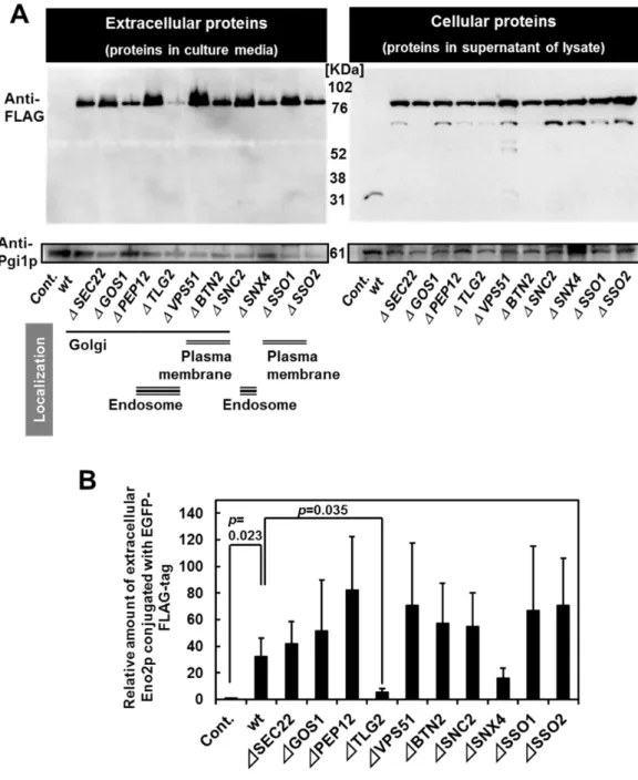
Previously, Morisaka and colleagues have developed a novel two-dimensional high-performance liquid chromatography (2D-HPLC)-based method that detects proteins on the living cell surface (Morisaka et al. 2012). Using this method, an overview of the proteins on the outside of the cell can be gained. In addition, glycolytic enzymes suitable for secretion analyses can be selected. In this study, I utilized enhanced green fluorescence protein conjugated with FLAG-tag (EGFP-FLAG)-tagged glycolytic enzymes to analyze the secretory pathway of glycolytic enzymes. Western blot analysis enabled detection of the secreted proteins in the culture media. Moreover, the use of plasmid-based protein expression facilitated uniform protein levels and analysis of the secreted proteins. Moreover, the secretory pathway was visualized and assessed with the aid of the conjugated fluorescent proteins (Hirschberg and Lippincott-Schwartz 1999, Huang and Shusta 2005).
Materials and methods Strains and media
Escherichia coli DH5 (F-, 80dlacZM15, (lacZYA-argF)U169, deoR, recA1, endA1,
hsdR17(rK-, mK+), phoA, supE44, -, thi-1, gyrA96, relA1) strain was used for host cells in the cloning experiments. The temperature-sensitive sec23-1 strain RSY282 (MATa, leu2 ura3
sec23-1) was kindly provided by Dr. Randy Schekman (Department of Molecular and Cell Biology
and Howard Hughes Medical Institute, University of California at Berkeley). The yeast strain BY4741 (MATa, his31, leu2 met15 ura3), and the derived deletion strains of SED1 (sed1),
SSO1 (sso1), SSO2 (sso2), SEC22 (sec22), SNC2 (snc2), TLG2 (tlg2), BTN2 (btn2), PEP12 (pep12), VPS51 (vps51), GOS1 (gos1), ATG1 (atg1), ATG8 (atg8), ATG11 (atg11), ATG17 (atg17), ATG20 (atg20), VAM3 (vam3), and GRH1 (grh1) were purchased from EUROSCARF (Frankfurt, Germany). The yeast GFP clones (Invitrogen, Carlsbad, CA, USA) with GFP-tagged endogenous proteins (Pma1p, Nup84p, Mae1p, Chs5p, Snf7p, Vrg4p, Pex11p, and Sec13p) and HIS3 marker in the parent BY4741 strain were used to determine the localization of proteins. E. coli was grown in lysogeny broth (LB) (1% (w/v) tryptone, 0.5% (w/v) yeast extract, 0.5% (w/v) sodium chloride, and 100 ng/mL ampicillin). The yeast cells were grown in yeast extract peptone dextrose (YPD) medium (1% (w/v) yeast extract, 2% (w/v) polypeptone, and 2% (w/v) glucose), SD+HM medium (0.67% (w/v) yeast nitrogen base without amino acids, 2% (w/v) glucose, 0.002%
30
L-histidine-HCl, and 0.003% L-methionine), SDC+HM medium (0.67% (w/v) yeast nitrogen base without amino acids, 2% (w/v) glucose, 0.002% L-histidine-HCl,0.003% L-methionine, 2% casamino acids (BD, Franklin Lakes, NJ), and 2% (w/v) agar), SC+ML medium (0.67% (w/v) yeast nitrogen base without amino acids, 2% (w/v) glucose, 0.003% L-methionine,0.003% L-leucine, 0.13% SD Multiple drop Out (-Ade, -His, -Leu, -Lys, -Trp, -Ura, Funakoshi Co., Ltd., Tokyo, Japan), 2% (w/v) agar), SD+ML medium (0.67% (w/v) yeast nitrogen base without amino acids, 2% (w/v) glucose, 0.003% L-methionine,0.003% L-leucine), or SDC+ML medium (SD+ML supplemented with 2% casamino acids).
Construction of S. cerevisiae expression plasmids
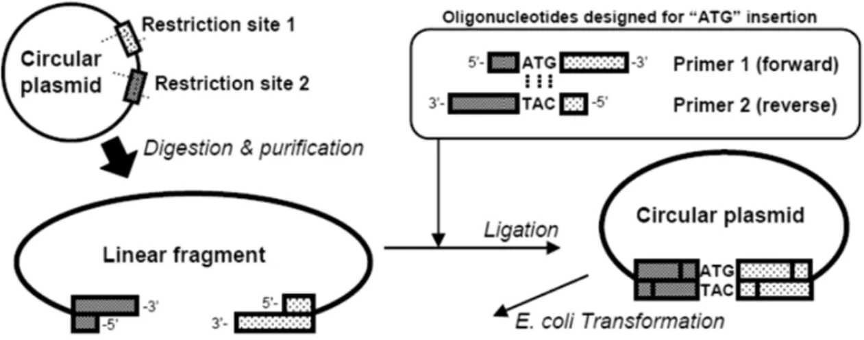
The plasmids were constructed using a conventional PCR-based method and our novel PCR-free method (one-step construction method for plasmids (OSCoM-P); Fig. 1). In addition, iProof DNA polymerase (Bio-Rad, Richmond, CA, USA), KOD-plus-DNA polymerase (Toyobo, Osaka, Japan), KOD-plus-Neo-DNA polymerase (Toyobo), Ligation High (Toyobo), and synthetic oligonucleotides (Japan Bio Services, Saitama, Japan) were used. All primers used in this study are shown in Table 1. The plasmids for the internal production of the recombinant proteins were constructed from pULSG1 (Matsui et al. 2009). The primers coding the ATG codon were mixed with the pULSG1 digest and inserted using the EcoR I and Xho I sites by OSCoM-P (Fig. 1); the resulting plasmid was named pUL-ATG-EGFP. The section of pUL-ATG-EGFP including the GAPDH promoter, the terminator, the FLAG-tag (DYKDDDDK; 21), and the EGFP sequence was amplified and added to the BamH I and Not I sites by PCR using the primers GAPDH promoter-F and GAPDH terminator-R, and inserted into the BamH I–Not I section of pRS423 (47, from ATCC); the resulting plasmid was named pRS423-ATG-EGFP. For constructing the plasmid pULGI2, OSCoM-P was also performed. Oligonucleotide fragments with several restriction sites were inserted into pULSG1 by using the
EcoR I and Xho I sites. The plasmids for the internal expression of the glycolytic enzymes
conjugated with EGFP-FLAG were constructed as follows. The yeast genomic DNA was extracted and purified from the S. cerevisiae BY4741 strain, and each gene coding a glycolytic enzyme was cloned using the appropriate primer set (Table 1). The fragments were digested and inserted into pULGI2 by using the BamH I and Xho I sites or the BamH I and Sac I sites. The internal expression vector without EGFP was constructed from pULSG1C (Matsui et al. 2009) and pWGP3 (Takahashi et al. 2001). The multi-cloning site followed by the GAPDH terminator sequence was amplified from pWGP3 and inserted into pULSG1C by using the Sac I and Kpn I sites; the resulting plasmid was named pULI1. For the construction of the plasmid for the intercellular production of enolase-EGFP-FLAG with the N-terminal peptide sequence (HA-tag), the HA-tag sequence was inserted into pULGI2 by using OSCoM; the resultant plasmid was named pULGI2-HA. The ENO2 coding sequence from pULGI2-ENO2 was inserted into pULGI2-HA, and the resultant plasmid was
31
named pULGI2-HA-ENO2. For the construction of plasmids to produce the red-fluorescent proteins, the Discosoma red fluorescent protein (DsRED) monomer with EcoR I and Xho I sites at the N-terminus and Sal I at the C-terminus was cloned from pKRD4 (Kuroda et al. 2009). The amplified fragments were digested with EcoR I and Sal I, and then inserted into the same site of pULSG1; the resultant plasmid was named pUL-ATG-DsRED. For production of the enolase fragments fused with EGFP or DsRED, amplified fragments were digested and inserted into pULSG1 or pUL-ATG-DsRED by using the EcoR I and Xho I sites (see Table 1). Plasmids for production of Tlg2p (p413-ADH-TLG2) were constructed as follows: TLG2-coding sequence was cloned from the genomic DNA extracted from S.cerevisiae BY4741 and inserted into MCS of p413-ADH (ATCC 87370) using EcoR I and Xho I sites. The plasmid construction was confirmed by DNA sequencing performed using a BigDye Terminator v3.1 cycle sequencing kit and an ABI PRISM 3100 Genetic Analyzer (Applied Biosystems, Foster City, CA, USA).
Fig. 1 Schematic illustration of the one-step construction method for plasmids (OSCoM-P) The digested linear fragments of the plasmids were ligated with the designed primers at a dry weight ratio of 1:1–1:10 (linear plasmid:each primer). The reaction solution was directly transformed into E.
coli DH5 competent cells (Toyobo). The purchased oligonucleotides were dissolved in dH2O at 10 ng/mL before use. TE buffer was not used because OSCoM-P was severely inhibited by the presence of TE buffer (data not shown). Maximum length of fragments to be inserted into plasmids using OSCoM-P is 45 bp (data not shown).
Production of recombinant proteins in yeast cells
The yeast cells were transformed with plasmids by using a Frozen-EZ Yeast Transformation II™ kit (Zymo Research, Orange, CA, USA) and grown on SDC+HM agar plates. The transformants were