はじめに 多発性硬化症(multiple sclerosis; MS)は中枢神経の代表的 な脱髄性疾患で,慢性炎症性自己免疫疾患であるが,病因は 未だに不明な点が多い.ウイルス感染や自己免疫応答性リン パ球が介在する仮説が提唱されているが解明には至っていな い1)~3).我々は幼少時に MS と診断され,脳静脈洞血栓症
(cerebral venous sinus thrombosis; CVT)を合併し,血栓症の 原因が antithrombin(AT)欠損症であった症例を経験した. 貴重な症例と考え報告する. 症 例 患者:25 歳,男性 主訴:腹痛・嘔吐 家族歴:母方祖父および叔父が脳梗塞. 内服薬:特記事項なし. 現病歴:6 歳時に左眼の球後視神経炎を発症した.12 歳で 右後頭葉に脱髄性病変が出現し,小児科で MS と診断された. 左側視野障害が残存していたが,問題なく通学していた.15 歳で右片麻痺が出現し,左頭頂葉に造影 MRI でリング状に造 影効果も認められ再発が認められた.メチルプレドニゾロン (mPSL)1 g/ 日,3 日間を 1 クールとして,4 クール施行され, 視野障害および右不全片麻痺が残存したが 6 週間で退院し た.1 年後(16 歳),頭痛が起こり,右前頭葉に浮腫を伴った 腫瘍様脱髄病変が出現した.この時も mPSL の同様 4 クール を行い改善した.再発防止のため,インターフェロン(IFN, interferon-β1b,800×104単位,隔日,皮下注射)が導入され, 2ヶ月で退院した.その後再発なく経過していたが,19 歳で 皮膚硬結の副作用が強くなり IFN は中止された.しかし,再 発なく順調に経過していた(Fig. 1).MS の分類は再発寛解 (relapseing-remitting; RR)型であり,Expanded Disability Status
Scale(EDSS)は 3.5 であった. 25歳の 12 月下旬に突然の腹痛と嘔吐が出現,近医で胃腸 炎として経過を見ていたが,頭痛が強くなり意識障害も認め, 発症 4 日目に当院に入院した.入院時理学的所見:血圧 122/73 mmHg,脈拍 66/ 分,体温 36.6°C.胸腹部に明らかな 異常所見は認めず.神経学的所見では,頭痛,軽度の意識障 害(JCSI-2)を認めるが髄膜刺激徴候なし.MS 後遺症の左同 名半盲と左上下肢不全麻痺,右上下肢腱反射亢進を認めたが, 新規の神経脱落所見は認めなかった.検査所見:末梢血で赤 血球,血小板は正常.白血球 9,770/μl 増加と炎症反応(CRP 6.8 mg/dl,fibrinogen 372 mg/dl)を認め,D-dimer(4.1 μg/dl) が上昇していた.その他電解質,肝機能,腎機能,甲状腺機 能などに異常はなかった. 脳 MRI では右小脳半球に T2強調,FLAIR 画像で高信号の
併発した多発性硬化症の 1 例
金谷 雄平
1)高松 和弘
1)下江 豊
1)仁井見英樹
2)北島 勲
2)栗山 勝
1)*
要旨: 症例は 25 歳の男性.6 歳で視神経炎,12∼16 歳で 3 回の大脳白質脱髄巣が出現した再発寛解型多発性 硬化症(multiple sclerosis; MS)である.25 歳で,嘔吐を伴う胃腸炎に罹患後,脳静脈洞血栓症(cerebral venous sinus thrombosis; CVT)を発症し,腰椎穿刺で悪化した.アンチトロンビン(AT)の低下を認め,AT 遺伝子エク ソン 5 番目のコドン 359 の CGA が TGA に置換したナンセンス変異の AT 欠損症ヘテロ接合体であった.MS に CVT が合併した 17 例が過去に報告されており,腰椎穿刺と高容量メチルプレドニゾロン(mPSL)投与が CVT を誘引した症例が多い.本例は mPSL 治療は行ってないが,AT 欠損症に,高ホモシステイン血症,感染,腰椎穿 刺が加わり発症増悪したと推測した. (臨床神経 2016;56:248-254) Key words: 多発性硬化症,アンチトロンビン欠損症,脳静脈洞血栓症,慢性脳脊髄循環不全 *Corresponding author: 脳神経センター大田記念病院脳神経内科〔〒 720-0825 広島県福山市沖野上町 3-6-28〕 1)脳神経センター大田記念病院脳神経内科 2)富山大学医学部附属病院検査部(Received August 17, 2015; Accepted February 8, 2016; Published online in J-STAGE on March 24, 2016) doi: 10.5692/clinicalneurol.cn-000796
先天性アンチトロンビン欠損症と脳静脈洞血栓症を併発した多発性硬化症 56:249 新病変を認めた(Fig. 2A, B).入院当日施行した腰椎穿刺に よる髄液検査では,蛋白や細胞数の増加は認めず,オリゴク ローナルバンド(等電点分画電気泳動)陰性,ミエリンベー シック蛋白 40 pg/ml 以下で,抗 AQP4 抗体も陰性であった. 経過と画像所見:入院 2 日目に明らかに意識レベル低下 (JCSII-20)を認め,視力障害も出現し,頭 MRI,FLAIR 画像 (Fig. 2C, D)で左右小脳半球,両側視床・左基底核に高信号 の病変を認め,頭 CT で直静脈洞,右 S 状静脈洞から横静脈 洞に高信号を認め,造影 MRI(Fig. 2E, F)で同部位の造強効 果が消失しており,CVT が疑われた.緊急脳血管撮影を行い, 右 S 状静脈洞から横静脈洞,直静脈洞および両側内大脳静脈, ガーレン大静脈の広範な静脈血栓症を確認された.重篤な静 脈血栓症であるため,ウロキナーゼ(UK,合計 48×104単位) の選択的脳血栓溶解療法を施行したが,再開通は得られず, その後はヘパリン(2×10⁴単位)による抗凝固療法を開始し た.徐々に意識状態は改善し,右麻痺は残存したがワルファ リン(Wa)内服に切り替えて,3 週間後に自宅退院した.そ の後外来で抗凝固療法を継続していたが,29 歳時,両下肢の しびれ感および歩行障害が増悪し再度入院,胸~腰部に MRI で多発性の造影効果を認める活動性脊髄病変を認めた.しか し,頭 MRI には新病巣は認めなかった.mPSL 大量療法を施 行し軽快した.再度抗 AQP4 抗体も検討したが陰性であった. また抗 MOG 抗体も測定し陰性であった. CVTの原因検索と遺伝子検査:抗核抗体および抗リン脂質 抗体症候群(ループスアンチコアグラント,抗カルジオリピ ン抗体,抗カルジオリピン β2GPI 複合体抗体)を含め自己抗 体(ds-DNA,抗 Sm,抗 SSA,抗 SSB,PR3-ANCA,MPO-ANCA)も検討したがすべて陰性であった.ホモシステイン がやや上昇(21.5 nmol/ml,正常値;3.7~13.5)していた.葉 酸(18.1 ng/ml),ビタミン B12(331 pg/ml)は正常.PT およ び APTT は正常範囲で,Protein C 活性 119%,Protein S 活性 67%も正常であったが,AT 活性値 30%(正常 88~116%)と 低下を認め,抗原量も 12.8 mg/dl(正常 23.6~33.5 mg/dl)と 低下していた.承諾のもと両親の AT 活性を測定したところ, 父親は AT 活性値 95%と正常であったが,母親は 53%と低下 を認めた.本人および両親の遺伝子解析を行った結果,本人 Fig. 1 Brain MRI performed 4 months before the onset of cerebral venous sinus thrombosis.
Multiple lesions, suggesting demyelinating plaques, were detected in the periventricular regions and subcortical white matter. Arrows indicate demyelinating plaques. A, B: Axial T2 weighted images (1.5 T, TR 3,800 ms, TE 90 ms), C, D: Axial FLAIR images (1.5 T, TR 9,000 ms, TE 87 ms).
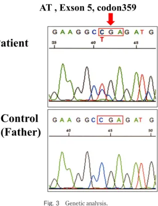
と母親で AT 遺伝子のエクソン 5 番目のコドン 359 の CGA が TGAに置換されたナンセンス変異のヘテロ接合体である事 が判明し,先天性 AT 欠損症 type 1 と診断した(Fig. 3). 考 察 先天性 AT 欠損症は常染色体優性遺伝疾患で,日本人の発 症頻度は 0.18%とされる稀な疾患である4).血栓症を発症す る危険率は健常人の 10~20 倍と高く,60 歳までに約 90%の AT欠損症の患者が血栓症を起こすといわれている.AT 活性 値と抗原量の両方が低下する type 1 と,抗原量は正常である が活性値が低下する type 2 に分類され5),本症例は type 1 で あった.AT 遺伝子は七つのエクソンと六つのイントロンで構 成され,染色体 1q23~25 上に 13.4 kb の長さで存在し4)5),多 くの遺伝子変異が知られている.Type 1 は古典的 AT 欠損症 として知られており,多種の変異が知られている6).本症例 の変異はエクソン 5 番目,コドン 359 における CGA が TGA に置換され終止コドンとなり AT 欠乏が生じた変異である. この変異は 1993 年にすでに見出されており7),新規のもので はなかった. CVT発症の危険因子としての,遺伝的血栓形成素因に関 しては,21 カ国 89 センターで共同して検討された CVT 624 症例の International Study on Cerebral Vein and Dural Sinus Thrombosis(ISCVT)の報告では,22.4%に認められている8). しかし,詳しい遺伝子変異を示す疾患の比率に関しては報 告されていない.本邦では,尾原らの 10 症例の CVT 中 1 例 が,AT 欠損症であり9),Shindo らの 22 例の報告では,7 例 (31.8%)が遺伝的血栓形成素因で,protein S の遺伝子異常が Fig. 2 Brain MRI on admission (A, B) and on the second day in the hospital (C–F).
On admission, the MRI scan showed the new lesion in the right cerebellar hemisphere (A, B). On the second day after admission to the hospital, the lesions extended to the left cerebellar hemisphere, bilateral thalamus, and left basal ganglia (C, D) and occlusions of the straight sinus, right lateral and sigmoid sinuses were detected by enhanced MRI, which suggested cerebral venous and sinus thrombosis. Arrows indicate infarct lesions with edema. The occlusions of the straight sinus (arrow heads) and right lateral sinus (broad arrow) are shown (E, F). A: Axial FLAIR image (1.5 T, TR 8,500 ms, TE 91 ms), B: Axial Diffusion image (1.5 T, TR 3,800 ms, TE 80 ms), C, D: Axial FLAIR images (1.5 T, TR 8,500 ms, TE 91 ms), E: Enhanced sagittal T1 weight image (1.5 T, TR 550 ms, TE 17 ms), F: Enhanced coronal T1 weight image (1.5 T, TR 635 ms, TE 17 ms).
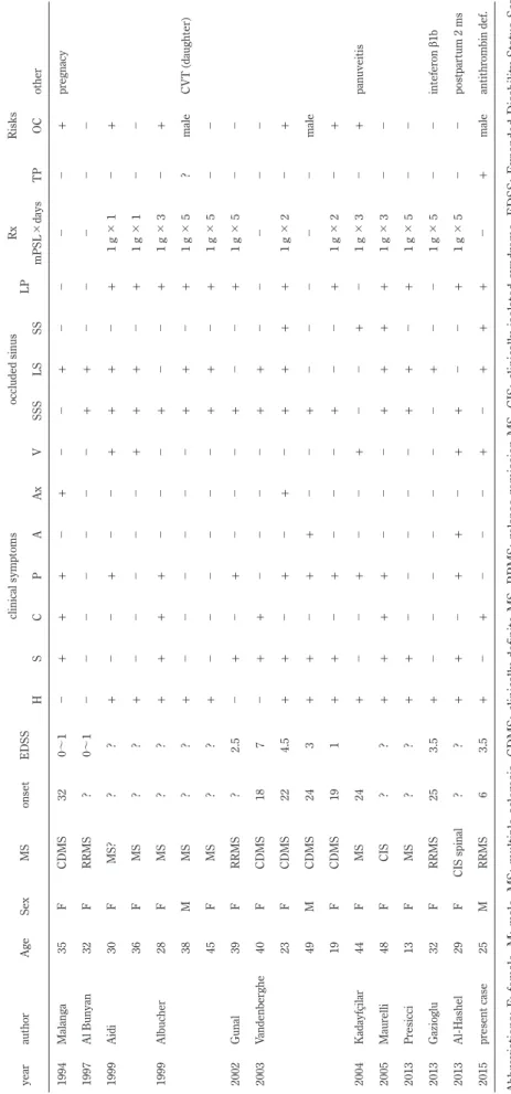
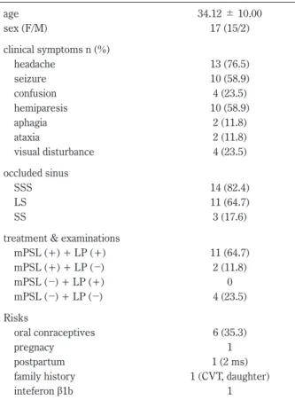
先天性アンチトロンビン欠損症と脳静脈洞血栓症を併発した多発性硬化症 56:251 4例(18.2%),protein C の遺伝子異常が 2 例(9.1%),ATIII 遺伝子異常が 1 例(4.5%)である10).日本人では CVT の危 険因子としては比較的頻度の低い疾患と思われる. MS患者に CVT が発症した症例は,文献検索した限り 1994 年 以来欧米諸国から,17 症例が報告されている11)~21)(Table 1). 年齢は 34.12±10.00 歳,女性 15 例,男性 2 例である.頭痛 13例(76.5%),けいれん 10 例(58.9%),意識障害・混迷 4 例(23.5%),運動麻痺 10 例(58.9%),失語 2 例(11.8%), 運動失調 2 例(11.8%),視力障害 4 例(23.5%)で,閉塞し た静脈洞は,上矢状静脈洞が 14 例(82.4%),横静脈洞 11 例 (64.7%),直静脈~深部脳静脈洞 3 例(17.6%)で,臨床症状 や閉塞静脈洞部位は,基本的には ISCVT 報告の CVT と変わ らないが,より若年で女性が多いようである.危険因子に関 しては,経口避妊薬使用が 6 例(35.3%)11)13)14)16)17),妊娠中 1例11),産後 2 ヶ月 1 例21),娘に CVT 発症 1 例14),危険因 子かは不明であるが IFNβ1b 使用中 1 例20)などが報告されて いる.問題とされている CVT 発症の機序として注目される点 は,髄液採取のための腰椎穿刺の後,mPSL 500~1,000 mg 投 与(1 日~5 日)後に生じている症例が 11 例(64.7%)に認 められたことである13)~21).終了翌日から数日以内が多く 2 週 間までに CVT が発生している.腰椎穿刺施行はなく mPSL 療 法後に生じた症例が 2 例(11.8%)17)20),腰椎穿刺も mPSL 療法 もなく発症した症例が 4 例(23.5%)11)12)16)である(Table 1, 2). 発症の機序は,腰椎穿刺により脳圧が低下し,脳内静脈の壁 は薄く脆弱なため拡張し,静脈流が遅くなる,さらに mPSL の大量を点滴注入することにより,凝固性が亢進することが CVTを引き起こす誘引となることが推測されている13)~21). さらに,Stolz らは 120 例の CVT 症例の危険因子を検討し, MS増悪期 4 例,視神経炎 2 例の 6 例が同様に 500~1,000 mg mPSL投与を 5 日間受け,終了後平均 4 日以内に発生してい る.そのうち 3 例が腰椎穿刺後に発生している.各症例の詳 細は不明であるが,3 例が経口避妊薬使用,4 例が喫煙で合併 する危険因子として加わったことが発症誘引と関連すると報 告されている22).Kalanie らは,増悪期の MS 症例に 1,000 mg mPSL 5日間投与の 188 例と同様 mPSL 投与に低分子ヘパリン (Enoxaparin)の皮下注射を加えた 178 例で無作為 2 群間比較 を行ったところ,前者で 3 例の下肢静脈血栓と 2 例の CVT が 発症し,後者では発症者はいなかった23).よって,MS 患者 に mPSL 大量療法を行う時には,予防的抗凝固薬の投与が重 要であるとしている22)23). 我々の症例は,25 歳時の入院時,数日前から胃腸炎に罹患 し嘔吐・頭痛があり,D-dimer の上昇も認められることから, 血栓形成が伺われるが,小脳の新病巣出に対しては CVT とは 診断できなかった.入院後腰椎穿刺後からさらに症状が悪化 し,入院 2 日目に画像を再度撮像し,病巣は拡大しており CVTを疑い,血管撮影を行い CVT と確定診断した.CVT の 原因は,AT 欠損症による血栓形成傾向が主因と思われるが, 発症時にはホモシステインの上昇も認められており,さらに 嘔吐による脱水傾向,腰椎穿刺による影響などが相加的に作 用して CVT を発症および増悪させたと推察した.本症例は MS の検査や治療上の留意点からも貴重な症例と考え報告した. 本論文の要旨は第 97 回日本神経学会中国・四国地方会(平成 26 年 12月 6 日)で発表した. 謝辞:抗 AQP4,抗 MOG 抗体の測定していただきました,東北大 学医学部神経内科高橋利幸先生に深謝いたします. ※本論文に関連し,開示すべき COI 状態にある企業,組織,団体 はいずれも有りません. 文 献
1) Compston A, Coles A. Multiple sclerosis. Lancet 2008;372: 1502-1517.
2) Nylander A, Hafler DA. Multiple sclerosis. J Clin Invest 2012; 122:1180-1188.
3) Ciccarelli O, Barkhof F, Bodini B, et al. Pathogenesis of multiple sclerosis: insights from molecular and metabolic imaging. Lancet Neurol 2014;13:807-822.
4) 阪田敏幸,松尾 汎,岡本 章ら.日本人における凝固制御 因子の異常と静脈血栓症との関連.静脈学 2003;14:339-345. 5) 辻 肇.先天性アンチトロンビン III 欠損症.血栓止血誌
2001;12:74-77.
6) Nakahara Y, Tsuji H, Nakagawa K, et al. Genetic analysis in Japanese kindreds of congenital type 1 antithrombin deficiency causing thrombosis. Thromb Haemost 1997;77:616-619. Fig. 3 Genetic analysis.
Genetic analysis revealed a single-base substitution (C>T) in the codon 359 (Arg to STOP) in the 5th exon portion of antithrombin gene, heterozygote. This mutation was observed in the patient and his mother, but not in his father. Therefore, this mutation was con-sidered to be inherited through his mother.
Table
1
The cases with multiple scler
osis associated with er
ebral venous sinus thr
ombosis. year author Age Sex MS onset EDS S clinical symptoms occluded sinus LP Rx Risks H S C P A Ax V SSS LS SS mPSL × days TP OC other 1994 Malanga 35 F CDMS 32 0~ 1 + + + + + + pr egnacy 1997 Al Bunyan 32 F RRMS ? 0~ 1 + + 1999 Aidi 30 F MS? ? ? + + + + + + 1 g × 1 + 36 F MS ? ? + + + + + 1 g × 1 1999 Albucher 28 F MS ? ? + + + + + + 1 g × 3 + 38 M MS ? ? + + + + 1 g × 5 ? male CVT (daughter) 45 F MS ? ? + + + + 1 g × 5 2002 Gunal 39 F RRMS ? 2.5 + + + + 1 g × 5 2003 Vandenber ghe 40 F CDMS 18 7 + + + + 23 F CDMS 22 4.5 + + + + + + + + 1 g × 2 + 49 M CDMS 24 3 + + + + + male 19 F CDMS 19 1 + + + + + 1 g × 2 + 2004 Kadayfçilar 44 F MS 24 + + + + 1 g × 3 + panuveitis 2005 Maur elli 48 F CIS ? ? + + + + + + + + 1 g × 3 2013 Pr esicci 13 F MS ? ? + + + + + 1 g × 5 2013 Gazioglu 32 F RRMS 25 3.5 + + 1 g × 5 intefer 2013 Al-Hashel 29 F CIS spinal ? ? + + + + + + + 1 g × 5 postpar 2015 pr esent case 25 M RRMS 6 3.5 + + + + + + + male antithr Abbr eviations; F: female, M:
male, MS: multiple scler
osis,
CDMS: clinically
definite MS, RRMS: r
elapse-r
emission MS, CIS:
clinically isolated syndr
ome, EDS S: Expanded Disability Status H: headache, S: seizur e, C: confusion, P: par esis, A: aphagia, Ax: ataxia, V : visual disturbance, S
SS: superior sagittal sinus,
LS: lateral sinus, SS: straight sinus, LP : lumbar punctur mPSL: methylpr ednisolone, TP : thr
ombophilia, OC: oral contraceptives, def
: deficiency
先天性アンチトロンビン欠損症と脳静脈洞血栓症を併発した多発性硬化症 56:253
7) Chowdhury V, Olds RJ, Lane DA, et al. Identification of nine novel mutations in type I antithrombin deficiency by heteroduplex screening. Br J Haematol 1993;84:656-661.
8) Ferro JM, Canhão P, Stam J, et al. Prognosis of cerebral vein and dural sinus thrombosis: results of the International Study on Cerebral Vein and Dural Sinus Thrombosis (ISCVT). Stroke 2004;35:664-670.
9) 尾原知行,山本康正,田中瑛次郎ら.脳静脈血栓症連続 10 症例の臨床像,画像所見の検討.脳卒中 2013;35:167-173. 10) Shindo A, Wada H, Ishikawa H, et al. Clinical features and
underlying causes of cerebral venous thrombosis in Japanese patients. Int J Hematol 2014;99:437-440.
11) Malanga GA, Gangemi E. Intracranial venous thrombosis in a patient with multiple sclerosis. A case report and review of
contraceptive alternatives in patients with disabilities. Am J Phys Med Rehabil 1994;73:283-285.
12) Al Bunyan M, Ogunniyi A. Incidental cerebral venous thrombosis in a patient with multiple sclerosis. J Neurol Sci 1997;149:191-194.
13) Aidi S, Chaunu MP, Biousse V, et al. Changing pattern of headache pointing to cerebral venous thrombosis after lumbar puncture and intravenous high-dose corticosteroids. Headache 1999;39:559-564.
14) Albucher JF, Vuillemin-Azaïs C, Manelfe C, et al. Cerebral thrombophlebitis in three patients with probable multiple sclerosis. Role of lumbar puncture or intravenous corticosteroid treatment. Cerebrovasc Dis 1999;9:298-303.
15) Gunal DI, Afsar N, Tuncer N, et al. A case of multiple sclerosis with cerebral venous thrombosis: the role of lumbar puncture and high-dose steroids. Eur Neurol 2002;47:57-58.
16) Vandenberghe N, Debouverie M, Anxionnat R, et al. Cerebral venous thrombosis in four patients with multiple sclerosis. Eur J Neurol 2003;10:63-66.
17) Kadayifçilar S, Gedik S, Eldem B, et al. Panuveitis associated with multiple sclerosis complicated by cerebral venous thrombosis. Ocul Immunol Inflamm 2004;12:153-157.
18) Maurelli M, Bergamaschi R, Candeloro E, et al. Cerebral venous thrombosis and demyelinating diseases: report of a case in a clinically isolated syndrome suggestive of multiple sclerosis onset and review of the literature. Mult Scler 2005;11:242-244. 19) Presicci A, Garofoli V, Simone M, et al. Cerebral venous
thrombosis after lumbar puncture and intravenous high dose corticosteroids: a case report of a childhood multiple sclerosis. Brain Dev 2013;35:602-605.
20) Gazioglu S, Solmaz D, Boz C. Cerebral venous thrombosis after high dose steroid in multiple sclerosis: a case report. Hippokratia 2013;17:88-90.
21) Al-Hashel JY, Ahmed SF, Alexnader KJ, et al. Cerebral venous thrombosis in a patient with clinically isolated spinal cord syndrome. Case Rep Neurol Med 2013;2013:364869.
22) Stolz E, Klötzsch C, Schlachetzki F, et al. High-dose corticosteroid treatment is associated with an increased risk of developing cerebralvenous thrombosis. Eur Neurol 2003;49: 247-248.
23) Kalanie H, Harandi AA, Alidaei S, et al. Venous thrombosis in multiple sclerosis patients after high-dose intravenous methylprednisolone: the preventive effect of enoxaparin. Thrombosis 2011;2011:785459.
Table 2 Multiple sclerosis associated erebral venous sinus thrombosis.
age 34.12 ± 10.00 sex (F/M) 17 (15/2) clinical symptoms n (%) headache 13 (76.5) seizure 10 (58.9) confusion 4 (23.5) hemiparesis 10 (58.9) aphagia 2 (11.8) ataxia 2 (11.8) visual disturbance 4 (23.5) occluded sinus SSS 14 (82.4) LS 11 (64.7) SS 3 (17.6)
treatment & examinations
mPSL (+) + LP (+) 11 (64.7) mPSL (+) + LP (︲) 2 (11.8) mPSL (︲) + LP (+) 0 mPSL (︲) + LP (︲) 4 (23.5) Risks oral conraceptives 6 (35.3) pregnacy 1 postpartum 1 (2 ms)
family history 1 (CVT, daughter)
inteferon β1b 1
Abbreviations; SSS: superior sagittal sinus, LS: lateral sinus, SS: straight sinus, LP: lumbar puncture, mPSL: methylprednisolone.
Yuhei Kanaya, M.D
1), Kazuhiro Takamatsu, M.D.
1), Yutaka Shimoe, M.D., Ph.D.
1),
Hideki Niimi, M.D., Ph.D.
2), Isao Kitajima, M.D., Ph.D.
2)and Masaru Kuriyama, M.D., Ph.D.
1) 1)Department of Neurology, and Department of Radiology, Brain Attack Center Ota Memorial Hospital2)Toyama University Hospital Clinical Laboratory
We report the case of a 25-year-old man with multiple sclerosis (MS) who had severe headache and unconsciousness.
He suffered from optic neuritis that had started at age 6. From the age of 12 years, he had suffered from multiple
sclerosis (MS) cerebral lesions that relapsed three times over for 5 years. At age 25, he showed a new lesion in the
cerebellar cortex, suggesting an exacerbation of the MS. However, magnetic resonance imaging findings the next day
showed cerebral venous sinus thrombosis. His laboratory findings showed low antithrombin activity. Genetic analysis
revealed a single-base substitution (C>T) at the codon 359 (Arg to STOP) in the 5th exon portion of the antithrombin
gene, heterozygote. In the literature review, 17 cases of multiple sclerosis associated with cerebral venous sinus
thrombosis, which occurred after the lumbar puncture and the treatment with high-dose methylpredonisolone in 11 of
these cases. In our case, antithrombin deficiency, hyperhomocystinemia, infection, and lumbar puncture were suggested
as the risk factors.
(Rinsho Shinkeigaku (Clin Neurol) 2016;56:248-254)
Key words: multiple sclerosis, antithrombim deficiency, cerebral venous sinus thrombosis, chronic cerebrospinal venous insufficiency