基礎疾患のない若年者に発症し肺結核と鑑別を要した
Actinomyces odontolyticus
による肺化膿症の 1 例
1兵頭健太郎
1, 2金澤 潤
1松村 聡介
1嶋田 貴文
1北岡 有香
1後藤 瞳
1笹谷悠惟果
1, 2中嶋 真之
1齋藤 武文
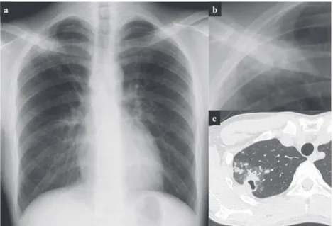
緒 言 一般に肺放線菌症は診断が困難なことが多いとされる が1),今回,画像上肺結核疑いで検査を施行した患者で, 肺結核や他の原因が検出されず,気管支肺胞洗浄液の嫌 気培養で Actinomyces odontolyticus が検出され診断をした 若年肺化膿症症例を経験した。肺放線菌症の起炎菌とし て頻度が高いのは A. israelii であるが2),今回 A. odontolyti-cusが同定され,菌種,診断方法,共に稀な症例であるた め報告する。 症 例 患 者:24 歳,男性。 主 訴:咳嗽。 既往歴:21 歳 気管支喘息(軽症,処方なし),22 歳 智歯を 4 本すべて抜歯,最近の齲歯治療なし。 喫煙歴:なし。 飲酒歴:ビール 350 ml 週 3 回。 内服薬:近医で処方されたアジスロマイシン(AZM) 鎮咳薬。 現病歴: 2 週間程度続く咳嗽のため,X−6 日に近医を 受診した。胸部画像上,空洞を伴う結節影,浸潤影を認 めた。AZM 処方を受けたが,画像上肺結核症も否定で きないため,X 日当科を紹介受診し,同日入院した。 入院時現症:血圧 122/66 mmHg,脈拍 60 bpm,体温 36.5℃,呼吸数 15 回 ⁄min,SpO2(室内気)98%,意識清 明,両肺でラ音を聴取せず。 入院時検査所見:胸部 X 線では右鎖骨と重なる付近に 胸膜と接する空洞を伴う径 24 mm 程度の結節影を認めた (Fig. 1a, b)。胸部 CT では右 S1領域を中心に含気を伴う 浸潤影,多巣性の結節影,空洞影,胸膜肥厚を認めた(Fig. 1c)。近接する胸膜や胸壁を巻き込む浸潤影や Air bron-chogram を伴った肺浸潤を認めた。Fig. 1 に示すように 胸水や,肺門縦隔リンパ節腫脹は明らかではなかった。 血液・尿検査では特記すべき所見を認めなかった(Table 1)。Table 1 に示すように,IGRA(Interferon-Gamma release1国立病院機構茨城東病院胸部疾患・療育医療センター内科診
療部呼吸器内科,2現:筑波大学大学院呼吸器内科
連絡先 : 兵頭健太郎,国立病院機構茨城東病院胸部疾患・療育 医療センター内科診療部呼吸器内科,〒 319 _ 1113 茨城県那珂 郡東海村照沼 825(E-mail: [email protected])
(Received 29 Jul. 2018 / Accepted 29 Nov. 2018)
要旨:症例は既往のない 24 歳男性。 1 週間持続する咳嗽を主訴に近医を受診し,胸部画像検査で右肺 上葉に空洞を伴う多発結節影・浸潤影を認めたため当院へ紹介受診となった。当初,肺結核を疑い喀 痰検査を行うも抗酸菌塗抹・結核菌 PCR は陰性で,また気管支洗浄液の一般細菌抗酸菌塗抹培養でも 菌の検出には至らなかった。 2 回目の気管支鏡検査では気管支肺胞洗浄を行い,得られた検体で前回 未実施だった嫌気培養を行った結果,放線菌属が検出され,肺放線菌症と診断した。後日,得られた 菌は放線菌属の中の Actinomyces odontolyticus と同定された。SBT/ABPC を 14 日間投与し,陰影の改善 がみられ,その後は AMPC 内服へ変更し,加療を継続した。肺放線菌症は基礎疾患のない者にも発症 することが知られている。本例では気管支肺胞洗浄液の嫌気培養を行ったことで診断に至ることがで きた,稀な A. odontolyticus の症例を報告した。
Table 1 Laboratory fi ndings on admission Fig. 1 Chest X ray (a, b), CT (c) on admission
WBC 6.6 ×10³/μμL Seg 71.6 % Lymph 21.3 % Mono 5.9 % Eosino 0.9 % Baso 0.3 % RBC 496 ×104/μμL Hb 14.8 g/dl Ht 43.7 % PLT 27.3 ×10⁴/μμL IGRAs (−) anti-gpl-core IgA antibodies for Mac (−) HBsAg (−) HCV (−) TPHA (−) RPR (−) TP 7.6 g/dl Alb 4.8 g/dl T-Bil 0.5 mg/dl AST 18 U/L ALT 22 U/L LDH 154 U/L ALP 375 U/L γ γ-GTP 19 IU/L CPK 77 U/L BUN 16.9 mg/dl CRE 0.78 mg/dl β βD Glu ≦ 5.0 Asper AG 0.1 (−) Candida AG (−) Crypt AG (−) Na 142 mEq/L K 4.4 mEq/L Cl 101 mEq/L Ca 10.1 mg/dl CRP 0.32 mg/dl BS 81 mg/dl HbA1c 4.9 % PT-INR 1.18 APTT 39.2 sec Fib 344 mg/dl CEA 1.1 ng/mL CYFRA 1.3 ng/mL C-ANCA <1.0 P-ANCA <1.0
Sputum GPC2+, GNR2+, phago (−), Eo (−), Neu (−)
Staphylococcus a. small amount, AFB smear (−), PCR (−), culture (−) Cytology class Ⅱ
blood culture negative
assay),マイコバクテリウム抗体キット,ββD グルカン, アスペルギルス抗原,カンジダ抗原,クリプトコッカス 抗原,喀痰検査では抗酸菌検査の塗抹,PCR(polymer-ase chain reaction),培養すべて陰性だった。Diff-Quik 染 色では喀痰中好酸球は認めなかった。 臨床経過:肺結核を第一に疑い気管支鏡検査を施行し (Table 2),病変部(右 B1a)でブラシ擦過施行したが,抗 酸菌塗抹は陰性だった。右 B1a,B2a で気管支洗浄を施 行したが抗酸菌塗抹,PCR,培養陰性だった。一般細菌 培養検査では口腔内常在菌以外は検出されなかった。細 胞診は class Ⅱで悪性所見なく,真菌を示唆する所見も認 めなかった。明らかな起炎菌の検出はなかった。抗酸菌, 真菌,腫瘍の診断つかず。菌検出の難しさおよび臨床所 見より肺放線菌症を疑った。原因検索のため,再度気管 支鏡検査を施行した(Table 2)。右 B1a より気管支肺胞洗 浄と,経気管支肺生検を施行した。気管支肺胞洗浄液の 回収率は39% で総細胞数は4.7×10⁵/ml,大食細胞が57%, リンパ球 21%,好中球 22%,好酸球 0 % と好中球数の上 昇を認めていた。CD 4/8 比は 2.28 と基準値内だった。気 管支肺胞洗浄液の嫌気培養から Actinomyces odontolyticus が検出された(Fig. 2)。経気管支肺生検の検体からは有 意な所見は認めなかった。気管支肺胞洗浄液の嫌気培養 から A. odontolyticus が検出されたことより,起炎菌と考 えた。A. odontolyticus の放線菌による肺化膿症と診断し,
Table 2 BFS
Fig. 2 Gram positive rod Actinomyces odontolyticus (Gram stain×1000)
Fig. 3 Treatment course SBT/ABPC: 2 weeks → AMPC: 3 months
[BFS (1st time)] Brush (rt-B1ai) ・AFB: smear negative
Washing (rt-B1a, B2a)
・AFB: smear negative, PCR (−), culture negative ・General bacteria: normal fl ora
・cytology: class Ⅱ, fungus (−) [BFS (2nd time)]
BAL (rt-B1a)
general bacteria: normal fl ora, AFB: negative
Anaerobic culture: Actinomyces odontolyticus positive, Veillonella sp. BAL fl uid: recovery rate 39%, CD4+/8+2.28
TCC 4.7×10⁵/ml, Mφ 57.0%, Lymph 21.0%, Neutl 22.0%, Eosino 0%
Cytology: class Ⅲ, fungus (−) TBLB (rt-B1a)
general bacteria: negative
Pathology: PAS, Gram, Grocott, Z-N stain negative
スルバクタム ⁄アンピシリン(SBT/ABPC)6 g ⁄日の 2 週 間点滴投与後,アモキシシリン(AMPC)1000 mg ⁄日の 内服に変更し加療を継続した。咳嗽は速やかに改善し胸 部 X 線上の陰影も数カ月の経過で徐々に改善した(Fig. 3)。AMPC は 4 カ月半内服継続した。 考 察 A. odontolyticus は 1958 年,齲歯より分離され初めて報 告された3)。グラム陽性,嫌気性,微好気性で,口腔内腸 管内に常在しているとされる。肺放線菌症のリスクファ クターは劣悪な口腔内衛生環境,糖尿病,アルコール多飲 と言われている。国内外で報告されている A. odontolyticus による肺放線菌症で PubMed で Actinomyces odontolyticus lung で検索したところ,12 編の文献が検出された。文献 内にみられた症例のうち,縦隔感染を除くと,特徴を確 認できた症例は 12 例だった(Table 3)。12 例の確定診断 の方法として,硫黄顆粒による診断(気管支洗浄液およ び喀痰の嫌気培養)が 1 例,肺組織内の放線菌顆粒およ び喀痰嫌気培養による診断が 1 例,胸水の嫌気培養が 4 例,肺膿瘍の嫌気培養が 2 例,胸壁生検の嫌気培養・気 管支鏡でのブラシ擦過検体の嫌気培養・気管支肺胞洗浄 液の嫌気培養による診断がそれぞれ 1 例だった7) 10) 13) 14)。 肺放線菌症は肺結核,真菌感染,細菌性の肺化膿症等 と似た臨床的特徴を共有し,また,菌の検出が困難で血 清抗原抗体検査等もない。悪性腫瘍と最も鑑別を要する と言われていた。画像的特徴としては,胸部 X 線では非 特異的であるが,胸部 CT では,浸潤影,多巣性の結節 影,空洞影,胸膜肥厚,近接する胸膜や胸壁を巻き込む 浸潤影や Air sign と呼ばれる Air bronchogram を伴った肺 浸潤影,胸水や,肺門縦隔リンパ節腫脹等を認めるとさ れる1)。肺放線菌症は肺腫瘍と誤診されることが最も多
いようであるが本症例では当院の施設バイアスにより, 肺結核との鑑別を第一に考えた。本症例では硫黄顆粒は
chest pain, cough, dyspnea
cavitary lesion Pneumonia 52/F Bronchiectasis Weight loss, fever LUL infi ltrate with
cavitation
Sputum culture, lung granule 11) Pneumonia, skin abscess 52/M Alcoholism, periodontal disease
Weight loss, fever, cutaneous drainage
Bilateral cavity apical infi ltrates, pleural thickening
Abscess culture 12)
Pneumonia, empyema 40/M Alcoholism, smoker Fever, chest pain,
productive cough RUL infi ltrate, pleural effusion Pleural fl uid culture 13) Empyema necessitatis 50/M S/P pneumonectomy for
aspergilloma, alcohol use, pulmonary TB
Fever, chest pain,
dyspnea Left pleural empyema Pleural fl uid culture 14) Pericardial and pleural
effusions 68/M S/P resection of gastric polyp Dyspnea on exertion, fever Pericardial and pleural effusions Pericardial fl uid culture 5) Empyema 59/M Bronchial asthma Cough, chest pain,
fever, foul smelling, mucopurulent expectoration
Obliteration of the right costophrenic angle
Pleural fl uid culture 7)
Disseminated
actinomycosis 52/M Heavy smoker, alcoholic Confusion Pseudo-tumoral right upper lobe pneumonia
Bronchoalveolar lavage culture 8) Lung abscess 64/F Periodontal disease Fever, right chest
pain, bloody sputum
Nodular shadow in the right middle lung fi elds
Culture of percutaneous needle aspiration biopsy
2)
S/P: status post LUL: left upper lobe RUL: right upper lobe
認めなかったが,A. odontolyticus が起炎菌の際は,硫黄顆 粒を形成しないとの報告もある2) 9)。本症例は気管支肺胞 洗浄液の嫌気培養より A. odontolyticus の存在を確認でき た。過去の報告では気管支肺胞洗浄液による診断は 1 例 しか確認できなかった。比較的稀な診断方法と考えられ た。文献を確認したところでは胸水からの検出の報告が 多かった。気管支鏡が診断に寄与していたのは 12 例中 3 例と比較的少なかった。放線菌症は硫黄顆粒が検出され た場合は積極的に診断することが可能かもしれないが, 硫黄顆粒が検出されないことが多い A. odontolyticus によ る放線菌症の場合は,除外診断の様相を呈すると考えら れた。本例でも今回,A. odontolyticus を起炎菌とした理 由としては画像所見をはじめとした臨床所見が合致する ことと,他の原因が証明されなかったことによる。当施 設では以前には A. odontolyticus がコンタミネーションと して検出された事例もある。 肺放線菌症の治療としてはペニシリン系が第一選択薬 のため,通常入院時等に使用される抗菌薬に感受性があ ると考えられ,肺炎,肺化膿症,胸膜炎と誤診されてい る症例もあると推測された。切除肺を後方視的に分析し, 肺癌疑いが実は放線菌だったという症例は比較的多いと 考えられた。本症例は 24 歳と若年者であった。文献検 索での 12 例のうちで平均年齢は 54.6 歳で,年齢の中央 値は 55 だった。 本症例は今回の報告症例の中では最も若いものであっ た。しかし,小児腹部放線菌症等の報告もあるため,報 告されていない若年者の肺放線菌症は潜在的に存在する と予想された。肺放線菌症は基礎疾患のない者でも疑う ことが必要と考えられた。 A. odontolyticus は歯牙の奥に存在し,本症例での菌の 肺への侵入経路としては血行性,経気道性が考えられた が,肺以外に明らかな播種所見がないこと,飲酒歴があ ること,B1a の気管支肺胞洗浄液より菌が検出されたこ とから,本例では,血行性やリンパ行性の菌の侵入では なく,睡眠時等の微量の不顕性誤嚥による経気道的感染 が第一に疑われた。気管支鏡検査で病変局所を生検でき
た場合,気管支鏡検査の有用性は高いと考えるが,経気 管支肺生検では診断に十分な組織検体を得難いことが多 いと考え,生検で所見が得られなかった理由もそこだと 考えた。口腔内からのコンタミネーションの可能性につ いては画像所見等を踏まえ,放線菌以外の原因を見出せ なかったので本症例は放線菌が原因と考えた。 結 語 われわれは,気管支肺胞洗浄液の嫌気培養から診断し えた A. odontolyticus による基礎疾患のない若年発症肺化 膿症の 1 例を報告した。画像上抗酸菌・真菌感染,腫瘍 が疑われても,診断がつかない際は放線菌症も疑う必要 があると考えられた。
著者の COI(confl icts of interest)開示:本論文発表内 容に関して特になし。
文 献
1 ) Mabeza GF, Macfarlane J: Pulmonary actinomycosis. Eur Respir J. 2003 ; 21 : 545 51.
2 ) Takiguchi Y, Terano T, Hirai A: Lung Abscess Caused by
Actinomyces odontolyticus. Intern Med. 2003 ; 42 : 723 5. 3 ) Batty I: Actinomyces odontolyticus, a new species of
acti-nomycete regularly isolated from deep carious dentine. J Pathol Bacteriol. 1958 ;75 : 455 9.
4 ) Baron EJ, Angevine JM, Sundstrom W: Actinomycotic pulmonary abscess in an immunocompromised patient. Am J Clin Pathol. 1979 ; 72 : 637 39.
5 ) Litwin KA, Jadbabaie F, Villanueva M: Case of Pleuro-pericardial Disease Caused by Actinomyces odontolyticus That Resulted in Cardiac Tamponade. Clin Infect Dis. 1999 ; 29 : 219 20.
6 ) Bellingan GJ: Disseminated actinomycosis. BMJ. 1990 ; 301 : 1323 24.
7 ) Ray P, Mandal J, Gautam V, et al.: A case of Pulmonary Actinomycosis Caused by Actinomyces odontolyticus from India. Indian J Med Res. 2005 ; 122 : 547 8.
8 ) Louerat C, Depagne C, Nesme P, et al.: Disseminated actinomycosis. Rev Mal Respir. 2005 ; 22 : 473 6.
9 ) Bassiri AG, Girgis RE, Theodore J: Actinomyces
odonto-lyticus thoracopulmonary infection. Two cases in lung and heart-lung transplant recipients and a review of the litera-ture. Chest. 1996 ; 109 : 1109 11.
10) Hooi LN, Na BS, Sin KS: A case of empyema thoracis caused by actinomycosis. Med J Malaysia. 1992 ; 47 : 311 15.
11) Verrot D, Disdier P, Harle JR, et al.: Actinomcose pulmo-naire: responsabilite d’actinomyces odontolyticus? Rev Med Intern. 1993 ; 14 : 179 81.
12) Dontfraid F, Ramphal R: Bilateral pulmonary infi ltrates in association with disseminated actinomycosis. Clin Infect Dis. 1994 ; 19 : 143 45.
13) Mateos Colino A, Monte Secades R, Ibáñez Alonso D, et al.: Actinomyces as the etiology of empyema. Arch Bronco-neumol 1995 ; 31 : 293 5.
14) Pérez-Castrillón JL, Gonzalez-Castâneda C, del Campo-Matias F, et al.: Empyema necessitatis due to Actinomyces
Abstract A previous healthy 24-year-old man came to our hospital. Chest image revealed nodule and infi ltrative shadows with cavity in the upper lobe of the right lung. In the sputum, the acid-fast bacterial smear and Mycobacterium
tuberculosis PCR were negative. In addition, it was not possible to detect bacteria even in smear and culture of general bacteria and acid-fast bacteria of bronchial lavage fl uid. In the second bronchoscopy, bronchoalveolar lavage was performed, and the obtained fl uid was anaerobically cultured which was not performed previously, and as a result, actinomycetes was detected. At a later date, the obtained bacteria were identi-fi ed as Actinomyces odontolyticus in the genus Streptomyces. SBT/ABPC was administered for 14 days, shading was im-proved, after that, it was changed to oral administration of AMPC, and treatment continued. It is known that pulmonary actinomycosis occurs also in people without basic disease. In this example, diagnosis could be reached by performing anaerobic culture of bronchoalveolar lavage fl uid. We reported
a case of rare A.odontolyticus lung disease.
Key words : Anaerobic culture, Actinomyces odontolyticus, Juvenile, Lung abscess, Pulmonary actinomycosis
1Department of Respiratory Medicine, The Center of Chest
Diseases and Severe Motor & Intellectual Disabilities, National Hospital Organization Ibarakihigashi National Hospital;
2Department of Respiratory Medicine, Graduate School of
Medicine, University of Tsukuba
Correspondence to: Kentaro Hyodo, Department of Respira-tory Medicine, The Center of Chest Diseases and Severe Motor & Intellectual Disabilities, National Hospital Orga-nization Ibarakihigashi National Hospital, 825, Terunuma, Tokai-mura, Naka-gun, Ibaraki 319_1113 Japan.
![Fig. 2 Gram positive rod Actinomyces odontolyticus (Gram stain×1000) Fig. 3 Treatment course SBT/ABPC: 2 weeks → AMPC: 3 months[BFS (1st time)]Brush (rt-B1ai)・AFB: smear negativeWashing (rt-B1a, B2a)・AFB: smear negative, PCR (−), culture negative・General](https://thumb-ap.123doks.com/thumbv2/123deta/8577620.1813512/3.892.484.782.781.1100/positive-actinomyces-odontolyticus-treatment-negativewashing-negative-negative-general.webp)
