Introduction
Previously, we reported that oral administered acetic acid contributed to suppression of lipogenesis in the liver and to the reduction of lipid accumulation in adipose tissue of Otsuka Long-Evans Tokushima Fatty (OLETF) rats, which exhibit hyper glycemic obesity with hyperinsulinemia and insulin resistance. (1) In addition, OLETF rats administered with acetic acid showed a higher rate of oxygen consumption and a smaller size of lipid droplets in white adipose
tissues (2). Under fed condition, orally administered acetic acid is immediately taken up from the digestive organs and excreted into the blood stream in rats (1, 3, 4). Then, acetic acid is absorbed by tissues such as skeletal muscle or liver, and converted to acetyl-CoA with concomitant formation of AMP by the catalytic activity of acetyl-CoA synthetase (AceCS) in the cytosol(5,6). We observed that the AMP/ATP ratio increased in the liver and muscle of acetic acid administered OLETF rats, which enhanced phosphorylation and
Effect of exercise training with intake of acetic acid on lipid metabolism
and endurance performance
AYA ARAKI*,YUKIHIRO YOSHIMURA**,YUMIKO YAMAGUCHI*,
HITOMI MARUTA*,MASUMI KIMOTO**,YOSHITAKA TAKAHASHI**,
HIROMI YAMASHITA**
* Graduate School of Health and Welfare Science, Okayama Prefectural University
** Department of Nutritional Science, Faculty of Health and Welfare Science, Okayama Prefectural University, 111 Kuboki, Soja-shi, Okayama 719-1197, Japan
Abstract Previously, we found that acetic acid had effects on lipid metabolism in skeletal muscles and
has functions that work against obesity and obesity-linked type 2 diabetes through the activation of AMP-activated protein kinase (AMPK). During exercise, AMPK is AMP-activated in skeletal muscle according to exercise intensity and it increases fatty acid oxidation. The purpose of this study was to investigate the interactive effects of chronic intake of acetic acid and exercise training on lipid metabolism and endurance performance. Six-week-old SD rats were randomly assigned to four groups: water-injected (rest-water), acetic acid-injected (rest-ace), exercise-trained after injection of water (water-ex), and exercise-trained after injection of acetic acid (ace-ex) for 4 weeks. Body weight (BW) in rest-ace and ace-ex groups was significantly lower than rest-water group. Exercise-training groups showed an increase of exercise capacity, by the addition of intake of acetic acid, lipid oxidation was promoted during exercise tolerance test. Skeletal muscle of rats treated with acetic acid and exercise training led to higher expressions of cytochrome c (cycs), and tended to stimulate expressions of peroxisome proliferator-activated receptor coactivator 1-α (PGC1-α ) and MHC1 genes than those of rest-water group. Those results indicate that treatments both of exercise training and intake of acetic acid contribute to enhancement of lipid metabolism and improvement of exercise capacity.
activation of AMPK in both tissues (1, 2).
AMPK is a heterotrimeric protein kinase which has been found to play a key role in regulation of whole-body energy by phosphorylating key metabolic enzymes in both biosynthetic and oxidative pathways (7). Various cellular or metabolic stresses that either inhibit ATP synthesis (e.g., heat shock (8), hypoxia (9), or glucose starvation (10)) or that enhance ATP consumption such as physical exercise (11, 12) increase the intracellular AMP/ATP ratio, leading to AMPK activation. Well known function of AMPK is an inactivation of acetyl-CoA carboxylase (ACC) which is the rate-limiting enzyme of fatty acid synthesis. Activated AMPK phosphorylates ACC, which leads to decrease of intracellular malonyl-CoA, block of fatty acid synthesis, activation of carnitine palmitoyl-CoA transferase I, increase of fatty acid oxidation to generate energy, and enhancement of energy expenditure system (7). In addition, activated AMPK phosphorylates peroxisome-proliferator-activated receptor γ coactivator 1 α (PGC-1 α ) and activates its downstream gene targets such as PGC-1α itself, GLUT4, cytochrome c and UCP-3 (13). Activated AMPK also activates the NAD+-dependent type III deacetylase sirtuin-1 (SIRT1) by increasing intracellular NAD+/NADH ratio via accelerating NAD+ synthesis (14), leading to deacetylation and activation of PGC-1 α (15). In skeletal muscle, activation of AMPK-SIRT1-PGC-1 α axis resulting from either muscle contraction or chemical stimulator (2, 16) leads to increase of slow twitch muscle fiber via enhancement of mitochondrial biogenesis, oxidative metabolism, and type I myofibers.
During exercise, AMPK is activated in skeletal muscle according to exercise intensity and it increases fatty acid oxidation. In this study, in order to investigate the interactive effects of acetic acid and exercise training on lipid metabolism and exercise capacity, we examined endurance performance and lipid metabolism during exercise in rats administered acetic acid with exercise training in the preliminary study.
Materials and Methods
As the experimental animals, 6 weeks old SD rats were used. Rats were fed normal laboratory diet for 1 week to stabilize the metabolic condition. Rats were randomly assigned to four groups: water-injected (rest-water group), acetic acid-water-injected (rest-ace group), exercise-trained after injection of water (water-ex group), exercise-trained after injection of acetic acid (ace-ex group). Rest-water group was given distilled water at 5 ml/kg of body weight, and acetic acid group was given 1% (v/v) acetic acid of 5 ml/kg body weight daily 5 days a week for 4 weeks.
During resting or exercise training, oxygen consumption of rats were measured by an O2/CO2
metabolism measuring system (Muromachi Kikai). This system monitors VO2 and VCO2 at 3 min
intervals and calculates the respiratory quotient (RQ) ratio (VCO2/VO2). Each rat was in a sealed
chamber with a constant air flow (of 1.5 l/min) for 24 h at 25˚C with free access to water and diet. Measurement was performed during the dark or light period. The consumed oxygen concentration (VO2), RQ ratio, and energy consumption were
calculated. To examine spontaneous physical activity, each rat was housed in sealed chamber equipped with an infrared sensor and the activity was measured using a Supermex system (Muromachi Kikai) concomitantly with measurement of VO2 and VCO2. The water-ex and
acetic acid-ex groups were exercise-trained by air-tight treadmill (Muromachi Kikai) for 30 min at 18 m/min after injection of water or acetic acid daily 5 days a week for 4 weeks.
The substrate utilization rate and energy production rate were calculated using the formula (17), that is, the rate of glucose oxidation (mg/min) = 4.55VCO2 (ml/min) - 3.21VO2 (ml/min) - 2.87N
(mg/min), the rate of lipid oxidation (mg/min)= 1.67(VO2-VCO2) - 1.92N, and the rate of energy
production (kcal/min)=(1.07 × RQ+3.98) × VO2 , in this
formula N is the rate of urinary nitrogen excretion used to estimate protein oxidation. However, the contributions of protein oxidation were ignored
as it was considering that only a small portion of resting and exercise energy expenditure arisen from protein oxidation.
To examine exercise capacity, we carried out exercise tolerance test. Rats were housed in an air-tight treadmill (Muromachi Kikai) and the rats were challenged at 18 m/min. The speed increased 1 m/min every min until exhaustion, which is defined as touching on the shocker in the rear part of treadmill more than 10 times for 1 min.
Food consumption and body weight were recorded every day. After one month of experiment, the rats were anesthetized by intraperitoneal injection of Nembutal, and abdominal, gastrocnemius, and soleus muscles were immediately isolated, weighed, frozen in liquid nitrogen, and stored at –80˚C for subsequent analysis of mRNA.
Quantitative RT- PCR analysis
Total RNA was prepared from isolated skeletal muscles by using Sepasol-RNA super I (Nacalai Tesque) and reverse-transcribed by using PrimeScript® RT reagent Kit with gDNA Eraser (Takara Bio) according to the manufacturer’ s instructions. To determine mRNA expression levels, quantitative real-time PCR analyses were performed by using the iQ5 (Bio-Rad) with KAPA SYBR Fast qPCR Kit (Nippon Genetics). The primer sequences used for the amplification were as follows: β -actin (actb), forward : 5’ -GGAGATTACTGCCCTGGCTCCTA-3’, reverse : 5’-GACTCATCGTACTCCTGCTTGCTG-3’, c y t o c h r o m e c ( c y c s ) , f o r w a r d : 5 ’ -AGCGGGACGTCTCCCTAAGA-3’, reverse : 5 ’ - C T T C C G C C C A A A C A G A C C A - 3 ’ , M H C 1 ( m y h 7 ) , f o r w a r d : 5 ’ -AGAGGAAGACAGGAAGAACCTAC-3’, reverse : 5’-GGCTTCACAGGCATCCTTAG-3’, MHC2b (myh4), forward : 5’-GAGGACCGCAAGAACGTG-3’, reverse : 5’-TGTGTGATTTCTTCTGTCACC-3’, P G C - 1 α ( p p a r g c 1 a ) , f o r w a r d : 5 ’ -GACCCCAGAGTCACCAAATGA-3’, reverse : 5’ -GGCCTGCAGTTCCAGAGAGT-3’. Data were normalized for actb mRNA and expressed relative
to that in muscles of rest-water group. Statistical analysis
Data are expressed as mean ± standard error (SE). Statistical differences were compared by one-way analysis of variance (ANOVA) followed by Tukey-Kramer post hoc analysis (Mulcell 2005). Differences between groups were considered statistically significant at p < 0.05.
Results
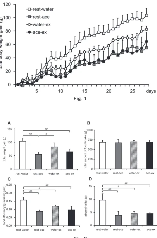
Body weight gain tended to be lower in rest-ace, water-ex, and ace-ex groups than that of rest-water group during experimental period (Fig. 1). Effects of treatments with acetic acid and exercise training on body weight gain, food intake, food efficiency, and abdominal fat content were shown in Fig. 2. The total body weight gain was significantly lower in ace and ace-ex groups than that of rest-water group. While, total amount of food intake was not significantly changed among 4 groups. Food efficiencies of rest-ace, water-ex, and ace-ex groups were significantly lower as compared with water group. Abdominal fat contents of rest-ace, water-ex, and ace-ex groups were significantly lower by about 60 %, 50 % and 50 %, respectively than that of the rest-water group.
Activation of AMPK by treatments of acetic acid and exercise training may increase lipid oxidation (2, 12). In order to determine whether acetic acid and exercise training change in the energy metabolic rate in the sedentary state, oxygen consumption was measured and RQ ratio was calculated (Fig. 3). Energy consumption rates in water-ex and ace-ex groups revealed significantly higher than that in rest-water group. Motor activity levels were significantly lower in ace, water-ex, and ace-ex groups than that in rest-water group.
In order to investigate the effect of acetic acid and exercise training on exercise capacity and fuel utilization during exercise, rats were started off running on a treadmill at 18 m/min, and then the speed was increased by the rate of 1 m/min
until exhaustion. Exercise capacity was shown to be increased in water-ex and ace-ex groups as compared with rest-water group (Fig. 4A). Running duration was significantly longer in water-ex and ace-ex groups than that in rest-water group (Fig. 4B).
To investigate the fuel utilization during exercise, VO2 and VCO2 were monitored simultaneously
until exhaustion (Fig. 5). Oxygen consumptions in all four groups were increased as speed increased until exhaustion. RQ ratios of all groups during
Fig. 2 Fig. 1
exercise were increased as the speed of treadmill increased, however the ratio in rest-ace and water-ex groups tended to be lower than that of rest-water group, and the RQ ratio in ace-ex group was significantly lower in 3, 9, 12 min after beginning of the run than that of rest-water group (Fig. 5). The calculated level of glucose oxidation during exercise tended to be higher in rest-water group, while glucose oxidation in ace-ex group was relatively lower, and it was significantly lower in
3 min and 12 min after start running than that of rest-water group (Fig. 6A). While, the calculated level of lipid oxidation during exercise tended to be higher in rest-ace, water-ex and ace-ex groups as compared with rest-water group, and it was significantly higher in water-ex and ace-ex groups in 9 min after start running than that in rest-water group (Fig. 6B).
To determine the effects of treatment of acetic acid and exercise training on mRNA levels related
Fig. 3
Fig. 5
to lipid metabolism in skeletal muscles, mRNA expressions of ppargc1a, cycs, myh4, and myh7 which were related to metabolism and muscle fiber types were analyzed by quantitative real-time RT-PCR. Transcript of PGC-1 α was tended to be increased in gastrocnemius muscle of ace-ex groups, whereas it was not changed by the treatment of acetic acid or excise in soleus muscle (Fig. 7A). Expressions of mRNA, cycs, myh4, and myh7 genes in gastrocnemius muscle were analyzed. Expression of cycs was tended to be increased in rest-ace and water-ex groups, and it was significantly induced higher in ace-ex group as compared with that in rest-water group (Fig. 7B). MHC1 expression, which is expressed much in slow twitch oxidative fiber, was significantly increased in rest-ace and ace-ex groups (Fig. 7C). On the other hand, the expression of MHC2b, which is expressed in fast twitch muscle fiber, was not changed among 4 groups (Fig. 7D).
Discussion
In our previous study, we showed that chronic intake of acetic acid induced gene expressions of myoglobin and GLUT4 and also increased energy consumption in rats (2). Orally administered acetic acid was taken up from the intestine, absorbed by liver and skeletal muscles, and it increased AMP/ ATP ratio in those cells (1, 2). An increase in the AMP/ATP ratio leads to the phosphorylation of AMPK. Treatment of acetic acid increased AMP/ ATP ratio and promoted phosphorylation of AMPK in skeletal muscle of rats. Physical exercise is well known factor to increase AMP/ATP ratio and to activate AMPK (11, 12). In this study, we examined the effects of acetic acid and exercise training on the endurance performance and lipid metabolism of rats during exercise. Treatments of chronic intake of acetic acid and exercise training led to the reduction of abdominal fat content and result in lower weight gain. Energy consumption in rats
treated with exercise training (water-ex and ace-ex groups) were higher, while, those motor activities were lower than control group, suggesting that energy consumption that may be derived from increased basal energy metabolism was stimulated by exercise training. Endurance performance was promoted in exercise training groups. During endurance exercise, glucose utilization was lower and lipid utilization was higher in rats treated with exercise training groups than that of ret-water group. These data indicate that the treatment both of chronic intake of acetic acid and exercise training increases fat oxidation and decrease glucose oxidation during endurance exercise and it is promoted in fiber type switching to oxidative fiber in gastrocnemius muscle.
Exercise training induce muscle remodeling and mitochondrial proliferation in skeletal muscle, resulting fiber-type switch from glycolytic to oxidative fibers and enhancing lipid oxidative capacity (18-21). Ppargc1a expression is increased in muscle by an acute exercise or long-term exercise training (22-27). Increased ppargc1a expression in muscle showed an enhanced ability to exercise and improvement of peak oxygen uptake (21). Furthermore, AMPK is also activated by exercise, and activated AMPK phosphorylates PGC-1 α, then its downstream genes targets are upregulated (13). In this study we observed that skeletal muscle of rats treated with acetic acid and exercise training induced mRNA expression of cytochrome c gene and tended to increase PGC1-α gene as compared with rest-water group. Exercise-trained rats showed an increase of exercise capacity, additionally with acetic acid treatment, glucose oxidation was reduced, and lipid oxidation was promoted during exercise tolerance test. Those results indicate that treatments both of acetic acid and exercise training would contribute to enhancement of lipid metabolism and improvement of exercise capacity, which may be through the activation of AMPK and PGC1-α. These treatments have a potential to prevent life-style related diseases and increase life span.
Acknowledgement
This work was supported by the JSPS KAKENHI Grant Number JP15K07437 for Grant-in-Aid for Scientific Research (C) and the JSPS KAKENHI under Grant Number JP26850087 for young scientist (B).
Figure legend
Figure 1, Total body weight gain.
Time course of body weight change of rats at 6 weeks of age administrated distilled water (rest-water) or acetic acid (rest-ace) during resting period or administrated distilled water (water-ex) or acetic acid (ace-ex) before exercise. Values are shown as arithmetic mean ± SE of 3 rats of each group. n=3
Figure 2, Effect of acetic acid ingestion on total weight gain, total amount of food intake, food efficiency, and abdominal fat content.
Total body weight gain (A), total amount of food intake (B), food efficiency (C), and abdominal fat content (D) of rats administered water or acetic acid during rest period and before exercise. Values are shown as arithmetic mean ± SE of 3 rats of each group. Statistical differences are shown as # P<0.05, ## P<0.01 by Tukey-Kramer’s post hoc test. n=3
Figure 3, Effect of acetic acid ingestion on oxygen consumption, RQ, energy consumption, and spontaneous motor activity in the sedentary state. Oxygen consumption and carbon dioxide production were monitored using on O2/CO2
metabolism measuring system for small animals in air tight chamber. The rats of energy consumption were calculated using energy consumption (kcal/ day/kg)= light cycle (kcal/h/kg)*12 + dark cycle (kcal/h/kg)*12 (A), oxygen consumption, (B), RQ, (C), energy consumption, and (D), spontaneous motor activity of rats.
Values are shown as arithmetic mean ± SE of 3 rats of each group. Statistical differences are shown as # P<0.05, ## P<0.01 by Tukey-Kramer’s
post hoc test. n=3
Figure 4, Effect of acetic acid ingestion on exercise tolerance.
Rats were administered water or acetic acid daily (5 days a week) for 4 weeks and then they were exercised by forced running on a treadmill at 18 m/min. The speed increased by 1 m/min every 1 min until exhaustion. Exhaustion is defined as touching on the shocker more than 10 times for 1 min. Exercise tolerance is shown as a Kaplan-Meier survival curve (A). Average running duration is shown in panel B. Values are shown as arithmetic mean ± SE of 4 rats of each group. Statistical differences are shown as # P<0.05 by Tukey-Kramer’s post hoc test.
Figure 5, Effect of acetic acid ingestion on the oxygen consumption and RQ during exercise tolerance test.
D u r i n g e x e r c i s e t o l e r a n c e t e s t , o x y g e n consumption and carbon dioxide production were monitored using on O2/CO2 metabolism measuring
system for small animals in air tight chamber. (A), Oxygen consumption. (B), RQ. Values are shown as arithmetic mean ± SE of 3-4 rats of each group. Statistical differences are shown as *P<0.05 **P<0.01 vs rest-water by student t-test. rest-water n=4, rest-ace n=4, water-ex n=3, ace-ex n=3
Figure 6, Effect of acetic acid ingestion on glucose and lipid oxidation during exercise tolerance test. Glucose (A) and lipid oxidation (B) during exercise tolerance test were calculated as described in “Materials and Methods”. Values are shown as arithmetic mean ± SE of 3-4 rats of each group. Statistical differences are shown as *P<0.05 **P<0.01 vs rest-water by student t-test. rest-water n=4, rest-ace n=4, water-ex n=3, ace-ex n=3
Figure 7, Effect of chronic intake of acetic acid and exercise training on gene expressions in skeletal muscles.
Quantitative RT-PCR analysis was performed
using total RNA isolated from gastrocnemius and soleus muscle and of rats administered distilled water or acetic aicd with sedentary or exercise training. (A), ppargc1a expression in gastrocnemius muscle and soleus muscle. (B-D), cycs, myh7, and myh4 expressions in gastrocnemius muscle. Values are shown as arithmetic mean ± SE of 3-5 rats of each group. Statistical differences are shown as # P<0.05 by Tukey-Kramer’s post hoc test.
References
1 )Yamashita H, Fujisawa K, Ito E, Idei S, Kawaguchi N, Kimoto M, Hiemori M, Tsuji H. 2007. Improvement of obesity and glucose tolerance by acetate in Type 2 diabetic Otsuka Long-Evans Tokushima Fatty (OLETF) rats. Biosci Biotechnol Biochem 71: 1236-1243.
2 )Yamashita H, Maruta H, Jozuka M, Kimura R, Iwabuchi H, Yamato M, Saito T, Fujisawa K, Takahashi Y, Kimoto M et al. 2009. Effects of acetate on lipid metabolism in muscles and adipose tissues of type 2 diabetic Otsuka Long-Evans Tokushima Fatty (OLETF) rats. Biosci Biotechnol Biochem 73: 570-576.
3 )Schmitt, M.G.JR., Soergel, K.H., and Wood, C.M. 1976. Absorption of short chain fatty acids from the human jejunum. Gastroenterology, 70, 211-215.
4 )Watson, A.J.M., Brennan, E.A., Farthing, M.J.G., and Fairclough, P.D., 1991.Acetate uptake by intestinal brush border membrane vesicles. Gut, 32, 383-385.
5 )Knowles SE, Jarrett IG, Filsell OH, et al. 1974. Production and utilization of acetate in mammals, Biochem. J. 142: 401-11
6 )Zydowo MM, Smoleński RT, Swierczyński J. 1993. Acetate-induced changes of adenine nucleotide levels in rat liver. Metabolism. 42: 644-8.
7 )Hardie DG, Sakamoto K. 2006. AMPK: a key sensor of fuel and energy status in skeletal muscle. Physiology 21: 48-60.
8 )Corton JM, Gillespie JG, Hardie DG. 1994. Role of the AMP-activated protein kinase in the
cellular stress response. Curr Biol 4: 315-324. 9 )Esumi H, Izuishi K, Kato K, Hashimoto K,
Kurashima Y, Kishimoto A, Ogura T, Ozawa T. 2002. Hypoxia and nitric oxide treatment confer tolerance to glucose starvation in a 5'-AMP-activated protein kinase-dependent manner. J Biol Chem 277: 32791-32798.
10 )Salt IP, Johnson G, Ashcroft SJ, Hardie DG. 1998. AMP-activated protein kinase is activated by low glucose in cell lines derived from pancreatic beta cells, and may regulate insulin release. Biochem J 335: 533-539.
11 )Hayashi T, Hirshman MF, Kurth EJ, Winder WW, Goodyear LJ. 1998. Evidence for 5' AMP-activated protein kinase mediation of the effect of muscle contraction on glucose transport. Diabetes 47: 1369-1373.
12 )Rasmussen BB, Winder WW. 1997. Effect of exercise intensity on skeletal muscle malonyl-CoA and acetyl-malonyl-CoA carboxylase. J Appl Physiol (1985) 83: 1104-1109.
13 )Jager S, Handschin C, St-Pierre J, Spiegelman BM. 2007. AMP-activated protein kinase (AMPK) action in skeletal muscle via direct phosphorylation of PGC-1alpha. Proc Natl Acad Sci U S A 104: 12017-12022.
14 )Fulco M, Cen Y, Zhao P, Hoffman EP, McBurney MW, Sauve AA, Sartorelli V. 2008. Glucose restriction inhibits skeletal myoblast differentiation by activating SIRT1 through AMPK-mediated regulation of Nampt. Dev Cell 14: 661-673.
15 )Canto C, Gerhart-Hines Z, Feige JN, Lagouge M, Noriega L, Milne JC, Elliott PJ, Puigserver P, Auwerx J. 2009. AMPK regulates energy expenditure by modulating NAD+ metabolism and SIRT1 activity. Nature 458: 1056-1060.
16 )Iwabu M, Yamauchi T, Okada-Iwabu M, et al. 2010 Adiponectin and AdipoR1 regulate PGC-1alpha and mitochondria by Ca(2+) and AMPK/ SIRT1. Nature. 464: 1313-1319
17 )Ferrannini E, 1988. The theoretical bases of indirect calorimetry: a review. Metabolism 37: 287-301.
18 )Chi MM, Hintz CS, Coyle, EF, Martin WH III, Ivy JL, Nemeth PM, Holloszy JO, Lowry OH. 1983. Effects of detraining on enzymes of energy metabolism in individual human muscle fibers. Am J Physiol Cell 244: C276-C278.
19 )Goodpaster BH, Katsiaras A, Kelley DE. 2003. Enhanced fat oxidation sensitivity in obesity. Diabetes 282: 2191-2197.
20 )Holloszy JO, Booth FW. 1976. Biochemical adaptations to endurance exercise in muscle. Annu Rev Physiol 38: 273-291.
21 )Calvo JA, Daniels TG, Wang X, Paul A, Lin J, Spiegelman BM, Stevenson SC, Rangwala SM. 2008. Muscle-specific expression of PPARg coactivator-1a improves exercise performance and increases peak oxygen uptake. J Appl Physiolo 104: 1304-1312.
22 )Baar K, Wende AR, Jones TE, Marison M, Nolte LA, Chen M, Kelly DP, Holloszy JO. 2002. Adaptations of skeletal muscle to exercise: rapid increase in the transcriptional coactivator PGC-1. FASAEB J 16: 1879-1886.
23 )Kuhl JE, Ruderman NB, Musi N, Goodyear LJ, Patti ME, Crunkhorn S, Dronamraju D, Thorell A, Nygren J, Ljungkvist O, Degerblad M, Stahle A, Brismar TB, Andersen KL, Sha AK, Efendic S, Bavenholm PN. 2006. Exercise training decreases the concentration of malonyl-CoA and increases the expression and activity of malonyl-CoA decarboxylase in human muscle. Am J Physiol Endocrinol Metab 290: E1296-E1303.
24 )Pilegaard H, Saltin B, Neufer PD. 2003. Exercise induce transient transcriptional activation of the PGC-1 alpha gene in human skeletal muscle. J Physiol 546: 851-858.
25 )Russell AP, Feilchenfeldt J, Schreiber S, praz M, Crettenand A, Gobelet C, Meier CA, Bell DR, Kralli A, Giacobino JP, Deriaz O. 2003. Endurance training in humans leads to fiber type-specific increases in levels of peroxisome proliferator-activated receptor-gamma coactivator-1 and peroxisome proliferator-activated receptor-alpha in skeletal muscle. Diabetes. 52: 2874-2881.
P. 2005. Regulation of metabolic transcriptional co-activatorfs and transcrition factors with acute exercise. FASEBJ. 19: 986-988.
27 )Short KR, Vittone JL, Bigelow ML, Proctor DN, Rizza RA, CoenenSchimke JM, Nair KS. 2003. Impact of aerobic exercise training on age-related changes in insulin sensitivity and muscle oxidative capacity. Diabetes. 52: 1888-1896.